BASIC RESEARCH
Zonguldak Karaelmas University, General Surgery – Turkey. [email protected]
Received for publication on October 03, 2007. Accepted for publication on October 25, 2007.
SODIUM NITROPRUSSIDE AS A NITRIC OXIDE
DONOR IN A RAT INTESTINAL
ISCHEMIA-REPERFUSION MODEL
Ali Emre, Orhan Bayram, Bulent Salman, Sevim Ercan, Ziya Anadol, Okhan Akin
Emre A, Bayram O, Salman B, Ercan S, Anadol Z, Akin O. Sodium nitroprusside as a nitric oxide donor in a rat intestinal ischemia-reperfusion model. Clinics. 2008;63(1):91-6.
AIM: The aim of this study was to investigate the efficacy of sodium nitroprusside in the reduction of the intestinal ischemia-reperfusion injury as a nitric oxide donor after intraperitoneal administration.
METHODS: The histopathological examinations and tissue malonyldialdehyde levels of 35 Wistar albino rats that were subjected to ischemia-reperfusion, were performed in 5 groups. The groups include Control, Ischemia -reperfusion, Sodium nitroprusside, NG-Nitro-L-Arginine Methyl Ester (L-NAME) and Sodium nitroprusside+L-NAME. Each rat was subjected to ischemia for 40 minutes and reperfusion for 30 minutes, except the control group. The medications were done intraperitoneally as saline 4 ml/kg, Sodium nitroprusside 5 mg/kg, L-NAME 10 mg/kg just before reperfusions.
RESULTS: Significant tissue injury in histological sections and an increase in tissue levels of Malonyldialdehyde was detected in the I/R group. The efficacy of intraperitoneal administration of Sodium nitroprusside in both Sodium nitroprusside alone and Sodium nitroprusside+L-NAME groups was found statistically significant for the reducing of injury scores (p<0.05). The difference between the Ischemia/reperfusion and Sodium nitroprusside groups was found statistically significant as in the Ischemia/reperfusion and Sodium nitroprusside+L-NAME groups due to the tissue Malonyldialdehyde levels (p<0.05). There was no statistical difference between Ischemia/reperfusion and L-NAME groups.
CONCLUSION: Ischemia/reperfusion induced injury might be reduced by the intraperitoneal administration of Sodium nitroprusside, even in the presence of L-NAME, in the rat intestinal model.
KEYWORDS: Ischemia. Reperfusion. Intestine. Sodium nitroprusside. Rat.
INTRODUCTION
It is a well known phenomenon that reperfusion is as dangerous for the tissues as ischemia, particularly in the heart and the intestines.1 Intestinal ischemia is a result of
systemic factors (hypovolemia, hypotension, hypoxia, sep-sis) or local factors (mechanical obstruction of the intes-tine or the intestinal vessels). The restored circulation re-sults in the formation of free oxygen radicals and other acute phase reactants. Resulting cellular death occurs via
the lipid peroxydation of the cell wall.2,3
The role of endogenous endothelin peptides in the in-testinal ischemia-reperfusion (I/R) model has been well studied.4 Although the exact mechanism is still not fully
understood, investigations on nitric oxide (NO), an endothe-lial-derived relaxation factor, revealed its importance in the management of I/R injury. 5,6 NO inhibits lipid peroxidation
via the lipophilic free radical effect.7 There are many
are the major products used.1,7
Sodium Nitroprusside (SNP) is another nitric oxide do-nor investigated for its intravascular and intraluminal ef-fects on ischemia reperfusion injury. We investigated the effects of NO in I/R injury with the commercially avail-able and inexpensive product sodium nitroprusside (SNP) administered intraperitoneally in a rat intestinal model.
MATERIALS AND METHODS
Ethics
This study was performed in the investigation labora-tories of Gazi University with the permission of the Gazi University Ethical Committee.
Study design
The study was conducted with 35 male Wistar albino rats weighing between 200-230 grams, divided into 5 groups. Rats were fed standard food until the last 12 hours of the study and were given water ad libitum. A midline incision was performed and the superior mesenteric artery was clamped for 40 minutes to create ischemia. Intestinal segments were taken into the abdominal cavity and a light source was used to avoid hypothermia. All medications were administered intraperitoneally (i.p) immediately be-fore removal of the superior mesenteric artery clamp, and the intestines were allowed to reperfuse for 30 minutes af-ter the abdominal wall was closed.
The groups are designed as:
In the SNP group, Sodium Nitroprusside (Nipruss, ADEKA) was given i.p. at 5 mg/kg; in the NG-Nitro-L
-Arginin Methyl Ester (L-NAME) group, L-NAME was given i.p. at 10 mg/kg (Sigma Chemical Co). In the SNP+L-NAME group, SNP and L-NAME were given i.p. at 5 mg/kg and 10 mg/kg, respectively
Intestinal tissue samples taken from the jejunum 30 minutes after the reperfusion were preserved in 10%
formaline solution for histopathological examinations and snap frozen in liquid nitrogen and then stored at –80°C for tissue MDA level determination.
Pathological examination
Tissue samples were examined under the light micro-scope after haemotoxylen-eosine (H&E) staining using the criteria reported by Park and Chiu.8 Briefly, [0] was
de-scribed as normal mucosa, [1] as subepithelial space at vil-lus tips, [2] as extension of subepithelial space with mod-erate lifting, [3] as massive lifting down sides of villi, with some denuded tips, [4] as denuded villi, dilated capillar-ies, [5] as disintegration of lamina propria, [6] as crypt layer injury, [7] as transmucosal infarction and [8] as transmu-ral infarction.
Tissue malonyldialdehyde (MDA) level
Tissue MDA levels were studied as reported by Uchiyama et al.9 Tissue samples were homogenized with
1.15% cold KCL, and then 1% phosphoric acid and 0.6% thiobarbituric acid were added sequentially. After boiling in a water bath for 45 minutes, n-buthanol was added. The material was centrifuged at 2500 rpm for 5 minutes and spectrophotometric determinations were performed at 535 and 520 nm. The results are given as nmol/gr of tissue.
Statistics
Tissue MDA levels and the results of the pathological examinations were subjected to the Kruskal-Wallis test. Post-hoc multiple comparison test (Bonferroni) was also performed in order to find significant differences between groups. Values of p<0.05 were accepted as significant.
RESULTS
There is a significant difference between groups accord-ing to Kruskal-Wallis variance analysis (p<0.001). The his-topathological scores are displayed in Table 1
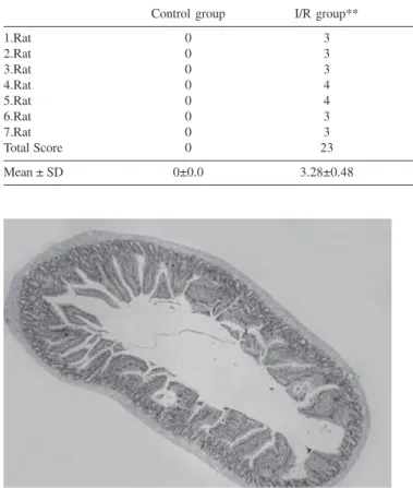
Control Group. The mean histopathological injury score in the control group is 0. The histological appear-ance of the control group is shown in Figure 1.
I/R Group. In the samples of the I/R group, the mean score of injury is 3.28 ± 0.48. Histopathological examina-tion reveals significant villous epithelial separaexamina-tion, lumi-nal epithelial seeding and naked subepithelial areas (Fig-ure 2).
SNP Group. In the SNP group, the mean injury score is 1.43 ± 0.53, and a contact epithelial lining, except with
Group 1 (Control) Laparatomy
Group 2 (I/R) Ischemia Saline Reperfusion
40 min. (4 ml/kg i.p.) 30 min
Group 3 (SNP) Ischemia SNP Reperfusion
40 min. (5 mg/kg i.p.) 30 min Group 4 (L-NAME) Ischemia L-NAME Reperfusion
40 min. (10 mg/kg i.p.) 30 min
Group 5 (SNP + Ischemia SNP Reperfusion
L-NAME) 40 min. (5 mg/kg i.p.) 30 min
some subepithelial spacings, is observed upon histopatho-logical examination (Figure 3).
L-NAME Group. In the L-NAME group, where endog-enous NO synthesis is thought to be inhibited, the mean score of injury is 3.28 ± 0.95, as in the I/R group. Similar to the I/R group, free mucosal cell particles can be seen in the lumen and epithelial layer separations are obvious (Fig-ure 4).
SNP+L-NAME Group. In the SNP+L-NAME group, the mean score of injury is 2.0 ± 0.81. Pathological exami-nation revealed an increase in the amount of subepithelial spaces and epithelial separations in this group (Figure 5).
Statistical analysis of the data with the Mann-Whitney U test shows that the I/R group is statistically significantly different than the SNP and the SNP+L-NAME groups
Table 1 - The histological injury scores according to the criteria of Park and Chiu.
Control group I/R group** SNP group* L-NAME group** SNP+L-NAME group*
1.Rat 0 3 2 2 3
2.Rat 0 3 2 4 3
3.Rat 0 3 1 2 2
4.Rat 0 4 1 4 1
5.Rat 0 4 1 3 2
6.Rat 0 3 1 4 2
7.Rat 0 3 2 4 1
Total Score 0 23 10 23 14
Mean ± SD 0±0.0 3.28±0.48 1.42±0.53 3.28±0.95 2.0±0.81
Figure 1 - Histological appearance of the control group (H&E x 40).
Figure 2 - Histological appearance of the I/R group. Villous epithelial separation, luminal epithelial seeding and naked subepithelial areas (H&E x100).
Figure 3 - Histological appearance of the SNP group. A contact epithelial lining except for some subepithelial spacings (H&E x 40).
(p< 0.05). The difference is not significant between either the I/R group and the L-NAME group or the SNP and the SNP+L-NAME group. There is also a statistically signifi-cant difference between the L-NAME group and the SNP group, and between the L-NAME group and the SNP+L-NAME group (p<0.05).
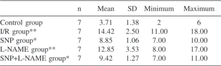
Tissue MDA levels are displayed in Table 2
In the Sham Group, the tissue MDA level is 3.71 ± 1.38 nmol/gr of tissue.
In the I/R Group, I/R injury to the intestinal tissues results in a significant increase in MDA levels. The mean MDA level in this group is 14.42 ± 2.50 nmol/gr of tissue.
In the SNP Group, the mean MDA level is significantly decreased to 8.85±1.06 nmol/gr of tissue. There is no dif-ference between the SNP and SNP+L-NAME groups, al-though the difference between the MDA levels of the SNP and L-NAME groups are again statistically significant (p<0.05).
In the L-NAME Group, the mean tissue MDA level is 12.85 ± 3.53 nmol/gr of tissue. There is no difference between the L-NAME and I/R groups.
In the SNP+L-NAME Group, the mean MDA level is 9.42 ± 1.27nmol/gr of tissue and the difference compared with the L-NAME group is statistically significant (p<0.05).
DISCUSSION
The deleterious effect of reperfusion on intestines and its exacerbation of the ischemic event is strongly sug-gested.10 The mechanism responsible for the injury caused
by reperfusion is the formation of free oxygen radicals and the oxidative stress generated by these reactive oxygen spe-cies. In fact, the mechanism of injury is not fully under-stood, as there are other factors involved such as activa-tion of phospholipase A2 and alteraactiva-tions of calcium flux. Chan et al. reported that third degree injury begins after only 15 mins of ischemia in the jejunum and after 60 mins in the ileum.11 Ischemia lasting more than 40 minutes can
cause irreversible damage in the small intestine. Kurose et al. reported that ischemia lasting over 30 minutes does not cause a change in reperfusion.12 In order to avoid such a
scenario, we used jejunal segments after 40 minutes of ischemia. As 40 minutes of ischemia is sufficient for cre-ating ischemic injury and is less than the time needed for irreversible transmural infaction, the maximum grade of injury was found to be 4 in all groups. In the I/R group, the mean level of injury was 3.28 ± 0.95, which is similar to the results reported by Kazez et al. with the same scor-ing system.13 Schoenberg et al. showed that ischemia itself
causes less injury within the same time period.3 Most of
the systems used for grading the injury were qualitative or semi-quantitative.8 Quadeckers et al. studied the different
grading methods and reported that the Park and Chiu method for grading injury has advantages over the others becasue it minimizes examiner-based differences in patho-logical examination and detailed structural aspects.8 Park’s
classification alone is not able to take the villous and crypt injury into account.8 Apoptosis is also a valuable area of
study for estimating tissue injury, and has become more popular in recent years.14
The histopathological results of the study are also sup-ported by the increase in the tissue MDA levels. The MDA levels of normal intestinal tissue in the sham group were found to be 3.71 ± 1.38 nmol/gr of tissue, which increased to 14.42 ± 2,50 nmol/gr of tissue in the I/R group. These results demonstrate the correlation of histological and bio-chemical markers of the tissue injury. Lipid peroxidation results from the reaction of reactive oxygen metabolites, especially the hydroxyl and hydroperoxyl radicals with the membrane bound polyunsaturated fatty acids with a loss of a carbon radical and its rearrangement for formation of a conjugated diene. This conjugated form reacts immedi-ately with oxygen to form peroxide radicals. Peroxide radi-cals initiate a chain reaction by removing a hydrogen atom from the other fatty acids. The end product of this reac-tion is tissue MDA and hydroperoxide.17
Table 2 - Tissue Malonyl dialdehyde levels of the groups (nmol/gr tissue).
n Mean SD Minimum Maximum
Control group 7 3.71 1.38 2 6
I/R group** 7 14.42 2.50 11.00 18.00
SNP group* 7 8.85 1.06 7.00 10.00
L-NAME group** 7 12.85 3.53 8.00 17.00
SNP+L-NAME group* 7 9.42 1.27 7.00 11.00
The spectrophotometric analysis of this tissue MDA is an important marker of in vivo lipid peroxidation. The other lipid peroxidation products used for the same purpose are conjugated dienes, endoperoxidases and hydrogen peroxi-dase. Many investigators used tissue MDA as a marker of lipid peroxidation in ischemic tissue,7,15,16 although there are
some limitations of the procedure. There are more specific assay methods including C11-BODIPY that may be stud-ied where available. Estimation of the tissue MDA react-ing with amine groups on the phospholipids by photomet-ric analysis of the fluorescent effect is the most frequently used method, as performed here. 18,19
Increasing the level of NO production is thought to de-crease the reperfusion injury of the post-ischemic intestine via the increase in the tissue perfusion or by protecting the mucosa from the high levels of the cytotoxic agent superoxide.20 Increasing the perfusion is an important
fac-tor, because dilution provides buffering of the acidic lamina propria of reperfused intestine and cleansing of the media from the toxins passing through the epithelium. Investiga-tions into the mechanism of the I/R effect on NO produc-tion on the post-ischemic intestine revealed that reperfusion following 20 minutes of occlusion of the superior me-senteric artery causes a decrease in nitrite/nitrate levels, which is explained by the inhibition or inactivation of the NOS.12 Increased production of superoxide in post-ischemic
tissue is also responsible for minimizing the effect of en-dothelium-derived NO. The half-life of NO is very short (about 6 seconds) and the basic mechanism of this short half-life is thought to be the reaction with superoxide, as the half-life of NO increases in samples treated with superoxide dismutase.21
As the endogenous production and the use of nitric ox-ide decreases after I/R, the importance of exogenous NO is better understood. Mason et al. reported the increase in free oxygen radicals after the decrease of tissue NO lev-els.22 Endogenous NO can inhibit or delay the injury caused
by the free oxygen radicals in the early periods of I/R. The results of the previous studies revealed that nitric oxide do-nors might cause vasodilatation and scavenge the free oxy-gen radicals, the major cause of injury in reperfusion in-jury. However, the function of NO is still not fully under-stood as NO reacts with superoxide anion to form peroxynitrite anion (ONOO2), a highly reactive oxidizing agent capable of causing tissue damage.
Payne and Kubes studied the permeability of intestines with I/R injury treated with SIN 1, CAS-754 and nitroprus-side, and reported a significant decrease in permeability
even in the presence of the NOS inhibitor L-NAME.20 NO
released by the donor compounds not only inactivates the superoxide radicals, the product of hypoxantine-xantine pathway, but also has a direct inhibitory effect on inflam-matory cells. The inhibitory effect of NO on neutrophils and mast cells occurs via the inhibition of neutrophil ad-hesion, migration and aggregation and blocking the hista-mine release from the mast cells.
SNP is a cost effective and easily accessible compound. In our study, intraperitoneal use of the compound resulted in a significant decrease in the level of tissue injury, as seen both histopathologically and by the changes in tissue MDA levels with respect to the control group. This significant decrease in both SNP and SNP+L-NAME groups convinced us that SNP administered intraperitoneally can supply enough NO concentration in the tissues. SNP acts not only by inactivating the superoxide radicals and suppressing the inflammatory response, but can also cause vasodilatation of the constricted vessels after ischemia. On the other hand, it should also be noted that SNP is a disodium salt of ni-troprusside (Na2[Fe(CN)5NO]·2H2O) with a central iron molecule surrounded by one nitric oxide and five cyanide ligands. Wang et al. presented evidence that SNP donates iron to cells. The increase in intracellular iron is another cause of oxidative injury.
L-NAME is a NOS inhibitor. It inhibits both Ca2+
de-pendent neuronal nitric oxide synthase (nNOS) and Ca2+
independent inducible nitric oxide synthase (iNOS).23 Luo
et al. reported that L-NAME reduces the I/R injury by in-hibiting the endogenous NO.24 Kurose et al. showed that
L-NAME causes the formation of platelet-leukocyte aggre-gates, and reported that decreased NO production caused an increase in aggregate formation.11 P-selectin specific
monoclonal antibodies were thought to be the main fac-tors of this aggregate formation. NO donors hinder plate-let-leukocyte aggregate formation by blocking P-selectin function on the I/R activated platelet surface.
We studied the L-NAME and L-NAME+SNP groups separately to compare the effects of exogenous NO. We did not find any difference neither between the I/R and L-NAME groups nor between SNP and SNP+L-L-NAME groups. This can be explained by a significant decrease in NO levels due to NOS inactivation and superoxide related NO inactivation after 40 minutes of ischemia followed by reperfusion.
REFERENCES
14. Shah KA, Shurey S, Green C. Apoptosis after intestinal ischemia-reperfusion injury: a morphological study. Transplantation. 1997;27;64:1393-1397.
15. Lai HS, Chen WJ, Chiang LY. Free radical scavenging activity of fullerenol on the ischemia-reperfusion intestine in dogs. World J Surg. 2000;24:450-454.
16. Lai HS, Chen Y, Chen WJ, Chang J, Chiang LY. Free radical scavenging activity of fullerenol on grafts after small bowel transplantation in dogs. Transplant Proc. 2000;32:1272-1274.
17. Nalini S, Mathan MM, Balasubramanian KA. Oxygen free radical induced damage during intestinal ischemia/reperfusion in normal and xanthine oxidase deficient rats. Mol Cell Biochem. 1993;124:59-66. 18. Czyrko C, Steigman C, Turley DL, Drott HR, Ziegler MM. The role of
reperfusion injury in occlusive intestinal ischemia of the neonate: malonaldehyde derived fluorescent products and correlation of histology. J Surg Res. 1991;51:1-4.
19. Otamiri TA. Influence of quinacrine on plasma malondialdehyde after small intestinal ischemia and reperfusion. Circ Shock. 1988;24:63-69. 20. Payne D, Kubes P. Nitric oxide donors reduce the rise in reperfusion-induced intestinal mucosal permeability. Am J Physiol. 1993;265 (1 pt 1):G189-95.
21. Wallace JL, Miller MJ. Nitric oxide in mucosal defence: a little goes a long way. Gastroenterology. 2000;119:512-520.
22. Mason RB, Pluta RM, Walbridge S, Wink DA, Oldfield EH, Boock RJ. Production of reactive oxygen species after reperfusion in vitro and in vivo: protective effect of nitric oxide. J Neurosurg. 2000;93:99-107. 23. Luo CC, Chen HM, Chiu CH, Lin JN, Chen JC. Effect of
N(G)-nitro-L-arginine methyl ester on intestinal permeability following intestinal ischemia reperfusion injury in a rat model. Biol Neonate. 2001;80:60-63.
24. Chen JC, Chen HM, Shyr MH, Fan LL, Chi TY, Chi CP, et al. Selective inhibition of inducible nitric oxide in ischemia-reperfusion of rat small intestine. J Formos Med Assoc. 2000;99:213-218.
1. Barie PS. Schemes against ischemia; solutions for reperfusion (injury)? Crit Care Med. 1999;27:684-685.
2. Horton JW, Walker PB. Oxygen radicals, lipid peroxidation, and permeability changes after intestinal ischemia and reperfusion. J Appl Physiol. 1993;74:1515-1520.
3. Schoenberg MH, Beger HG. Reperfusion injury after intestinal ischemia. Crit Care Med. 1993;21:1376-1386.
4. Anadol AZ, Bayram O, Dursun A, Ercan S. Role of endogenous endothelin peptides in intestinal ischemia-reperfusion injury in rats. Prostaglandins Leucot Essent Fatty Acids. 1998;59:279-283. 5. Nijkamp FP, Folkerts G. Nitric oxide: initiator and modulator. Clin Exp
Allergy. 1997;27:347-350.
6. Vallance P, Hingorani A. Endothelial nitric oxide in humans in health and disease. Int J Exp Pathol. 1999;80:291-303.
7. Van Ye TM, Roza AM, Pieper GM, Henderson J Jr, Johnson CP, Adams MB Inhibition of intestinal lipid peroxidation does not minimize morphologic damage. J Surg Res. 1993;55:553-558.
8. Quaedackers JS, Beuk RJ, Bennet L, Charlton A, oude Egbrink MG, Gunn AJ, et al. An evaluation of methods for grading histologic injury following ischemia/ reperfusion of the small bowel. Transplant Proc. 2000;32:1307-1310.
9. Uchiyama M, Mihara M. Determination of malonaldehyde precursor in tissues by thiobarbituric acid test. Anal Biochem. 1978;86:271-8. 10. Sies H, de Groot H. Role of reactive oxygen species in cell toxicity.
Toxicol Lett. 1992;64-65 Spec No:547-551.
11. Chan KL, Zhang XH, Fung PC, Guo WH, Tam PK. Role of nitric oxide in intestinal ischemia-reperfusion injury studied using electron paramagnetic resonance. Br J Surg. 1999;86:1427-1432.
12. Kurose I, Wolf R, Grisham MB, Granger DN. Modulation of ischemia/ reperfusion- induced microvascular dysfunction by nitric oxide. Circ Res. 1994;74:376-382