Received from the Hospital Lifecenter, Belo Horizonte, MG, Brazil.
1. Professor, Physician; Coordinator of the Serviço de Anestesiologia of Hospital Municipal Odilon Behrens; Responsible for the CET-HC-UFMG, Anesthesiologist of Hospital Lifecenter-BH
2. Anesthesiologist of Hospital Lifecenter-BH
3. Neurosurgeon; Chief of the Serviço de Neurocirurgia of Hospital Lifecenter-BH; Chief of the Serviço de Neurocirurgia of Santa Casa de Misericórdia de Minas Gerais
Submitted on August 1, 2010. Approved on January 4, 2011.
Corresponce to:
Dra. Walkíria Wingester Vilas Boas Rua São Romão, 343/701 Santo Antônio
30330120 – Belo Horizonte, MG, Brazil E-mail: [email protected] scientific article
Hydroelectrolytic Balance and Cerebral Relaxation with
Hypertonic Isoncotic Saline
versus
Mannitol (20%) During
Elective Neuroanesthesia
Walkíria Wingester Vilas Boas, TSA
1, Mirna Bastos Marques
2, Atos Alves
3Summary: Vilas Boas WW, Marques MB, Alves A – Hydroelectrolytic Balance and Cerebral Relaxation with Hypertonic Isoncotic Saline versus
Mannitol (20%) During Elective Neuroanesthesia.
Background and objectives: Cerebral relaxation during intracranial surgery is necessary, and hiperosmolar therapy is one of the measures used to this end. Frequently, neurosurgical patients have sodium imbalances. The objective of the present study was to quantify and determine cerebral relaxation and duration of hydroelectrolytic changes secondary to the use of mannitol versus hypertonic isoncotic solution (HIS) during neurosurgery.
Methods: Cerebral relaxation and hydroelectrolytic changes were evaluated in 29 adult patients before de beginning of infusion, and 30 and 120 minutes after the infusion of equiosmolar loads of approximately 20% mannitol (250 mL) or HIS (120 mL). The volume of intravenous fluids infused and diuresis were recorded. A p < 0.05 was considered significant.
Results: A statistically significant difference in cerebral relaxation between both groups was not observed. Although several changes in elec-trolyte levels and acid-base balance with mannitol or HIS reached statistical significance only the reduction in plasma sodium 30 minutes after infusion of mannitol, mean of 6.42 ± 0.40 mEq.L-1, and the increase in chloride, mean of 5.41 ± 0.96 mEq.L-1 and 5.45 ± 1.45 mEq.L-1 30 and 120 minutes after infusion of HIS, caused a transitory dislocation of serum ion levels from normal range. The mannitol (20%) group had a significantly greater diuresis at both times studied compared with HIS group.
Conclusions: A single dose of hypertonic isoncotic saline solution [7.2% NaCl/6% HES (200/0.5)] and mannitol (20%) with equivalent osmolar loads were effective and safe in producing cerebral relaxation during elective neurosurgical procedures under general anesthesia.
Keywords: Saline Solution, Hypertonic; Mannitol; Water-Electrolyte Imbalance; Neurosurgery.
[Rev Bras Anestesiol 2011;61(4): 456-468] ©Elsevier Editora Ltda.
INTRODUCTION
Cerebral relaxation is essential in anesthesia for intracranial surgery, mandatory in cases of intracranial hypertension, and of great interest for other neurosurgical approaches. It has been considered a neuroprotective measure as it can redu-ce surgical compression, local hypoperfusion, and redu-cerebral ischemia 1. After opening of the dura-mater intracranial
pres-sure (ICP) is virtually zero but a non-relaxed brain can reduce the working conditions of neurosurgeons 2. Administration of
osmotherapy at the onset of craniotomy before opening the
dura-mater is one of the interventions used to produce ce-rebral relaxation in elective neurosurgeries. Osmolality is the primary determinant of water movements through the intact blood-brain barrier (BBB), and it is predictable. If we incre-ase serum osmolality, normal brain tissue would dehydrate, and the cerebral volume, as well as the intracranial pressure, would be reduced 3. On the other hand, hyperosmotic therapy
produces hydroelectrolytic changes that may be a confoun-ding factor in the management of neurosurgical patients who frequently have water and sodium imbalances usually attribu-ted to cerebral salt wasting syndrome, inappropriate antidiure-tic hormone secretion, and diabetes insipidus 4. Mannitol has
become the traditional basis of hyperosmolar therapy 5.
Ho-wever, it can be associated with severe adverse effects such as intravascular volume depletion, rebound ICP elevation, and renal failure 6. Although it also has potential side effects,
hypertonic saline solutions (HS) have gained renewed inte-rest as an alternate therapy and recently have been use in neurosurgical patients 7. Several clinical studies comparing
the effects of mannitol and HS on intracranial pressure have suggested that HS is as effective as mannitol if not better for treating intracranial hypertension 8. The objective of the
versus mannitol (20%) in patients undergoing craniotomy for elective neurosurgical procedures.
MATERIAL AND METHODS
Cerebral relaxation and hydroelectrolytic balance after in-travenous administration of HIS [NaCl (7.2%) in HES (6%)] (Hyper HAES, Fresenius Kabi AG) or mannitol (20%) were evaluated in 29 adult patients undergoing elective craniotomy and cerebral aneurism clipping, arteriovenous malformations, or cerebral tumors. Both groups mannitol (20%) and HIS were composed of 17 and 12 patients respectively.
Exclusion criteria were as follows: age < 21 years, initial sodium < 130 mEq.L-1 or > 150 mEq.L-1, metabolic disorders,
treatment with hyperosmotic solution up to 24 hours before surgery, or history of heart or renal failure.
Total intravenous anesthesia with propofol (serum target of 2-4 µ.mL-1) and remifentanil (mean dose 0.15-0.35 µ.kg-1.
min-1) was the anesthetic technique of choice. All patients were
intubated and volume-controlled mechanical ventilation with a mixture of oxygen and room air was initiated. Cisatracurium was the muscle relaxant used. Standard monitoring included electrocardiogram, peripheral oxygen saturation (SpO2), cen-tral temperature (target 36-37°C), capnography (end-expira-tory carbon dioxide – ETCO2 – target of 35-40 mmHg),
invasi-ve blood pressure (mean arterial pressure – MAP – target of 70 mmHg), and diuresis.
Hypotension was treated with a vasopressor (bolus of 100 µg of phenylephrine). If necessary, inhalational anesthe-sia with sevoflurane, at a minimal alveolar concentration of 0.5-0.75, was added to manage hypertension. Normovolemia was maintained by the intravenous administration of 0.9% saline.
After incision of skin, the parameters evaluated were recor-ded; they included: hemodynamic variables (heart rate – HR, and MAP), central temperature, and laboratorial data (arterial blood gases, and plasma electrolytes, glucose, and osmolali-ty). Hypertonic isoncotic saline or mannitol (20%) was infused through a peripheral vein with an infusion pump at a rate of 360 mL.h-1 or 750 mL.h-1, respectively, for 20 minutes. The
total volume of 120 mL of HIS has an osmolar load close to 250 mL of mannitol (20%) 6. The same variables were then
evaluated and recorded 30 and 120 minutes after the end of the infusion of hyperosmolar therapy besides the intrave-nous volume administered and dieresis at the beginning of the hyperosmolar infusion, and 30 and 120 minutes after its termination.
Cerebral relaxation was evaluated by the same surgeon who was blind to the hyperosmolar therapy used after its ad-ministration upon opening of the dura-mater, in a four-point scale:
1) Perfect relaxation 2) Satisfactory relaxation 3) Firm brain
4) Swollen brain
The statistical software Graphpad PRISM, version 4.03, was used for statistical analysis. Gaussian distribution of va-riables was assessed by Shapiro normalcy test. Results were reported as mean ± standard deviation. ANOVA followed by Bonferroni test was used to compare the intragroup means, while the Student t test was used to compare intergroup me-ans. A p < 0.05 was considered significant.
RESULTS
A total of 29 patients were included in the study according to the inclusion and exclusion criteria to receive mannitol (20%) or HIS as hyperosmolar therapy aiming at achieving cerebral relaxation during elective neurosurgery.
In the mannitol (20%) group, 12 patients were schedule for elective craniotomy for cerebral aneurism, one for arte-riovenous malformation, and four for cerebral tumor. The de-mographic data of this group included: age = 44 ± 3.34 years, weight = 72 ± 2.81 kg, gender M/F = 8/9, ASA I/II = 4/13.
In the HIS group, six patients were scheduled for elective craniotomy for cerebral aneurism, one for arteriovenous mal-formation, and five for cerebral tumor. The demographic data of this group included: age = 49.5 ± 4.52, weight = 70.75 ± 3.81, gender M/F = 6/6, ASA I/II = 2/10.
Statistically significant differences in demographic data be-tween both groups were not observed.
MANNITOL (20%) GROUP
Changes in serum sodium concentration: Serum sodium le-vels showed a significant reduction 30 minutes after infusion of mannitol (20%) (p < 0.001), a mean of 6.42 ± 0.40 mEq.L-1.
After 120 minutes, serum sodium concentration increased (p < 0.01) when compared to its level 30 minutes after infu-sion, but it did not reach the levels prior to infusion of mannitol (20%) (p < 0.001) (Table I).
Changes in serum chloride concentration: Serum chloride levels showed a significant reduction 30 minutes after infusion of mannitol (20%) (p < 0.01), but it returned to baseline levels 120 minutes after infusion (p < 0.01) (Table I).
Changes in serum calcium levels: Serum calcium levels showed a reduction 30 minutes after infusion of mannitol (20%) (p < 0.01); 120 minutes after infusion, serum calcium concentration remained reduced when compared to baseline levels (p < 0.01) (Table I).
Changes in serum potassium levels: Serum potassium le-vels were increased by 0.45 ± 0.08 mEq.L-1 30 minutes after
infusion of mannitol (20%) (p < 0.01), and 120 minutes after infusion it remained elevated when compared to baseline le-vels (p < 0.01) (Table I).
Changes in BE (base excess), HCO3-2, AG (anion gap), PaCO2: Base excess 120 minutes after infusion of mannitol (20%) differed from baseline levels (p < 0.001) and from the le-vels 30 minutes after infusion (p < 0.01). A difference between the baseline level of BE and that 30 minutes after infusion was not observed. A significant reduction in HCO3-2 was observed
only 120 minutes after infusion of mannitol (20%) (p < 0.01). Mean levels of AG, as well as mean levels of PaCO2, did not
show a significant difference during the study, (Table I). Diuresis and intravenous volume administered intraope-ratively: Diuresis during the period immediately before the administration of mannitol (20%) until 30 minutes after admi-nistration was 376.5 ± 62.44 mL, and the intravenous fluid administrated (250 mL of 20% mannitol + 0.9% saline) in the same period was 516.7 ± 0.49 mL. Between 30 and 120 minutes after administration of mannitol (20%), diuresis was 598.7 ± 110.2 mL, while the intravenous volume (0.9% saline) administered was 1165 ± 122 mL. A positive correlation was observed between the volume of diuresis from the infusion of mannitol (20%) up to 30 minutes after infusion and the serum sodium concentration 30 minutes (r = 0.74, p = 0.0006) after administration. The intravenous volume infused between the beginning of the infusion of mannitol (20%) and the second data collection, 30 minutes after infusion, did not show a signi-ficant correlation with serum sodium, potassium, and chloride concentrations, but it showed a negative correlation with se-rum calcium levels (r = -0.65, p = 0.005) and a positive corre-lation with hemoglobin variation (r = -0.58, p = 0.02). On the other hand, the volume infused between 30 and 120 minutes after infusion of mannitol (20%) did not show a correlation with any changes in serum ion levels, but it did show a correlation with the volume of diuresis (r = 0.73, p = 0.006).
Cerebral relaxation according to the four-point scale: 13 patients had a score of 1 (perfectly relaxed brain) and four (23.5%), a score of 2 (satisfactory relaxed brain).
HYPERTONIC ISONCOTIC SOLUTION GROUP (7.2% NACL IN 6% HES)
Changes in serum sodium levels: Mean baseline sodium le-vels were 136.4 ± 0.64 mEq.L-1, with significant changes 30
minutes (140.9 ± 0.97 mEq.L-1, p < 0.01) and 120 minutes
(140.1 ± 0.97 mEq.L-1, p < 0.01) after infusion of HIS
(Ta-ble II). Serum sodium levels showed a mean increase of 4.56 ± 0.69 mEq.L-1 and 3.75 ± 0.90 mEq.L-1 30 and 120
mi-nutes after infusion respectively.
Changes in serum osmolality: Baseline serum osmolality was 292.3 ± 3.05 mOsm.kg-1, with significant changes 30
minutes (303.2 ± 3.2 mOsm.kg-1, p < 0.01) and 120
minu-tes (305.1 ± 2.89 mOsm.kg-1, p < 0.01) after infusion of HIS
(Table II). Serum osmolality showed a mean increase of 10.89 ± 1.62 mOsm.kg-1 and 11.5 ± 1.88 mOsm.kg-1 30 and
120 minutes after infusion, respectively.
Changes in other serum electrolytes: Mean baseline cal-cium serum levels were 1.08 ± 0.02 mmol.L-1, and a
signifi-cant change in serum levels 30 and 120 minutes after infusion of HIS was not observed (Table II). Mean potassium serum levels were 3.63 ± 0.11 mEq.L-1, and it showed a significant
change 30 minutes (3.80 ± 0.13 mEq.L1, p < 0.01) and 120
mi-nutes (4.06 ± 0.14 mEq.L-1, p < 0.01) after infusion (Table II).
Between 30 and 120 minutes after infusion of HIS, a signi-ficant difference in mean serum potassium concentrations were also observed (Table II). Mean baseline chloride serum levels were 109 ± 1.4 mEq.L-1, and they showed a significant
change 30 minutes (114.4 ± 1.19 mEq.L-1, p < 0.01) and 120
minutes (113.8 ± 1.7 mEq.L-1, p < 0.01) after infusion of HIS
(Table II).
Changes in hemoglobin concentration: Mean baseline he-moglobin concentration was 12.2 ± 0.59 g.L-1, with a significant
change 30 minutes (11.36 ± 0.52 g.L-1, p < 0.01) and 120
minu-tes (11.56 ± 0.50 g.L-1, p < 0.01) after infusion of HIS (Table II).
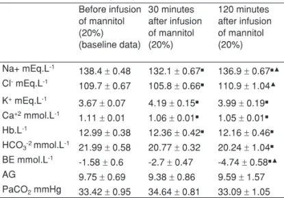
Table I – Changes in Hydroelectrolytic and Acid-Base Balance after Mannitol (20%)
Before infusion
of mannitol (20%) (baseline data) 30 minutes after infusion of mannitol (20%) 120 minutes after infusion of mannitol (20%) Na+ mEq.L-1
138.4 ± 0.48 132.1 ± 0.67■ 136.9 ± 0.67■▲ Cl- mEq.L-1
109.7 ± 0.67 105.8 ± 0.66■ 110.9 ± 1.04▲ K+ mEq.L-1
3.67 ± 0.07 4.19 ± 0.15■ 3.99 ± 0.19■ Ca+2 mmol.L-1
1.11 ± 0.01 1.06 ± 0.01■ 1.05 ± 0.01■ Hb.L-1
12.99 ± 0.38 12.36 ± 0.42■ 12.16 ± 0.46■ HCO3-2 mmol.L-1 21.99 ± 0.58 20.77 ± 0.32 20.24 ± 1.04■ BE mmol.L-1
-1.58 ± 0.6 -2.7 ± 0.47 -4.74 ± 0.58■▲ AG 9.75 ± 0.69 9.38 ± 0.86 9.59 ± 1.57 PaCO2 mmHg 33.42 ± 0.95 34.64 ± 0.81 33.09 ± 1.05 ■ p < 0.01 when compared to baseline data;▲ p < 0.01 when compared to 30 minutes after infusion.
Table II – Changes in Hydroelectrolytic and Acid-Base Balance after Hypertonic Isoncotic Saline Solution
Before infusion (baseline data) 30 minutes after infusion 120 minutes after infusion Osmolality
mOsm.kg-1 292.3 ± 3.05 303.2 ± 3,2
■ 305.1 ± 2.89■ Na+ mEq.L-1
136.4 ± 0.64 140.9 ± 0.90■ 140.1 ± 0.97■ Cl- mEq.L-1
109 ± 1.4 114.4 ± 1.19■ 113.8 ± 1.7■ K+ mEq.L-1
3.63 ± 0.11 3.80 ± 0.13■ 4.06 ± 0.14■▲ Ca+2 mmol.L-1
1.08 ± 0.02 1.04 ± 0.02 1.05 ± 0.02 Hb.L-1
12.2 ± 0.59 11.36 ± 0.52■ 11.56 ± 0.50■ HCO3-2 mmol.L-1 22.95 ± 0.53 21.63 ± 0.62 20.65 ± 0.76■ BE mmol.L-1
Changes in BE (base excess), HCO3-2, AG (anion gap), PaCO2: Mean baseline BE was -1.26 ± 0.67 mmol.L-1, sho-wing a significant change 30 minutes (-2.48 ± 0.81 mmol.L-1,
p < 0.01) and 120 minutes (-4.22 ± 0.81 mmol.L-1, p < 0.01)
after infusion of HIS (6%). A statistically significant differen-ce was also observed in BE 30 and 120 minutes after in-fusion of HIS (Table II). Mean baseline serum HCO3-2 was
22.95 ± 0.53 mmol.L-1, with significant changes 120 minutes
(20.65 ± 0.76 mmol.L-1, p < 0.01) after infusion of HIS (Table
II). Mean baseline AG was 7.97 ± 2.1 and a statistically sig-nificant difference 30 and 120 minutes after infusion of HIS was not observed (Table II). PaCO2 did not show a significant change during the study (Table II).
Diuresis and intraoperative intravenous volume admi-nistration: Diuresis during the period immediately before HIS administration and 30 minutes after administration was 127.5 ± 26.94 mL, while the intravenous volume administe-red (120 mL of HIS + 0.9% saline) in the same period was 504.2 ± 109.8 mL. Between 30 and 120 minutes after the ad-ministration of HIS the diuresis was 302.7 ± 83.06 mL, whi-le the intravenous volume administered (0.9% saline) was 1323 ± 192.4 mL. A negative correlation (r = -0.70, p < 0.01) was observed between the intravenous volume administe-red 30 and 120 minutes and BE 120 minutes after infusion of HIS.
Cerebral relaxation according to the four-point scale: 10 (83.4%) patients had a score = 1 (perfectly relaxed brain), while two patients (16.6%) received a score = 2 (satisfactorily relaxed brain).
MANNITOL (20%) GROUP VERSUS HS (7.2%) IN HES (6%) GROUP
Regarding cerebral relaxation the neurosurgeon did not con-sider the brain of any patient inadequate, and statistically significant differences between both groups were not obser-ved. Thirty minutes after hyperosmolar therapy a significant difference was observed in the direction of serum sodium and chloride changes between the mannitol (20%) and HIS groups (Table III). While a reduction was observed in serum
sodium and chloride levels in the mannitol (20%) group, there was an increase in the HIS group. During that time, although a significant increase in serum potassium levels was obser-ved in both groups, this increase was more significant in the mannitol (20%) group (Table III). One hundred and twenty minutes after hyperosmolar therapy, the direction in serum sodium changes remained different in both groups (Table III). Serum potassium levels remained elevated 120 minutes after administration of mannitol (20%) and HIS, but without a signi-ficant difference between both types of hyperosmolar therapy (Table III). The direction in serum chloride change was the same 120 minutes after administration of mannitol (20%) and HIS, but the increase was significant only in the latter group (Tables II and III); 30 and 120 minutes after hyperosmolar therapy, BE and HCO3-2 did not show differences between
both groups. The intravenous volume administered was not different between both groups, but diuresis was. The mannitol (20%) group had a significantly greater diuresis in both study times than the HIS group (Table III).
DISCUSSION
The present study results showed that hyperosmolar thera-py with HIS, composed of 7.2% NaCl in 6% HES (200/0.5), and mannitol (20%) is effective in producing cerebral relaxa-tion during elective neurosurgical procedures under general intravenous anesthesia. And although statistically significant differences in diuresis and serum electrolyte and acid-base changes are observed in the first two hours after the adminis-tration of both types of hyperosmolar therapies, and among them most of these changes are within or very close to normal range, they are apparently not clinically harmful.
The effects of hypertonic saline (HS) in the brain of pa-tients without intracranial hypertension have been investiga-ted in patients undergoing elective craniotomies for several surgical procedures 7,9,10. Using a cerebral relaxation scale
similar to the one we used, Gemma et al. 9 reported
satisfac-tory cerebral relaxation in all cases when different osmolar loads but similar volume of HS (7.5%) or mannitol (20%) were administered. On the other hand, Rozet et al. 10 observed a
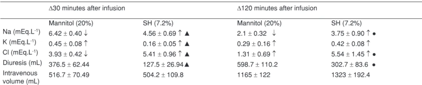
Table III – Variation in Hydroelectrolytic Balance after Mannitol (20%) and versus Hypertonic Isoncotic Saline Solution
∆30 minutes after infusion ∆120 minutes after infusion
Mannitol (20%) SH (7.2%) Mannitol (20%) SH (7.2%)
Na (mEq.L-1)
6.42 ± 0.40 ↓ 4.56 ± 0.69 ↑▲ 2.1 ± 0.32 ↓ 3.75 ± 0.90 ↑●
K (mEq.L-1)
0.45 ± 0.08 ↑ 0.16 ± 0.05 ↑▲ 0.29 ± 0.16 ↑ 0.42 ± 0.08 ↑ Cl (mEq.L-1)
3.93 ± 0.42 ↓ 5.41 ± 0.96 ↑▲ 1.31 ± 0.69 ↑ 5.54 ± 1.45 ↑●
Diuresis (mL) 376.5 ± 62.44 127.5 ± 26.94▲ 598.7 ± 110.2 302.7 ± 83.6 ● Intravenous
volume (mL) 516.7
± 70.49 504.2 ± 109.8 1165 ± 122 1323 ± 192.4
similar effect on cerebral relaxation when equiosmolar solu-tions of mannitol and HS were used. The main mechanism of action of hyperosmolar solutions is the generation of an osmolar gradient across the blood brain barrier (BBB), due to its impermeability to solutes (sodium and mannitol) 10,
lea-ding to contraction of cerebral tissue (were the BBB is intact) and therefore reducing the ICP. The effectivity of hyperos-molar solutes depends on their reflection coefficient, which determines the relative impermeability of BBB to the solute where 1 (one) means a solute for which the barrier is abso-lutely impermeable and 0 (zero) means a solute for which it is completely permeable 11. The reflection coefficient of the
membrane for sodium is 1 (different from the reflection coeffi-cient for mannitol, which is 0.9) 12. In the periphery (muscles,
lungs, and other tissues) the reflection coefficient of the en-dothelial membrane for sodium is only 0.1 meaning that the intracellular, and not interstitial, fluid represents the majority of fluid mobilized after HS administration 13. Thus, in the
peri-phery water movement from interstitial space to intravascular space is governed by the serum concentration of large mole-cules (oncotic gradient). In contrast with peripheral capillaries in cerebral capillaries (when the BBB is intact) the endothelial membrane is impermeable to sodium, and water movements across BBB is determined by the total osmotic gradient ge-nerated by both large molecules and small ions 14, while fluid
mobilization after the HS administration is from intracellular and interstitial spaces to intravascular space. Since there are very few protein molecules when compared to the number of inorganic ions, its effect on osmolality is minimal (normal POC ~ 20 mmHg ~ 1 mosm.kg-1) 14. These differences explain why
the administration of large volumes of isotonic crystalloids with dilutional reduction of colloid oncotic pressure (POC) results in peripheral edema, but it does not increase cerebral water content and/or ICP 14. When serum osmolality increases the
osmotic gradient drives water out of brain tissue. In our study a mean increase in serum osmolality of only 10.89 ± 1.62 and 11.5 ± 1.88 was measure 30 and 120 minutes, respectively, after infusion of HIS. Serum osmolality was always below the safe threshold proposed (320 mOsm.kg-1) during the use of
HS by the Brain Trauma Foundation 15. Although in our study
increases in osmolality with HIS were small, it has been des-cribed that even small changes (< 5%) can modify cerebral water content and ICP 16. Harutjunyan et al. 17, using 7.2%
NaCl/HES 200/0.5 (1.4 mL.kg-1) in the treatment of
intracra-nial hypertension observed an increase in serum osmola-lity from 284 (272-300) mOsm.kg-1 to 300 (284-319 mOsm.
kg-1), corresponding to reductions in ICP from 22 (19-31) to
15 (8-18) mmHg, and increases in cerebral perfusion pres-sure from 60 mmHg to 72 mmHg in neurosurgical patients. Schwarz et al. 18, in a study that evaluated HS and mannitol in
patients with elevated ICP after cerebral infarction, demons-trated that the serum osmolarity increased from a baseline level of 310.1 ± 5.1 mOsm.L-1 to 320.5 ± 4.6 mOsm.L-1 (after
15 minutes) and to 316.6 ± 4.8 mOsm.L-1 (after 60 minutes),
which correlated with a reduction in ICP. Therefore, although small, the changes in serum osmolality after HS in our study probably contributed to the perfect or satisfactory cerebral
re-laxation observed by the neurosurgeon. This study used 7.5% NaCl/6% HES (200/0.5) because HS concentrations above 10% can open tight junctions in BBB 13. The effect of HS
alo-ne is short-acting 13. With the addition of a colloid, its clinical
effect can be prolonged for 2-4 hours 13. In addition to
crea-ting an osmolar gradient across BBB, the reduction in CSF production, improvement in blood rheology, and anti-inflam-matory properties of HS and mannitol seem to have a role in bran therapy 10,18,19. An impaired BBB has the potential to leak
out osmotic substances into the brain parenchyma causing a reverse osmotic effect 13. The combination of HS and colloid
could increase concerns about its safe use. However, in elec-tive neurosurgeries BBB would be intact in most part of the brain. Austrian physicians have extensive experience with the use of a large number of solutions composed of 7.2% to 7.5% NaCl with dextran or 6% to 10% HES and for almost a decade their routine with these solutions indicate a low potential for complications 20. In theory the most serious obstacle in the
treatment with HS would be the development of neurologic complications secondary to osmotic demyelination syndrome (ODS) or central pontine myelinolysis (CPM). The literature on correction of hyponatremia in prospective animal studies and case reports in humans recommend that serum sodium should not be increased above 10-20 mEq.day-121. In our
stu-dy, we observed a mean increase in serum sodium levels of 4.56 ± 0.69, 30 minutes, and 3.75 ± 0.90, 120 minutes after administration of HIS (Table III). Besides, other human triages with HS have not observed very elevated and fast increases in serum sodium nor ODS 19. Patients have tolerated an
acu-te increase in serum sodium levels up to 155-160 mEq.L-1,
apparently without any harm 9,14. There is still the
hypothe-tic risk that acute brain dehydration could cause mechanical stretching of ligating blood vessels with the consequent su-barachnoid hemorrhage 13. Hypertonic isoncotic saline and
0.9% saline infused during our study are hyperchloremic 22,
which can increase serum chloride above normal levels such as observed in our study. HCO3-2 and BE were reduced 30
and 120 minutes after infusion of HIS, which has been ob-served in other studies 23, and in all of these most likely as a
consequence of the dilutional effects of hyperchloremic acido-sis. Hypertonic saline has a tendency to reduce the difference in strong ions leading to metabolic acidosis with normal AG (anion gap) 3. One hundred and twenty minutes after infusion
of HIS a negative correlation (r = -0.70, p < 0.01) was obser-ved between BE and the volume infused between 30 and 120 minutes; most likely, the large amount of 0.9% NaCl infused in this period produced dilutional effects or hyperchloremic acidosis without significant changes in AG. The impact that hyperchloremic acidosis could have on prognosis remains controversial and may produce diagnostic confusion 24.
Chan-ges in hemoglobin levels with HIS reached statistical signifi-cance, but which was not clinically significant, and they pro-bably were a consequence of HIS- and 0.9% NaCl-induced dilution. Considerable blood loss was not observed in any of the study groups.
neurosurgeries are directly related to intravascular content lution. Among these changes we should mention transient di-lutional hyponatremia which oftentimes can reach levels below the lower limit of normalcy was also reported in other studies10.
The correlation between hyponatremia and increased diuresis should not be forgotten in the period shortly after infusion of mannitol (20%), therefore avoiding diagnostic and therapeutic confusion. The diuretic effects of mannitol tend to normalize sodium serum levels, initially reduced, which would be de-monstrated by the presence of a positive correlation (r = 0.74, p < 0.01) between serum sodium levels 30 minutes after man-nitol administration and diuresis in that period. Sodium serum levels before 30 minutes after mannitol administration would, most likely, be even lower. Rozet et al. 10 demonstrated lower
sodium serum levels 15 and 30 minutes after mannitol admi-nistration. Changes in serum levels of chloride, calcium, and hemoglobin after mannitol administration are most likely dilutio-nal with longer lasting calcium and hemoglobin changes since solutions containing calcium of red blood cells were not admi-nistered. The development of hypokalemia with administration of HIS and mannitol has been reported 10,13,19,23,25, most likely
as a result of dilutional effects or urinary losses, but the exact mechanism is unknown. Our results revealed increased serum potassium levels, which was statistically significant, but without clinical significance both with mannitol and HIS. And the sug-gested mechanisms include outflow of cellular potassium with water as a result of hyperosmolar conditions; dilutional acidosis due to expansion of intracellular fluid and dilution of
bicarbona-te; or reduction in the difference of strong ions 25. In general,
electrolytic changes associated with the use of mannitol (20%) were short-lived, since 120 minutes after its infusion serum ions were within normal limits close to pre-infusion levels, although still statistically different. Even the hyponatremia observed 30 minutes after mannitol (20%) infusion was not present 120 mi-nutes later. Regarding the duration of electrolytic changes asso-ciated with HIS, 120 minutes after infusion hyperchloremia still persisted most likely related not only to the amount of chloride in HIS, but also the saline solution used in maintenance infusion and volemic replacement. Hypertonic saline and mannitol have similar neurologic effects, but the fact that HS does not produce an immediate diuretic effect, as observed in our study, simplifies the intraoperative management of fluids 14, although for Huang
et al. 23, HS is a diuretic 2.
A single dose of hypertonic isoncotic saline [7.2% NaCl/6% HES (200/0.5)] and mannitol (20%) with equivalent osmolar lo-ads were proven to be effective and safe in producing cerebral relaxation during elective neurosurgical procedures under ge-neral anesthesia. Although several differences in electrolytes and acid-base balance reached statistical significance with the use of mannitol or HIS, only the reduction in serum sodium le-vels, mean of 6.42 ± 0.40 mEq.L-1 30 minutes after the infusion
of mannitol, and an increase in chloride serum levels, mean 5.41 ± 0.96 mEq.L-1 and 5.45 ± 1.45 mEq.L-1 30 minutes and
01. Hans P, Bonhomme V – Why we still use intravenous drugs as the basic regimen for neurosurgical anaesthesia. Curr Opin Anaesthesiol, 2006;19:498-503.
02. Randell T, Niskanen M – Management of physiological variables in neuroanaesthesia: maintaining homeostasis during intracranial sur-gery. Curr Opin Anaesthesiol, 2006;19:492-497.
03. Tommasino C, Picozzi V – Volume and electrolyte management. Best Pract Res Clin Anaesthesiol, 2007;21:497-516.
04. Tisdall M, Crocker M, Watkiss J et al. – Disturbances of sodium in critically ill adult neurologic patients: a clinical review. J Neurosurg Anesthesiol, 2006; 18:57-63.
05. Kofke WA, Stiefel M – Monitoring and intraoperative management of elevated intracranial pressure and decompressive craniectomy. An-esthesiol Clin, 2007;25:579-603.
06. Himmelseher S – Hypertonic saline solutions for treatment of intracra-nial hypertension. Curr Opin Anaesthesiol, 2007;20:414-426.
08. Bentsen G, Breivik H, Lundar T et al. – Hypertonic saline (7.2%) in 6% hydroxyethyl starch reduces intracranial pressure and improves hemodynamics in a placebo-controlled study involving stable patients with subarachnoid hemorrhage. Crit Care Med, 2006;34:2912-2917. 09. Gemma M, Cozzi S, Tommasino C et al. – 7.5% hypertonic saline
versus 20% mannitol during elective neurosurgical supratentorial pro-cedures. J Neurosurg Anesthesiol, 1997;9:329-334.
10. Rozet I, Tontisirin N, Muangman S et al. – Effect of equiosmolar solu-tions of mannitol versus hypertonic saline on intraoperative brain re-laxation and electrolyte balance. Anesthesiology, 2007;107:697-704. 11. Tommasino C – Fluids and the neurosurgical patient. Anesthesiol Clin
North America, 2002;20:329-346.
12. Qureshi AI, Suarez JI – Use of hypertonic saline solutions in treat-ment of cerebral edema and intracranial hypertension. Crit Care Med, 2000;28:3301-3313.
13. Rocha-e-Silva M, Poli de Figueiredo LF – Small volume hypertonic resuscitation of circulatory shock. Clinics (Sao Paulo), 2005;60:159-172.
14. Strandvik GF – Hypertonic saline in critical care: a review of the litera-ture and guidelines for use in hypotensive states and raised intracra-nial pressure. Anaesthesia, 2009;64):990-1003.
15. The Brain Trauma Foundation. The American Association of Neu-rological Surgeons. The Joint Section on Neurotrauma and Critical Care. Guidelines for cerebral perfusion pressure. J Neurotrauma, 2000;17:507-511.
16. Tommasino C, Moore S, Todd MM - Cerebral effects of isovolemic hemodilution with crystalloid or colloid solutions. Crit Care Med, 1988;16:862-868.
17. Harutjunyan L, Holz C, Rieger A et al. – Efficiency of 7.2% hypertonic saline hydroxyethyl starch 200/0,5 versus mannitol 15% in the treat-ment of increased intracranial pressure in neurosurgical patients – a randomized clinical trial [ISRCTN62699180]. Crit Care, 2005;9:R530-R540.
18. Schwarz S, Schwab S, Bertram M et al. – Effects of hypertonic saline hydroxyethyl starch solution and mannitol in patients with increased intracranial pressure after stroke. Stroke, 1998;29:1550-1555.
19. Tyagi R, Donaldson K, Loftus C M et al. – Hypertonic saline: a clinical review. Neurosurg Rev, 2007;30:277-289.
20. Schimetta W, Schochl H, Kroll W et al. – Safety of hyperoncotic solu-tions: a survey from Austria. Wien Klin Wochenschr, 2002;114:89-95. 21. Sterns RH, Riggs JE, Schochet SS Jr – Osmotic demyelination
syn-drome following correction of hyponatremia. N Engl J Med, 1986, 314:1535-1542.
22. Morris CG, Low J – Metabolic acidosis in the critically ill: part 1. Clas-sification and pathophysiology. Anaesthesia, 2008;63:294-301.
23. Huang SJ, Chang L, Han YY et al. – Efficacy and safety of hypertonic saline solutions in the treatment of severe head injury. Surg Neurol, 2006;65:539-546.
24. Morris CG, Low J – Metabolic acidosis in the critically ill: part 2. Causes and treatment. Anaesthesia, 2008;63:396-411.
25. Flynn BC – Hyperkalemic cardiac arrest with hypertonic mannitol infusion: the strong ion difference revisited. Anesth Analg, 2007;104:225-226.
Resumen: Vilas Boas WW, Marques MB, Alves A – Equilibrio Hidro-electrolítico y Relajación Cerebral con Salino Isoncótico-Hipertónico
versus Manitol (20%) durante Neuroanestesia Electiva.
Justificativa y objetivos: La relajación cerebral es necesaria du-rante la cirugía intracraneana, y la terapia hiperosmolar es una de las medidas para su producción. Los pacientes neuroquirúrgicos a menudo presentan disturbios del sodio. El objetivo del trabajo fue cuantificar y determinar la relajación cerebral y la duración de las alteraciones hidroelectrolíticas provenientes del uso del manitol versus solución isoncótica hipertónica (SIH), durante la neuro-cirugía.
Método: Se evaluaron la relajación cerebral y las alteraciones hidro-electrolíticas de 29 pacientes adultos antes, 30 y 120 min después del término de la infusión de carga aproximadamente equiosmolar de manitol 20% (250 mL) o SIH (120 mL). Se registró el volumen de los líquidos intravenosos infundidos y la diuresis. El P < 0,05 fue considerado significativo.
Resultados: No hubo ninguna diferencia estadística significativa en-tre los dos grupos en cuanto a la relajación cerebral. Aunque varias diferencias en los electrólitos y el equilibrio ácido-básico con el uso de manitol o SIH, hayan alcanzado una significancia estadística, so-lamente la reducción del sodio plasmático 30 min después del uso del manitol, como promedio de 6,42 ± 0,40 mEq.L-1, y la elevación
del cloro como promedio 5,41 ± 0,96 mEq.L-1 y 5,45 ± 1,45 mEq.L-1,
30 y 120 min respectivamente después de la SIH, alteraron transito-riamente los niveles séricos de esos iones del rango de la normalidad laboratorial. El grupo del manitol (20%) tuvo una diuresis significati-vamente mayor en los dos tiempos estudiados en comparación con el grupo de la SIH.
Conclusiones: La solución salina isoncótica-hipertónica [NaCl 7,2%/ HES (200/0,5) 6%] y manitol (20%), en dosis única con carga osmo-lar equivalente, fueron efectivos y seguros para generar la relajación cerebral durante los procedimientos neuroquirúrgicos electivos bajo la anestesia general.