Acta Cir. Bras. 2018;33(10):924-934 DOI: http://dx.doi.org/10.1590/s0102-865020180100000007
Gémerson GabiattiI, Tomaz de Jesus Maria Grezzana-FilhoII, Carlos Thadeu Schmidt CerskiIII, Carlos
BofillIV, Stella ValleV, Carlos Otávio CorsoVI
Topical hepatic hypothermia associated with ischemic
preconditioning. Histopathological and biochemical
analysis of ischemia reperfusion damage in a 24 hour
model
1Abstract
Purpose: To develop a new 24 hour extended liver ischemia and reperfusion (LIR) model analyzing the late biochemical and histopathological results of the isolated and combined application of recognized hepatoprotective mechanisms. In addition, we used a new stratification with zoning to classify the histological lesion.
Methods: A modified animal model of severe hepatic damage produced through 90 minutes of segmental ischemia (70% of the organ) and posterior observation for 24 hours of reperfusion, submitted to ischemic preconditioning (IPC) and topical hypothermia (TH) at 26ºC, in isolation or in combination, during the procedure. Data from intraoperative biometric parameters, besides of late biochemical markers and histopathological findings, both at 24 hours evolution time, were compared with control (C) and normothermic ischemia (NI) groups.
Results: All groups were homogeneous with respect to intraoperative physiological parameters. There were no losses once the model was stablished. Animals subjected to NI and IPC had worse biochemical (gamma-glutamyl transpeptidase, alkaline phosphatase, lactate dehydrogenase, aspartate aminotransferase, alanine aminotransferase, direct bilirubin, and total bilirubin) and histopathological scores (modified Suzuki score) compared to those of control groups and groups with isolated or associated TH (p < 0.05).
Conclusion: The new extended model demonstrates liver ischemia and reperfusion at 24 hour of evolution and, in this extreme scenario, only the groups subjected to topical hypothermia, combined with ischemic preconditioning or alone, had better outcomes than those subjected to only ischemic preconditioning and normothermic ischemia, reaching similar biochemical and histopathological scores to those of the control group.
Key words: Ischemic Preconditioning. Ischemia. Reperfusion. Liver. Hypothermia. Rats.
IFellow PhD degree, Postgraduate Program of Surgical Sciences, Universidade Federal do Rio Grande do Sul (UFRGS),
Porto Alegre-RS, Brazil. Conception and design of the study, technical procedures, acquisition and analysis of data, manuscript writing.
IIPhD, Liver Transplantation Surgeon, Hospital de Clinicas de Porto Alegre (HCPA), Department of Surgery, UFRGS, Porto
Alegre-RS, Brazil. Conception and design of the study, analysis of data, manuscript writing.
IIIPhD, Associate Professor, Department of Pathology, UFRGS, Porto Alegre-RS, Brazil. Histopathological examinations. IVGraduate student, Faculty of Medicine, UFRGS, Porto Alegre-RS, Brazil. Technical procedures, acquisition of data. VCoordinator, Laboratory of Veterinary Clinical Analysis (LACVet), Porto Alegre-RS, Brazil. Acquisition of data.
VIPhD, Associate Professor, Postgraduate Program of Surgical Sciences, Department of Surgery, UFRGS, Porto Alegre-RS,
either method alone in an LIR model with
extreme injury, monitoring intraoperative
physiological parameters19 and their possible histopathological20 and biochemical21 consequences measured at 24 hours of
reperfusion after ischemic insult. The application of TH, alone or in conjunction with IPC, has already been evaluated by our group in an acute animal model with partial 90-minute liver ischemia followed by 120 minutes of
reperfusion and immediate euthanasia, with
the demonstration of its protective effects through the reduction of inflammatory markers such as tumor necrosis factor-α (TNF-α) and interleukin (IL)-1, IL-6, and IL-12; an increase in anti-inflammatory cytokines such as IL-10; increased levels of nitric oxide; and elevated expression of Nrf2, Keap1, and NQO1 genes; all are related to antioxidant effects22-24.
Several modifications have been made to the original partial ischemia model,
especially related to the anesthesia method,
surgical incision, and intra- and post-operative
care, thus allowing an experimental model
with extended observation time of 24 hours of
reperfusion, enabling the analysis of the late
effects of different cytoprotective methods (IPC and TH). The combined application of IPC and TH in a model with this time duration, both
with ischemia and reperfusion, has not been
described to date; however, it is fully justified considering the therapeutic plateau reached regarding the pursuit of proven effective options related to the control of experimental LIR. It should be noted that it carries a greater likelihood of clinical applicability, possibly the critical point of the current studies related to LIR, namely the application and reproduction of
experimental studies in clinical care models25-27.
■
Methods
This study, identified by number 08486, was approved by the Bioethical Committee
■
Introduction
Liver ischemia and reperfusion (LIR)
has been the subject of intense study in
recent decades. LIR is involved in many
healthcare scenarios, including trauma,
hemorrhagic shock and resuscitation, septic shock, cardiac failure, respiratory insufficiency,
hepatectomy and liver transplantation1.
With the introduction of surgical control techniques of the hepatic vascular pedicle, today eventually still necessary during hepatic
surgical procedures2, ischemia and reperfusion
have been considered key elements in postoperative morbidity and mortality since severe complications such as liver dysfunction
and/or failure can result from this reperfusion process and injure other organs and systems3.
The major mechanisms of such injuries are the generation of reactive oxygen species (ROS) and reactive nitrogen species (RNS)4 together with the infiltration of polymorphonuclear
leukocytes among other mechanisms of
cellular injury5. In normothermic conditions,
the liver can tolerate clamping of the vessels of
its pedicle for approximately 1 hour when the
liver parenchyma is normal, whereas additional protection is required with diseased livers6.
To minimize the effects of LIR, several
methods have been studied7-9, including
ischemic preconditioning (IPC)10,11 and
topical hypothermia (TH)12. The reduction of
oxidative stress with the application of IPC is multifactorial13,14, with the activation of specific
genes, production of antioxidant enzymes and production of nitric oxide, the latter being a potent vasodilator with important antagonist effects during the ischemia
phase15,16. Hypothermia achieves its objectives
by inducing hypometabolism via suppressing mitochondrial activity, as an essential part of its cytoprotective effect17,18.
and funded by the Post-Graduation Group of the HCPA-UFRGS, which adheres to the rules of the Council for International Organization of Medical Sciences (CIOMS).
Thirty-three male Wistar rats (mean weight, 279g; mean age, 65 days) were divided into five groups: control (C; n = 5), normothermic ischemia (NI = 7), topical hypothermia at 26ºC (TH; n = 7), ischemic preconditioning (IPC; n = 7), and ischemic preconditioning + topical hypothermia at 26ºC (IPC+TH; n = 7).
Anesthesia protocol
The animals were subjected to inhalation anesthesia administered through a
facial cannula connected to a respirator, which
maintained a constant oxygen flow of 0.5 L
per minute and a mixture of anesthesia with
isoflurane 1–2%. The upper portion of the abdomen and the right cervical region were trichotomized and prepared with aqueous iodine solution.
Monitoring was performed through
a catheter installed in the right carotid artery (PE-50; Portex, Kent, England) to control the mean blood pressure (MBP) during the entire procedure. In addition, oximetry was
performed through an external sensor applied to the animal`s tail, that enabled also the
recording of heart rate (HR). Saline solution (NaCl 0.9%, 2 mL/kg/h) was continuously infused into the carotid artery to replace the basal needs, compensate for hydroelectrolytic losses and maintain catheter patency. The body
temperature was monitored by a rectal digital
thermometer and maintained at 36–37.5ºC with the aid of a heating device of the surgical table and by external lamp heating.
Surgical procedure
Access to the abdominal cavity was attained through a median laparotomy. In group C, the hepatic lobes were isolated
without any additional procedure. In the
groups with ischemia (NI, IPC, TH, and IPC+TH), the liver was isolated in the operative field and an atraumatic neurosurgical clip was used to interrupt the portal and arterial blood flow to the left and middle lobes (70% of the hepatic mass) for 90 minutes, while the right and caudate lateral lobes remained uninterrupted. In the groups with IPC, the left and median lobes of the liver were isolated in the operative field and submitted to 10 minutes of ischemia,
followed by reperfusion for another 10 minutes
before the final clamping for 90 minutes. In the hypothermia groups, the hepatic lobes were isolated in the operative field using a concha-like thermo-insulating styrofoam device and cooled with cold physiological solution dripped onto the surface of the ischemic lobes during the 90-minute ischemia period. The excess liquid was removed by aspirators, preventing the freezing liquid from penetrating the abdominal cavity and causing
systemic hypothermia in the animals, similar
to the model developed by Biberthaler et al.28. A flexible microprobe (TI-23; Braintree
Scientifics) was implanted into the left lobe and the intrahepatic temperature (IHT) was monitored continuously to reach 26ºC locally (Thermalert Monitoring Thermometer TH-5; Boston Scientifics). In the other groups, IHT was also recorded at the same insertion point and with the same measurement duration.
During the entire procedure, at 15-minute intervals, vital physiological parameters such as HR, MBP, respiratory rate (RR), systemic temperature (ST), and intrahepatic temperature (IHT) were recorded. In all groups, the same parameters were recorded 1 minute after declamping of the pedicle (91-minute procedure). The percentage of isoflurane in the inhalational anesthetic mixture was also recorded during
Post-anesthesia procedures and sampling
After 90 minutes of ischemia, the metal
clips of the pedicle were removed, and the organ was reperfused. The abdominal wall was closed, the surgical incision infiltrated with 0.1
mL of 0.5% bupivacaine solution in 0.9 mL of
saline solution, and the animal was transferred to an incubator for anesthesia recovery.
At the end of 24 hours, the rats were anesthetized again with isoflurane inhalation as described in the previous procedure. The abdomen was opened and the animals were sacrificed by cardiac puncture and exsanguinating blood aspiration. The liver was removed and stored; a fraction was fixed in formalin and another fraction was frozen in nitrogen and kept in a freezer at -80ºC. The
blood was immediately centrifuged and the
plasma obtained frozen in liquid nitrogen and stored in a freezer at -80ºC. The liver samples were identified from the area in which they originated in each experiment: ischemic (IL), belonging to the intervention zone (left lobe and median), and non-ischemic (NIL), belonging to the hepatic segment that was protected
against ischemia by pedicular clamping and the
application of hypothermia (right lobe).
Plasma analysis
Serum biochemistry of the stored
material was performed by the certified and specialized laboratory of the Faculty of Veterinary Medicine of the UFRGS using a WIENER-CM200 with imported reagents specific for the animal used (WIENER and RANDOX). The levels of aspartate aminotransferase (AST), alanine aminotransferase (ALT), ammonia (AMO), total bilirubin (TB) and direct bilirubin (DB), alkaline phosphatase (AP), gamma-glutamyl transpeptidase (GGT), glucose (GLU), lactate (LAC), and lactate dehydrogenase (LDH) were assessed.
Histopathological analysis
The liver samples, divided into IL and NIL from the same animal, were fixed in 10% formalin and stained with hematoxylin-eosin. The blinded evaluation of each specimen was performed by the pathology service at the Hospital de Clínicas de Porto Alegre using an optical microscope and assigning a value to the morphological changes observed following the original model proposed by Suzuki et al.29 to
examine the findings of sinusoidal congestion (SC), hydropic degeneration (HD), neutrophilic leukocyte infiltrate (NLI), and hepatocellular necrosis. The rating of steatosis (S) was
added to these parameters and the analysis
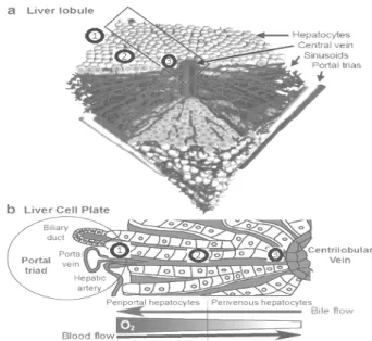
in the necrosis area was improved (Modified Histopathological Classification of Suzuki [MHCS]), performed in a stratified way using liver zoning (Figure 1)30,in which each lobe is
divided into three zones: zone 1 (NZ1), starting from the area next to the triad portal; Zone 2 (NZ2), passing by the sinusoidal intermediate region; and Zone 3 (NZ3), in the region near the central lobular vein (Table 1).
Figure 1 - Schematic representation of
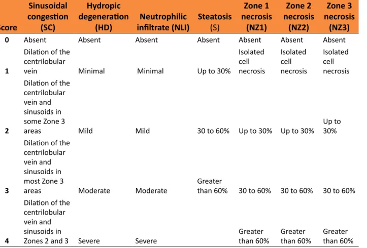
Table 1 - Modified Histopathological Classification of Suzuki (MHCS).
Score
Sinusoidal
congestion
(SC)
Hydropic
degeneration
(HD)
Neutrophilic
infiltrate (NLI) Steatosis(S)
Zone 1 necrosis
(NZ1)
Zone 2 necrosis
(NZ2)
Zone 3 necrosis
(NZ3)
0 Absent Absent Absent Absent Absent Absent Absent
1
Dilation of the
centrilobular
vein Minimal Minimal Up to 30%
Isolated cell necrosis
Isolated cell necrosis
Isolated cell necrosis
2
Dilation of the
centrilobular
vein and
sinusoids in
some Zone 3
areas Mild Mild 30 to 60% Up to 30% Up to 30%
Up to 30%
3
Dilation of the
centrilobular
vein and
sinusoids in
most Zone 3
areas Moderate Moderate
Greater
than 60% 30 to 60% 30 to 60% 30 to 60%
4
Dilation of the
centrilobular
vein and
sinusoids in
Zones 2 and 3 Severe Severe Greater than 60% Greater than 60% Greater than 60% Note: MSS, modified Suzuki score; SC, sinusoidal congestion;
HD: hydropic degeneration; NLI, neutrophilic leukocyte infiltrate; NZ1, necrosis of Zone 1; NZ2, necrosis of Zone 2; NZ3: necrosis of Zone 3.
The final histological score (Modified Suzuki Score [MSS] was calculated using Formula 1:
All data are presented as mean and standard deviation and were analyzed statistically using the Statistical Package for Social Sciences (SPSS) version 17 and Statistical Analysis System (SAS) version 9.4 assuming a significance of 0.05 for the tests employed (mixed-model repeated-measures analysis of variance [ANOVA], generalized linear model with a gamma distribution to evaluate the
effect of groups and Kruskal-Wallis test for independent samples).
■
Results
Intraoperative biometric data
The analysis of intraoperative vital signs (mixed-model repeated-measures ANOVA)
confirmed the homogeneity of the parameters
other parameters between the intraoperative physiological data of the different groups. The percentage of isoflurane anesthetic mixture
remained stable between groups in the
different assessed moments without intergroup differences. The variables of weight, age, and
surgical room temperature also showed no
significant intergroup differences.
Biochemical analysis of plasma after 90 minutes of ischemia and 24 hours of reperfusion
The analysis of biochemical data
performed through the adjustment of a
generalized linear model with a gamma distribution to evaluate the effect of groups (for asymmetric data) revealed significant differences (p<0.05) in serum GGT, AP, LDH, AST, ALT, DB, and TB (Figures 2 and 3) for the IPC and NI groups. AMO, LAC, and GLU levels
were not significantly different between
groups.
Figure 2 - Levels of DB, TB and GGT in 24-hour plasma
for all groups: Control (C), Normothermic Ischemia (NI), Ischemic Preconditioning (IPC), Topical Hypothermia (TH), and Ischemic Preconditioning + Topical Hypothermia (IPC+TH). Data are presented as mean and standard deviation converted to log 10. *Generalized linear model with gamma distribution to assess the group effect revealed p<0.05 for NI and IPC groups.
Figure 3 - Levels of AP, LDH, AST and ALT in 24-hour
plasma for all groups: Control (C), Normothermic Ischemia (NI), Ischemic Preconditioning (IPC), Topical Hypothermia (TH), and Ischemic Preconditioning + Topical Hypothermia (IPC+TH). Data are presented as mean and standard deviation. *Generalized linear model with gamma distribution to assess the group effect revealed p <0.05 for NI and IPC groups.
Histological analysis after 90 minutes of ischemia and 24 hours of reperfusion
The analysis of the histopathological
data (through adjustment of the generalized linear model with a gamma distribution to evaluate the effect of groups) revealed significant differences (p<0.05) for the MSS in IL samples of the NI and IPC groups (Figure
4) compared to those of the other groups.
Comparing individual items of the MSS among
the groups, using the Kruskal-Wallis test for independent samples, significant differences
were observed (p<0.05) for items NZ2 and NZ3 and for NLI in IL samples of the NI group.
The other individual aspects of histopathological scores evaluated (SC, HD, NZ1, and S) showed no significant differences between the IL specimens. Under the point of view of analysis of the method applied in the experiment, the comparative analysis of the NIL of all groups showed no statistically significant differences in any comparisons.
When analyzed pairwise, the MSS between samples of the IL and NIL zone
statistical significance for groups NI (p = 0.028) and IPC (p = 0.046), but in groups C (p = 1.0), TH (p = 0.75), and IPC+TH (p = 0.470) there were no significant differences between the IL and NIL specimens.
Figure 4 - MSS for ischemic liver samples of all
groups: Control (C), Normothermic Ischemia (NI), Ischemic Preconditioning (IPC), Topical Hypothermia (TH), and Ischemic Preconditioning + Topical Hypothermia (IPC+TH). Data are presented as mean and standard deviation. *Generalized linear model with a gamma distribution to assess the group effect revealed p<0.05 for NI and IPC groups.
■
Discussion
Here we evaluated the effects of LIR monitored during its implementation through intraoperative physiological parameters and 24 hours after the initial insult with assessments of
biochemical and histopathological parameters
using different combinations of protective mechanisms (TH, IPC, IPC+TH), associating the principle of zoning in hepatic histopathological analysis (Formula 1, Table 1). Given the already
expected serious injury produced by 90 minutes
of liver ischemia in this type of model, the
obtained biochemical and histopathological
findings indicated that, in this extreme scenario, only the animals subjected to TH, with or without the application of IPC, displayed histopathological findings with obvious cytoprotection compared to those in the other groups (Figure 4). Animals subjected to NI with
or without IPC displayed worse results in the
MSS compared to the animals subjected to TH, with or without IPC. Considering the findings of biochemical analyses in relation to the findings of the MSS in each group, the ALT, AST, DB, TB, LDH, AP, and GGT levels were different in groups C, TH, and IPC+TH compared to those in groups NI and/or IPC, i.e., higher MSS scores
were associated with higher biochemical
alterations of these markers.
No references were found in the
literature with an LIR model that combined 90 minutes of ischemia associated with
the application of hypothermia and/or preconditioning and subsequent evolution of 24 hours during the reperfusion phase. So we can only infer some considerations of our findings and those available in other LIR
models in which the ischemia phase and/or
the reperfusion phase and/or the protective mechanisms tested were distinct and/or occurred at different times. In an IPC model
associated with 90 minutes of IR associated
with the combined prior infusion of several
substances, Peralta et al.31 reported a protective
effect of IPC in the liver and the lungs. In an IPC model consisting of 60 minutes of ischemia
and up to 24 hours of reperfusion, Jin et al.32
showed a protective effect of IPC with reduced enzyme levels, a reduction in apoptosis, and improved NLI. In an IPC model consisting of 60 minutes of ischemia and 3 hours of reperfusion, Adam et al.33 also demonstrated
a beneficial effect of IPC with elevated levels
of hepatic glycogen compared to the group
(minocycline) with 90 minutes of ischemia and
up to 24 hours of reperfusion, Li et al.34 showed
a peak of enzymatic activity (AST, ALT, LDH) at 6 hours of reperfusion, marked histopathological changes from 6 hours of reperfusion, a progressive decline in these markers after 24 hours of evolution and a statistically significant cytoprotective effect in the group subjected to IPC. In our study, biochemical markers including bilirubin (DB and TB), transaminases (ALT and AST), alkaline phosphatase (AP), and GGT indicated significant organ lesions in the normothermic groups (NI and IPC) compared to those of the hypothermic groups (TH and IPC+TH) (Figures 2 and 3).
When analyzed separately, in this specific model, the mechanism of IPC was ineffective at promoting hepatic cytoprotection
since the scores of animals subjected to IPC
were similar to those of the NI group, through
the histopathological assessment and the
intraoperative biochemical and physiological effects. Similar findings were described by Steenks et al.35 in an animal model comparing
IPC with intermittent ischemia in steatotic livers, in which the authors described a smaller protective effect of IPC compared to that of intermittent or continued ischemia and argued that such findings might be explained by the negative effects of cytokines and other inflammatory molecules released by
the ischemic lobe subjected to IPC onto the
untreated lobes. Chu et al.36, in a systematic
review, described conflicting findings of some studies that evaluated the effect of IPC on steatotic livers submitted to LIR, suggesting that future experimental studies should attempt to better emulate the usual clinical scenario to provide an accurate understanding of the mechanisms and effects of IPC and identify a particular group of patients that can benefit from IPC. The scenario in which this method is most effective, likely does not include ischemia periods as long as the one used in this study.
It must be also highlighted that randomized studies in humans have failed to demonstrate these beneficial effects of IPC identified in
experimental models37,38.
The MHCS and the MSS, variants developed from the initial proposal of their
authors, were used in this project to grade
and compare the histopathological findings, permitting, with this expansion of its original
indicators, especially those related to the
division of necrotic areas in zones from the prior knowledge concerning the functional zoning of
this organ, the obtaining of more accurate data
in relation to the effects of the LIR and each protective mechanism tested, thus calculating a more comprehensive and more detailed injury score upon separate analysis of the different histological areas of necrosis. Originally, the table of Suzuki et al.29considered a single value for the item of necrosis and did not include the
degree of S. There was no significant finding for NZ1 in all groups, but the findings for NZ2 and NZ3 in isolation are significant for group NI. The same significant finding alone in Group NI is repeated with the NLI item of the score. It is speculated that the lack of significance for the findings of NZ1 in this study was due to the proximity of this zone with the triad portal, a zone with greater tissue perfusion.
The complementary analyses of the intraoperative physiological parameters of different groups (p > 0.05) between the NIL samples of different groups (p > 0.05) and the paired analysis between the IL and NIL samples within each group (p < 0.05) using the MHCS and MSS scores, making each animal as
a control of oneself and the others, highlight the methodological rigor employed since the
LIR model28, especially related to the anesthetic
method, as already reported by Steenks et al.35,
in addition to the surgical incision and intra- and postoperative care, allowed for a viable
extended LIR experimental model with 24
hours of evolution after severe original ischemic insult, thus enabling the analysis of the effects of different methods of cytoprotection cited previously compared between themselves and jointly in this scientific project.
■
Conclusions
In this extreme scenario of LIR of
this new model, evaluated with 24 hours of evolution after reperfusion, we concluded that only the groups submitted to intraoperative
topical hypothermia demonstrated an
expected hepatoprotective effect. This LIR model performs well and opens possibilities to test other tools to get better understanding and
control of this important pathological process.
■
References
1. Abu-Amara M, Yang S, Tapuria N, Fuller B, Davidson B, Seifalian A. Liver ischemia/reperfusion injury: Processes in inflammatory networks-A review. Liver Transpl Surg. 2010;16(9):1016-32. doi: 10.1002/lt.22117.
2. Bismuth H, Eshkenazy R, Arish A. Milestones in the Evolution of Hepatic Surgery. Rambam Maimonides Med J. 2011;2(1):1-11. doi: 10.5041/RMMJ.10021.
3. Jochmans I, Meurisse N, Neyrinck A, Verhaegen M, Monbaliu D, Pirenne J. Hepatic
ischemia/reperfusion injury associates with
acute kidney injury in liver transplantation: prospective cohort study. Liver Transpl. 2017;23(5):634-44. doi: 10.1002/lt.24728. 4. Prieto I, Monsalve M. ROS homeostasis,
a key determinant in liver ischemic-preconditioning. Redox Biol. 2017;12:1020-5. doi: 10.1016/j.redox.2017.04.036.
5. Quesnelle K, Bystrom P, Toledo-Pereyra L. Molecular responses to ischemia and reperfusion in the liver. Arch Toxicol.
2015;89(5):651-7. doi: 10.1007/s00204-014-1437-x.
6. van Riel W, van Golen R, Reiniers M, Heger M, van Gulik T. How much ischemia can the liver tolerate during resection? Hepatobiliary Surg Nutr. 2016;5(1):58-71. doi: 10.3978/j. issn.2304-3881.2015.07.05.
7. Patel R, Lang J, Smith A, Crawford J, J. Redox therapeutics in hepatic ischemia reperfusion injury. World J Hepatol. 2014;6(1):1-8. doi: 10.4254/wjh.v6.i1.1.
8. Cannistra M, Ruggiero M, Zullo A, Gallelli G, Serafini S, Maria M, Naso A, Grande R, Serra R, Nardo B. Hepatic ischemia reperfusion injury: a systematic review of literature and the role of current drugs and biomarkers. Int J Surg. 2016;33 Suppl 1:57-70. doi: 10.1016/j.ijsu.2016.05.050.
9. Talebi Z, Peyvandi H, Dabbagh A.
Cellular and molecular mechanisms in
perioperative hepatic protection: a review of current interventions. J Cell Mol Anesth. 2017;2(2):82-93. doi: 10.22037/jcma. v2i2.14839.
10. Jaeschke H, Woolbright B. Current strategies to minimize hepatic ischemia-reperfusion injury by targeting reactive oxygen species. Transplant Rev (Orlando). 2012;26(2):103-14. doi: 10.1016/j.trre.2011.10.006.
11. Suyavaran A, Thirunavukkarasu C.
Preconditioning methods in the management of hepatic ischemia reperfusion-induced injury: update on molecular and future perspectives. Hepatol Res. 2017;47(1):31-48. doi: 10.1111/hepr.12706.
12. Abdo EE, Figueira ERR, Rocha-Filho JA, Chaib E, D’Albuquerque LAC, Bacchella T. Preliminary results of topical hepatic hypothermia in a model of liver ischemia/ reperfusion injury in rats. Arq Gastroenterol. 2017;54(3):246-9. doi: 10.1590/s0004-2803.201700000-30.
13. Khodosovsky MN. Effects of hypoxic preconditioning on the mechanisms of oxygen transport and oxidative damage
during hepatic ischemia-reperfusion
syndrome in rabbits. Int J Phys Pathophys.
2017;8(2):165-75. doi: 10.1615/
IntJPhysPathophys.v8.i2.70.
J. 2017;16:483-96. doi: 10.17179/ excli2017-166.
15. Song X, Zhang N, Xu H, Cao L, Zhang
H. Combined preconditioning and
postconditioning provides synergistic
protection against liver ischemic reperfusion injury. Int J Biol Sci. 2012;8(5):707-18. doi: 10.7150/ijbs.4231.
16. Reyes-Gordillo K, Shah R, Muriel P. Oxidative stress and inflammation in hepatic diseases: Current and future therapy. Oxid Med Cell Longev. 2017;2017:1-2. doi: 10.1155/2017/3140673.
17. Olthof PB, Reiniers MJ, Dirkes MC, Van Gulik TM, Heger M, Van Golen RF. Protective mechanisms of hypothermia in liver surgery and transplantation. Mol Med. 2015;21:833-46. doi: 10.2119/molmed.2015.00158. 18. Tapuria N, Junnarkar S, Abu-Amara M,
Fuller B, Seifalian AM, Davidson BR.
Haemoxygenase modulates cytokine
induced neutrophil chemoattractant in hepatic ischemia reperfusion injury. World J Gastroenterol. 2016;22(33):7518-35. doi: 10.3748/wjg.v22.i33.7518.
19. Lentsch AB. Regulatory mechanisms of injury and repair after hepatic ischemia/ reperfusion. Scientifica (Cairo). 2012;2012:1-14. doi: 10.6064/2012/513192.
20. Peralta C, Jimenez-Castro MB, Gracia-Sancho J. Hepatic ischemia and reperfusion injury: effects on the liver sinusoidal milieu. J Hepatol. 2013;59(5):1094-106. doi: 10.1016/j.jhep.2013.06.017.
21. Alkozai E, Lisman T, Porte R, Nijsten M. Early elevated serum gamma glutamyl transpeptidase after liver transplantation is associated with better survival. F1000Res. 2014;3(85):1-11. doi: 10.12688/ f1000research.3557.
22. Grezzana Filho Tde J, Mendonca TB, Gabiatti G, Kruel CD, Corso CO. Topic liver hypothermia and ischemic preconditioning:
a new model of ischemia and reperfusion
in rats. Acta Cir Bras. 2009;24(4):262-6. doi: 10.1590/S0102-86502009000400004. 23. Grezzana Filho Tde J, Mendonca TB,
Gabiatti G, Rodrigues G, Marroni NA, Treis L, De Rossi SD, Corso CO. Topical hepatic hypothermia plus ischemic preconditioning: analysis of bile flow and ischemic injuries after initial reperfusion in rats. Acta Cir Bras. 2011;26(3):194-201. doi:
10.1590/S0102-86502011000300007.
24. Longo L, Sinigaglia-Fratta LX, Weber GR, Janz-Moreira A, Kretzmann NA, Grezzana-Filho Tde J, Possa-Marroni N, Corso CO, Schmidt-Cerski CT, Reverbel-da-Silveira T, Alvares-da-Silva MR, dos-Santos JL. Hypothermia is better than ischemic preconditioning for preventing early hepatic ischemia/reperfusion in rats. Ann Hepatol. 2016;15(1):110-20. doi: 10.5604/16652681.1184285.
25. McCafferty K, Forbes S, Thiemermann C, Yaqoob MM. The challenge of translating ischemic conditioning from animal models to humans: the role of comorbidities. Dis Model Mech. 2014;7(12):1321-33. doi: 10.1242/dmm.016741.
26. van Golen RF, Reiniers MJ, Heger M, Verheij J. Solutions to the discrepancies in the extent of liver damage following ischemia/ reperfusion in standard mouse models. J Hepatol. 2015;62(4):975-7. doi: 10.1016/j. jhep.2014.12.014.
27. Nadarajah L, Yaqoob MM, McCafferty K. Ischemic conditioning in solid organ transplantation: is it worth giving your right arm for? Curr Opin Nephrol Hypertens.
2017;26(6):467-76. doi: 10.1097/
mnh.0000000000000367.
28. Biberthaler P, Luchting B, Massberg S, Teupser D, Langer S, Leiderer R, Messmer K, Krombach F. The influence of organ temperature on hepatic ischemia-reperfusion injury: a systematic analysis. Transplantation. 2001;72(9):1486-90. doi: 10.1097/00007890-200111150-00003. 29. Suzuki S, Toledo-Pereyra LH, Rodriguez
FJ, Cejalvo D. Neutrophil infiltration as an important factor in liver ischemia and reperfusion injury. Modulating effects of FK506 and cyclosporine. Transplantation. 1993;55(6):1265-72. PMID: 7685932.
30. Colnot S, Perret C. Liver zonation. In: Monga SPS, editor. Molecular pathology of liver diseases. Boston, MA: Springer US; 2011. p.7-16. doi: 10.1007/978-1-4419-7107-4_2. 31. Peralta C, Prats N, Xaus C, Gelpi E, Rosello-Catafau J. Protective effect of liver ischemic preconditioning on liver and lung injury induced by hepatic ischemia-reperfusion in the rat. Hepatology. 1999;30(6):1481-9. doi: 10.1002/hep.510300622.
Li H, Zheng SS. Ischemic preconditioning attenuates morphological and biochemical changes in hepatic ischemia/reperfusion in rats. Pathobiology. 2010;77(3):136-46. doi: 10.1159/000292647.
33. Adam AN. Some mechanisms of the protective effect of ischemic preconditioning on rat liver ischemia-reperfusion injury. Int J Gen Med. 2014;7:483-9. doi: 10.2147/IJGM. S66766.
34. Li Y, Li T, Qi H, Yuan F. Minocycline protects against hepatic ischemia/reperfusion injury in a rat model. Biomed Rep. 2015;3(1):19-24. doi: 10.3892/br.2014.381.
35. Steenks M, van Baal MC, Nieuwenhuijs VB, de Bruijn MT, Schiesser M, Teo MH, Callahan T, Padbury RT, Barritt GJ. Intermittent ischaemia maintains function after ischaemia reperfusion in steatotic livers. HPB (Oxford). 2010;12(4):250-61. doi: 10.1111/j.1477-2574.2010.00160.x. 36. Chu MJ, Vather R, Hickey AJ, Phillips
AR, Bartlett AS. Impact of ischaemic preconditioning on experimental steatotic livers following hepatic
ischaemia-reperfusion injury: a systematic review. HPB (Oxford). 2015;17(1):1-10. doi: 10.1111/ hpb.12258.
37. O’Neill S, Leuschner S, McNally SJ, Garden OJ, Wigmore SJ, Harrison EM. Meta-analysis of ischaemic preconditioning for liver resections. Br J Surg. 2013;100(13):1689-700. doi: 10.1002/bjs.9277.
38. Selzner N, Boehnert M, Selzner M.
Preconditioning, postconditioning,
and remote conditioning in solid organ transplantation: basic mechanisms and
translational applications. Transplant
Rev (Orlando). 2012;26(2):115-24. doi: 10.1016/j.trre.2011.07.003.
■
Acknowledgements
All members of this research group, in addition to the personnel of the Animal Experimentation Unit of the HCPA, for their support during the study execution and data analysis.
Correspondence:
Gémerson Gabiatti Rua Botafogo 1246/1002
90150-052 Porto Alegre – RS Brasil [email protected]
Received: June 07, 2018 Review: Aug 10, 2018 Accepted: Sept 09, 2018
Conflict of interest: none Financial source: none
1Research performed at Experimental Surgery
Unit, Hospital de Clinicas de Porto Alegre (HCPA), Universidade Federal do Rio Grande do Sul (UFRGS), Porto Alegre-RS, Brazil. Part of
PhD degree thesis, Postgraduate Program in