1MD, PhD, Cognitive and Behavioral Neurology Unit-INDC/UFRJ.2MD, PhD, Neuroradiology Unit-INDC/UFRJ, Radiology Unit of PróCardíaco-RJ Hospital.3MD, PhD, Coordinator of CDA-IPUB/UFRJ.
Dr. Eliasz Engelhardt – Av. Nossa Senhora de Copacabana, 749/708 - 22050-000 Rio de Janeiro RJ - Brazil. E-mail: eliasz @centroin.com
Vascular dementia and the
cholinergic pathways
Eliasz Engelhardt
1, Denise Madeira Moreira
2, Jerson Laks
3Abstract – Vascular cognitive impairment/vascular dementia have been the subject of a large number of stud-ies, due to their high prevalence and broad preventive and compensatory therapeutic potential. The knowledge of the cerebral anatomy correlated to the vascular territories of irrigation enables understanding of clinical manifestations, as well as classification into the several types of syndromic presentations. The central choliner-gic system exercises important neuromodulatory functions on cerebral circuits related to cognitive and behav-ioral integration, as well as on vasomotor control related to cerebral blood flow adjustments. The acquisition of data on the anatomy of the cholinergic pathways, including the localization of the nuclei of the basal prosen-cephalon and the routes of their projections, established an important milestone. The knowledge of the vascu-lar distribution and of the trajectories of the cholinergic pathways allows identification of the strategic points where a vascular lesion can cause interruption. The ensuing denervation leads to cholinergic hypofunction in the involved territories. This information proves important to better evaluate the sites of vascular lesions, emphasizing their strategic localizations in relation to the cholinergic pathways, and offering more robust foundations for treatment aiming at enhancing cholinergic activity.
Key words:anatomy, vascular dementia, cognitive impairment, cholinergic fibers.
Demência vascular e as vias colinérgicas
Resumo – Comprometimento cognitivo vascular/demência vascular vem sendo objeto de numerosos estu-dos, levando em conta sua alta prevalência e as amplas possibilidades terapêuticas preventivas e compen-satórias. O conhecimento da anatomia cerebral correlacionado ao dos territórios vasculares de irrigação per-mite a compreensão das manifestações clínicas, assim como a classificação dos diversos tipos de apresentações sindrômicas. O sistema colinérgico central exerce funções neuromoduladoras importantes dos circuitos rela-cionados à integração cognitiva e comportamental, além do controle vasomotor relacionado aos ajustes do fluxo sanguíneo cerebral. A obtenção de dados sobre a anatomia das vias colinérgicas, incluindo a localização dos núcleos do prosencéfalo basal e os trajetos das suas projeções, estabeleceu um marco importante. O con-hecimento da distribuição vascular e do percurso das vias colinérgicas permite identificar pontos estratégicos onde a lesão vascular pode causar sua interrupção. A desnervação que se segue causa hipofunção colinérgica dos territórios acometidos. Essas informações são importantes para melhor avaliar os locais das lesões vascu-lares, enfatizando suas localizações estratégicas em relação às vias colinérgicas, oferecendo, desse modo, bases mais sólidas para o tratamento que visa aumentar a atividade colinérgica.
Palavras-chave:anatomia, demência vascular, comprometimento cognitivo, fibras colinérgicas.
The study of cholinergic hypofunction in Alzheimer’s
disease (AD) is already more then two decades old, and
has recently been extended to other dementing illnesses,
such as the Lewy body diseases (dementia with Lewy
bodies, dementia and Parkinson’s disease) and vascular
dementia (VaD). This knowledge underpins the widely
known cholinomimetic treatment strategy, with the
effi-cacious use of cholinesterase inhibitors
1-4. Degeneration
of the cholinergic nuclei of the basal prosencephalon
(BP) and the derangement of their projections making
up the cholinergic pathways can be seen in several
pri-mary dementing diseases.
same can be seen in mixed presentations, the most
com-monly described being AD+CVD and MD (AD+VaD)
5.
The knowledge of the cholinergic system, both in
normal and pathological states, is important to fully
un-derstand how the cholinergic treatment strategy works in
VaD and what benefits it offers.
The cholinergic nuclei of
the basal prosencephalon
The central cholinergic system is made up of several
clusters of neurons distributed across different levels of
the brain. The BP lies in the basal part and comprises
four clusters or groups of cholinergic neurons, the large
nucleus basalis of Meynert (nbM) being among them.
The others include the medial nucleus of the septum
(nmS) and the nuclei of the diagonal band of Broca,
along with the vertical (ndbBvl) and the horizontal
(ndbBhl) limbs
6-7.
The groups of cholinergic neurons in these nuclei are
named according to the Ch nomenclature, and are found
in nmS (Ch1), in ndbBvl (Ch2), in ndbBhl (Ch3), and in
nbM (Ch4)
6,8.
The nmS plus ndbBvl comprise about 20 000
neu-rons, with 3 200 cholinergic neuneu-rons, in each
hemisphe-re
9-10. The nbM has about 200 000 neurons in each
hemi-sphere, subdivided into sectors related with particular
cortical areas, in approximately a mediolateral and
an-teroposterior topography
11-13,6,14-15.
All cholinergic neurons express acetylcholinesterase
(AChE) and choline acetyltransferase (ChAT). The
Ch1-Ch4 clusters differ by the presence of neurons (about
90%) containing the nerve growth factor receptor
(NGFr), tirosine kinase (TRKa) and the neurotrophine
receptor (p75NTR), not found in cholinergic neurons at
other levels
6.
The cholinergic system and its functions
The central cholinergic system exercises important
functions including neuromodulation of brain circuits
related to cognitive and behavioral integration
16-20and to
vasomotor control.
Vasomotor control is related to modulation of brain
blood flow, exerted through two mechanisms:
(i) circumscribed enhancement of perfusion related to
increased neural activity in a given area caused by
cholinergic stimulation, corresponding to ‘functional
hyperemia’ resulting from neurovascular metabolic
coupling
21and
(ii) vasodilator action on arteries of varied caliber, mainly
on terminal ramifications (arterioles, capillaries)
ac-complished through muscarinic receptors localized
close to astrocytic terminations (gliovascular
com-plexes) with liberation of nitric oxide to the smooth
muscular fibers and pericytes
22-25.
Vasomotor control has been studied in animal models,
where vasodilatation was shown by cholinergic
stim-ulation
22,26-29. An increase of perfusion was also seen in
normal subjects and patients with AD or VaD with PET
and SPECT imaging related to cholinergic intervention
(use of cholinesterase inhibitors)
5,30-34.
Thus, this double activity, tissular and vascular, makes
the cholinergic system important in normal functional
condition. On the other hand, its hypofunction becomes
an important target for interventions aiming to enhance
its modulatory activity.
The anatomy of the cholinergic pathways
The projections from the BP cholinergic groups are
directed toward several subcortical and cortical brain
regions
6,8-10,12,35-39.
The projections to the hippocampal formation and
entorhinal cortex originate mainly from Ch1-Ch2 and
have a route that accompanies the fornix. The terminals
reach mainly the CA2-CA4 sectors of the hippocampus
and the dentate gyrus, with a lesser density to sector CA1
and subiculum.
The Ch3 group is directed to olfactory areas, reached
through the medial prosencephalic fascicle.
The projections to other regions of the cortex
origi-nate in the Ch4 group and constitute two bundles, the
medial and the lateral. Fibers detach from these bundles
and supply subcortical regions and cerebral cortex.
The
medial cholinergic pathway
originates from the
nbM, passes through the white matter of the straight and
medial orbital gyri, around the rostrum of the corpus
cal-losum and accompanies the cingulum bundle until the
splenium, where it continues to the retrosplenial white
matter. This pathway supplies ramifications to the medial
orbitofrontal, subcallosal, cingulate, pericingulate, and
retrosplenial cortical regions.
The
lateral cholinergic pathway
arises from the nbM
and forms a compact bundle that subdivides in the
capsu-lar and perisylvian divisions that run through the external
capsule and the claustrum, ramify widely in the centrum
semiovale and subcortical white matter, and distribute
fibers to the inferior frontal, frontoparietal operculum,
temporal, insular, and para-hippocampal neocortex. The
amygdala also receives fibers from the lateral pathway.
the cholinergic axons is higher in the more superficial
cortical layers (I, II, and superior parts of layer III). There
is a significant difference in the global density of the
cho-linergic axons among the several cytoarchitetonic regions.
The highest fiber density is observed in the central limbic
structures, such as the hippocampal formation and
amyg-dala, followed by the cortical paralimbic areas, entorhinal
and cingulate cortex; the cholinergic innervation of the
unimodal and heteromodal associative areas is of
inter-mediary density, while that of the primary sensory areas is
the lowest
6,12,18(Table 1, Figures 1 and 2). The cortical
cholinergic axons are mainly amyelinic and establish
symmetric and asymmetric synapses with a large number
of cortical and subcortical neurons. It is likely that part of
the released ACh and the action it exerts is extra-synaptic,
reaching neurons and neuroglia relatively distant from
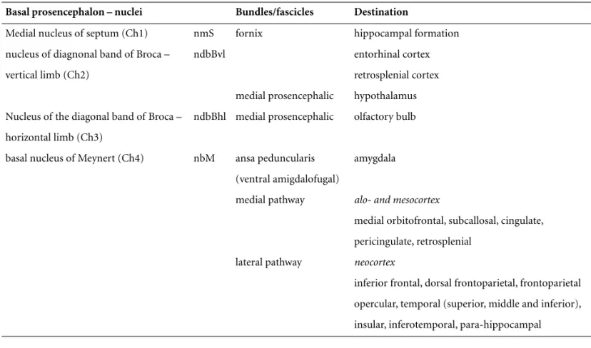
Table 1.Brain cholinergic system – cholinergic groups, main projections and most important destinations of the basal prosen-cephalon.
Basal prosencephalon – nuclei Bundles/fascicles Destination
Medial nucleus of septum (Ch1) nmS fornix hippocampal formation nucleus of diagnonal band of Broca – ndbBvl entorhinal cortex vertical limb (Ch2) retrosplenial cortex
medial prosencephalic hypothalamus Nucleus of the diagonal band of Broca – ndbBhl medial prosencephalic olfactory bulb horizontal limb (Ch3)
basal nucleus of Meynert (Ch4) nbM ansa peduncularis amygdala (ventral amigdalofugal)
medial pathway alo- and mesocortex
medial orbitofrontal, subcallosal, cingulate, pericingulate, retrosplenial
lateral pathway neocortex
inferior frontal, dorsal frontoparietal, frontoparietal opercular, temporal (superior, middle and inferior), insular, inferotemporal, para-hippocampal
(A) Sagital scheme of the brain to localize the nuclei of BP and their main projections. (B) Coronal schema with localization of medial and lateral cholinergic pathways. (C) Axial schemata, two levels (c1, basal ganglia level; c2, supracallosal level), with localization of medial and lateral cholinergic pathways. 1=nmS (Ch1)+ndbBvl (Ch2); 2=nbM (Ch4). a) lateral pathway projection to the amygdala; b) lateral pathway, initial part of the main pro-jection; c) medial pathway and its course in the cingulum; d) septo-hippocampal propro-jection; e) lateral path (black-continuous) in its external capsule-claustrum course; f) medial path (black-interrupted) in its cingulum course.
ACA, anterior cerebral artery; MCA, middle cerebral artery; PCA, pos-terior cerebral artery; ACoA, anpos-terior communicating artery; AChA, anterior choroidal artery.
Figure 2.Coronal schema of the brain – the cholinergic path-ways (left side) (medial path=black-interrupted; lateral path= black-continuous) and the limits of the main vascular territo-ries (right side).
the site of neurotransmitter release by diffusion (volume
transmission)
40-43.
The cholinergic pathways and the
cerebral vascular territories
The cholinergic projections of the septo-hippocampal
path present a fairly compact constitution, running
through the fornix to reach the hippocampal formation.
The Meynert-cortical projections present a relatively
com-pact origin, but once outside the basal ganglia territory, at
the level of the centrum semiovale, the lateral pathway
presents a fanlike distribution to reach their destination
areas, while the medial pathway runs mainly through the
cingulum and distributes ramifications along its route
38.
The main cerebral arteries – anterior cerebral artery
(ACA), middle cerebral artery (MCA), anterior
commu-nicating artery (ACoA), posterior commucommu-nicating artery
(PCoA), anterior choroidal artery (AChA) – provide
irri-gation of the territories where the cholinergic projections
travel
5,46-51(Table 2, Figure 2).
The cholinergic pathways and
cerebrovascular disease
Ischemic or hemorrhagic processes represent the
sev-eral cerebrovascular pathologies that can cause tissue
damage and interruption of the cholinergic pathways.
The ischemic processes cause territorial infarcts,
water-shed infarcts, lacunes, white matter demyelination,
affect-ing areas of varied size
52-56. It is possible to localize the
points where lesions can interrupt these pathways by
con-sidering the routes of the cholinergic pathways
38and the
vascular territories (Table 2, Figure 2, Figure 3).
The lesions of the BP, severely affecting the septal area
and/or the nbM, can occur due to ischemia in the
territo-ries of the ACA, ACoA and MCA. The projection of the
septal area to the hippocampal formation, via the fornix,
can be interrupted by lesions in the territory of the
ACoA. The interruption of the wide Meynert-cortical
projections may stem from a variety of lesion sites. The
main medial pathway can be interrupted at any point of
its route in the cingulum due to pathology in the ACA
and ACoA territories, while the ramifications of this
pathway, with a more radiating distribution, can be
injured in territories of the same arteries at a variety of
points. The main lateral pathway can be affected in its
sublenticular and paralenticular route (external capsule
and claustrum) due to lesions in the territories of the
ACA, ACoA and MCA, and its wide and fanlike course
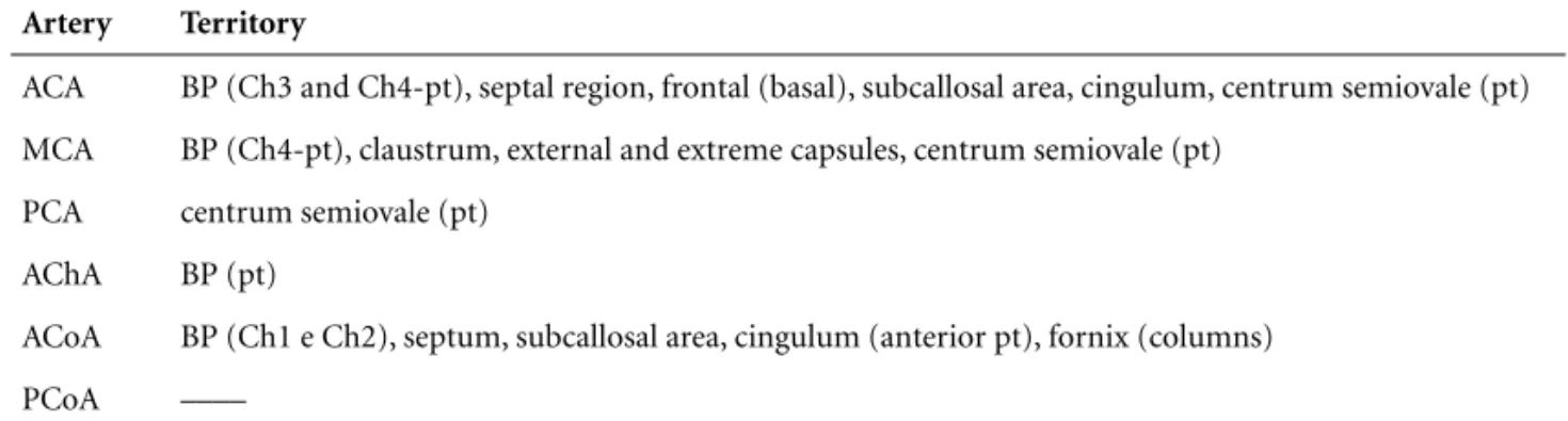
Table 2.Territories of the cerebral arteries related to cholinergic structures or their routes.
Artery Territory
ACA BP (Ch3 and Ch4-pt), septal region, frontal (basal), subcallosal area, cingulum, centrum semiovale (pt) MCA BP (Ch4-pt), claustrum, external and extreme capsules, centrum semiovale (pt)
PCA centrum semiovale (pt) AChA BP (pt)
ACoA BP (Ch1 e Ch2), septum, subcallosal area, cingulum (anterior pt), fornix (columns) PCoA ––––
Figure 3.RM-FLAIR. Axial sections of the brain of a case with extensive subcortical de-myelination. The cholinergic pathways are depicted to the left side (medial path= white -interrupted, lateral path=white continu-ous) on two levels (similar to Figure 1C).
can be affected in the white matter of the centrum
semio-vale, irrigated mainly by the ACA, MCA and PCA (Figure
2C, Figure 3).
Two neuropathologic studies indicate a relationship
between CVD and the interruption of these pathways,
besides the anatomic relations between the cholinergic
pathways and the vascular territories.
One of these studies was conducted using brain tissue
from a patient with CADASIL (cerebral autosomic
domi-nant arteriopathy with subcortical infarcts and
leucoen-cepalopathy), a disease that can be considered a model for
pure CVD (VaD). The material was stained with a
histo-chemical technique to show AChE revealing cholinergic
denervation in several cortical areas, except for the
hip-pocampal formation and entorhinal cortex. Even in the
more affected areas a number of AChE positive fibers were
seen. The cholinergic neurons of the nbM were
undam-aged, as verified by techniques for NGFr and AChE
57.
The other study was performed using brain tissue of
patients with VaD of the Binswanger subtype. The
mate-rial was stained with histochemical and
imunnohisto-chemical techniques to show AChE and ChAT. This
mat-erial revealed severe reduction of AChE and ChAT
posi-tive fibers in the external capsule and claustrum, in
com-parison to controls. The nbM had large neurons
pre-served, but showed some chromatolytic changes and
numerical reduction.
A neuroimage-neuropathological correlation was
possible for some of the patients. MRI showed
hyperin-tensities in the frontal periventricular white matter,
ex-tending to the subinsular white matter (where the
exter-nal capsule is found). The brains of the same patients at
autopsy showed loss of myelin in the corresponding
regions
58. Therefore, underpinned by these two
paradig-matic studies, we can state that CVD may cause
interrup-tion of segments of the cholinergic pathways, leading to
denervation and consequent cholinergic hypofunction of
the affected territories.
Cholinergic hypofunction, variable according to the
lesioned segment of the cholinergic pathways, causes
integrative dysfunction of the target brain structures and
disturbances of vasomotor control with consequent
reduction in brain blood flow of the affected areas
5,59.
These functional data gave rise to the proposal of a
‘cholinergic neurovascular hypothesis
22.
Recently, two studies were dedicated to the
relation-ship between the cholinergic pathways and the white
matter hyperintensities, correlated to the clinical
mani-festations of VCI/VaD, with the aim of staging scales.
These proposals relate the white matter lesions with their
localization in relation to the cholinergic pathways. The
staging was graduated according to the visually evaluated
extension, and number of lesions localized, along the
anatomical known routes of the cholinergic pathways.
One of these rating scales classified the lesions in the
cholinergic pathways as minimal (absence of lesions in
nbM and absence of hyperintensities in medial pathway
or external capsule), moderate (lesions in external
cap-sule plus in lateral pathway) and severe (nbM infarction
or external capsule plus lateral pathway hyperintensities
or large hyperintensities in lateral pathway or
hyperinten-sities in both lateral and medial pathways)
60. The other,
choliner-gic fibers as they project and fan out in the white
matter
61. The results of these studies suggest that the
localization of the hyperintensities in the white matter
holds special importance, considering that some of these
may occur at strategic points and may be related to
meas-urable clinical manifestations
60,61.
Thus, the knowledge of the anatomy of the
choliner-gic pathways and their relation to those vascular
territo-ries where an interruption can occur, allied to the
conse-quent clinical manifestations, enable better evaluation of
CVD clinical expression. It may also be able to lend a
more solid basis for treatment strategies, such as the
cholinergic approach.
Conclusion
CVD can cause clinical symptoms defining VCI/VaD
according to its extension and localization. Two
mecha-nisms play a role: one corresponding to tissue lesions of
cortical areas and subcortical regions, including white
matter responsible for disconnection related
manifesta-tions, while the other is related to the interruption of the
cholinergic pathways at various localizations along their
routes, producing manifestations consequent to
choliner-gic denervation which result in a hypocholinercholiner-gic state of
the affected territories.
Knowledge of cognitive-behavioral and vasomotor
functions of the cholinergic system, allied to that of the
anatomical localization of the course of its pathways, is
important to better assess the sites of vascular lesion.
Such knowledge permits strategic points of the
choliner-gic pathways to be highlighted and provides more solid
bases for use of cholinergic therapeutic strategies.
Acknowledgement –
To Luzinete N.O. Alvarenga for
editorial assistance.
References
1. Bartus RT, Dean RL, Beer, Lippa AS. The cholinergic hypothesis of geriatric memory dysfunction. Science 1982; 217:408-417.
2. Bartus RT. On neurodegenerative diseases, models, and treatment strategies: lessons learned and lessons forgotten a generation following the cholinergic hypothesis. Exp Neurol 2000;163:495-529.
3. Birks J. Cholinesterase inhibitors for Alzheimer’s disease. Cochrane Database Syst Rev; 2006.
4. Engelhardt E, Brucki SM, Cavalcanti JLS, Forlenza OV, Laks J, Vale FAC. Tratamento da doença de Alzheimer: recomen-dações e sugestões do Departamento Científico de Neuro-logia Cognitiva e do Envelhecimento da Academia Brasileira de Neurologia. Arq Neuropsiquiatr 2005;63:1104-1112.
5. Román GC, Kalaria RN. Vascular determinants of choliner-gic deficits in Alzheimer’s disease and vascular dementia. Neurobiol Aging (in press).
6. Mesulam MM. Structure and function of cholinergic path-ways in the cerebral cortex, limbic system, basal ganglia, and thalamus of the human brain. In: Bloom FE, Kupfer DJ, edi-tors. Psychopharmacology: The Fourth Generation of Pro-gress. New York: Raven Press; 1995:135-146.
7. Mufson EJ, Ginsberg SD, Ikonomovic MD et al. Human cholinergic basal forebrain: chemoanatomy and neurologi-cal dysfunction. J Chem Neuroanat 2003;26:233-242. 8. Nieuwenhuys R. Chemoarchitecture of the brain. Berlin:
Springer-Verlag; 1985:7-11.
9. Gertz HJ, Cervos-Navarro J, Ewald V. The septo-hippocam-pal pathway in patients suffering from senile dementia of Alzheimer’s type. Evidence for neuronal plasticity? Neurosc Lett 1987;76:228-232.
10. Fujishiro H, Umegaki H, Isojima D et al. Depletion of cholinergic neurons in the nucleus of the medial septum and the vertical limb of the diagonal band in dementia with Lewy bodies. Acta Neuropathol 2006;111:109-114.
11. Arendt T, Bigl V, Tennstedt A et al. Neuronal loss in different parts of the nucleus basalis is related to neuritic plaque for-mation in cortical target areas in Alzheimer's disease. Neuroscience 1985;14:1-14.
12. Geula C, Mesulam MM. Cholinergic Systems in Alzheimer’s Disease. In: Terry RD, Katzman R, Bick KL, Sisodia SS, edi-tors. Alzheimer Disease, 2nd ed, Philadelphia: Lippincott Williams & Wilkins; 1999:269-292.
13. McGeer PL, McGeer EG, Suzuki J et al. Aging, Alzheimer's disease, and the cholinergic system of the basal forebrain. Neurology 1984;34:741-745.
14. Mesulam M, Geula C. Nucleus Basalis (Ch4) and cortical cholinergic innervation in the human brain: observations based on the distribution of acetylcholinesterase and choline acetyltransferase. J Comp Neurol 1988;275:216-240. 15. Page KJ, Sofroniew MV. The ascending basal forebrain
cholinergic system. Prog Brain Res 1996;107:513-22. 16. Everitt B, Robbins TW. Central cholinergic systems and
cog-nition. Ann Rev Psychol 1997;48:649-684.
17. Gold PE. Acetylcholine modulation of neural systems involved in learning and memory. Neurobiol Learn Mem 2003;80:194-210.
18. Mesulam M. The cholinergic lesion of Alzheimer’s disease: pivotal factor or side show. Learn Mem 2004;11:43-49. 19. Minger SL, Esiri MM, McDonald B, et al. Cholinergic
deficits contribute to behavioral disturbances in patients with dementia. Neurology 2000; 55:1460-1467
21. Iadecola C. Neurovascular regulation in the normal brain and in Alzheimer’s disease. Nat Rev Neurosci 2004;5:347-360.
22. Claassen JAHR, Jansen WMM. Cholinergically mediated augmentation of cerebral perfusion in Alzheimer’s disease and related cognitive disorders: The cholinergic-vascular hypothesis. J Gerontol Med Sci 2006;61:267-271.
23. Elhusseiny A, Cohen Z, Olivier A, et al. Functional acetyl-choline muscarinic receptor subtypes in human brain microcirculation: identification and cellular localization. J Cer Bl Flow Metabol 1999;19:794-802.
24. Luiten PGM, de Jong Givan, der Zee EA, van Dijken H. Ultrastructural localization of cholinergic muscarinic re-ceptors in rat brain cortical capillaries. Brain Res 1996; 720: 225-229.
25. Tong XK, Hamel E. Regional cholinergic denervation of cor-tical microvessels and nitric oxide synthase-containing neu-rons in Alzheimer’s disease. Neuroscience 1999;92:163-174. 26. Barbelivien A, Bertrand N, Besret L, et al. Neurochemical
stimulation of the rat substantia inominata increases cere-bral blood flow (but not glucose use) through the parallel activation of cholinergic and non-cholinergic pathways. Brain Res 1999; 840:115-124.
27. Grantham C, Geerts H. The rationale behind cholinergic drug treatment for dementia related to cerebrovascular dis-ease. J Neurol Sci 2002;203-4:131-136.
28. Sato A, Sato Y, Uchida S. Regulation of regional cerebral blood flow by cholinergic fibers originating in the basal forebrain. Int J Dev Neuroscience 2001;19:327-337. 29. Scremin OU, Li MG, Scremin AME et al. Cholinesterase
inhibition improves blood flow in the ischemic cerebral cor-tex. Brain Res Bull 1997;42:59-70.
30. Blin J, Ivanoiu A, Coppens A, et al. Cholinergic neurotrans-mission has different effects on cerebral glucose consump-tion and blood flow in young normals, aged normals, and Alzheimer’s disease patients. Neuroimage 1997;6:335-343. 31. Ceravolo R, Volterrani D, Tognoni G, et al. Cerebral
perfu-sional effects of cholinesterase inhibitors in Alzheimer’s dis-ease. Clin Neuropharmacol 2004; 27:166-170.
32. Lojkowska W, Ryglewicz D, Jedrzejczak T, et al. The effect of cholinesterase inhibitors on the regional blood flow in patients with Alzheimer’s disease and vascular dementia. J Neurol Sci 2003;216:119-126.
33. Lipczynska-Lojowska W, Ryglewicz D, Jedrzejczak T, et al. The effect of rivastigmine on cognitive functions and region-al cerebrregion-al blood flow in Alzheimer’s disease and vascular dementia: follow-up for 2 years. Neurol Neuroch Polska 2004;38:471-481.
34. Nobili F, Koulibaly M, Vitali P et al. Brain perfusion follow-up in Alzheimer’s patients during treatment with acetyl-cholinesterase inhibitors. J Nucl Med 2002;43:983-990.
35. Kitt CA, Mitchell SJ, DeLong MR et al. Fiber pathways of basal forebrain cholinergic neurons in monkeys. Brain Res 1987; 406:192-206.
36. Mesulam MM, Hersh LB, Mash DC, Geula C. Differential cholinergic innervation within functional subdivisions of the human cerebral cortex: a choline acetyltransferase study. J Comp Neurol 1992;318:16-28.
37. Paré D, Smith Y, Parent A, Steriade M. Projections of brain-stem core cholinergic and non-cholinergic neurons of cat to intralaminar and reticular thalamic nuclei. Neuroscience 1988;25:69-86.
38. Selden NR, Gitelman DR, Salamon-Murayama N et al. Tra-jectories of cholinergic pathways within the cerebral hemi-spheres of the human brain. Brain 1998;121:2249-2257. 39. Steriade M, Paré D, Parent A, Smith Y. Projections of
cholin-ergic and non-cholincholin-ergic neurons of the brainstem core to relay and associational thalamic Nuclei in the cat and macaque monkey. Neuroscience 1988;25:47-67.
40. Descarries L, Gisiger V, Steriade M. Diffuse Transmission by Acetylcholine in the CNS. Progr Neurobiol 1997; 53:603-625.
41. Sykova E. Extrasynaptic volume transmission and diffusion parameters of the extracellular space. Neuroscience 2004; 129: 861-876.
42. Sykova E. Glia and volume transmission during physiologi-cal and pathologiphysiologi-cal states. J Neural Transm 2005;112:137-147.
43. Zoli M, Agnati LF. Wiring and volume transmission in the central nervous system: the concept of closed and open synapses. Progr Neurobiol 1996; 49:363-380.
44. Engelhardt E, Moreira DM, Nacif MS, Moscovici M. As vias colinérgicas e a demência vascular. Rev Bras Neurol 2006; 42:43-52.
45. Shute CC, Lewis PR. The ascending cholinergic reticular system: neocortical, olfactory and subcortical projections. Brain 1967; 90:497-520.
46. Adams RD, Victor M. Cerebrovascular diseases. In: Adams RD, Victor M. Principles of neurology. 5th ed, New York: McGraw-Hill, 1993:669-748.
47. Biller J. Vascular syndromes of the cerebrum. In: Brazes PW, Masseur JC, Biller J, editors. Localization in clinical neurol-ogy. 2nd ed, Boston: Little, Brown and Co., 1990:429-455. 48. Krayenbühl HÁ, Yasargil MG. Cerebral angiography. 2th ed.
Londres: Butterworths; 1968:20-84.
49. Salamon G. Atlas de la vascularisation arterielle du cerveau chez l’homme. Paris: Sandoz Editions; 1973.
50. Serizawa T, Saeki N, Yamaura A. Microsurgical anatomy and clinical significance of the anterior communicating artery and its perforating branches. Neurosurgery 1997;40:1211-1216. 51. Woolsey AT, Hanaway J, Gado MH. The brain atlas. 2nd ed.,
52. Chemerinski E, Robinson RG. The neuropsychiatry of stroke. Psychosomatics 2000;41:5-14.
53. Engelhardt E, Laks J, Cavalcanti JLS, Moreira DM, Madalen C. Demência vascular. Rev Bras Neurol 2004;40:5-26. 54. Erkinjuntti T. Clinicopathological study of vascular
demen-tia. In: Prohovnik I, Wade J, Knezevic S, Tatemichi T, Erkinjuntti T, editors. Vascular dementia: current concepts, Chichester: Wiley; 1996:73-112.
55. Jellinger KA. The pathology of ischemic-vascular dementia: an update. J Neurol Sci 2002;203-204:153-157.
56. Markus HS. Cerebral perfusion and stroke. J Neurol Neurosurg Psychiatry 2004;75:353-361.
57. Mesulam M, Siddique T, Cohen B. Cholinergic denervation in a pure multi-infarct state. Neurology 2003; 60:1183-1185.
58. Tomimoto H, Ohtani R, Shibata M, Nakamura N. Loss of cholinergic pathways in vascular dementia of the Bins-wanger type. Dement Geriatr Cogn Disord 2005;19:282-288.
59. Román GC. Cholinergic dysfunction in vascular dementia. Curr Psychiatry Rep 2005;7:18-26.
60. Swartz RH, Sahlas DJ, Black SE. Strategic involvement of cholinergic pathways and executive dysfunction: does loca-tion of white matter signal hypertensities matter? J Stroke Cerebrovasc Dis 2003;12:29-36.