Relationship between structural
abnormalities in the cerebellum and
dementia, posttraumatic stress disorder
and bipolar disorder
Leonardo Baldaçara1,2, João Guilherme Fiorani Borgio1, Célia Araújo1,
Fabiana Nery-Fernandes3, Acioly Luiz Taveres Lacerda1, Walter André dos Santos Moraes1,
Maria Beatriz Marcondes Macedo Montaño1, Marlos Rocha3, Lucas C. Quarantini3, Aline Schoedl1,
Mariana Pupo1, Marcelo F. Mello1, Sergio B. Andreoli1, Angela Miranda-Scippa3, Luiz Roberto Ramos1, Jair J. Mari1, Rodrigo Affonseca Bressan1, Andrea Parolin Jackowski1
ABSTRACT. New evidence suggests that the cerebellum has structural and functional abnormalities in psychiatric disorders.
Objective: In this research, the goal was to measure the volume of the cerebellum and its subregions in individuals with psychiatric disorders and to relate these findings to their symptoms. Methods: Patients with different degrees of cognitive impairment (Epidemiology of the Elderly - UNIFESP) and patients with post-traumatic stress disorder (PTSD) from population studies were analyzed. Also, patients with bipolar disorder from an outpatient clinic (Center for the Study of Mood and Anxiety Disorders, Universidade Federal da Bahia) were recruited for this study. All subjects underwent a 1.5T structural magnetic resonance scan. Volumetric measures and symptom measurements, by psychometric scales, were performed and compared between patients and controls. Results: The cerebellum volume was reduced in patients with cognitive impairment without dementia and with dementia, in patients with PTSD, and in patients with bipolar disorder compared to controls. In dementia and PTSD, the left cerebellar hemisphere and vermis volume were reduced. In bipolar disorder, volumes of both hemispheres and the vermis were reduced. In the first two studies, these cerebellar volumetric reductions correlated with symptoms of the disease. Conclusion: The exact nature of cerebellar involvement in mental processes is still not fully understood. However, abnormalities in cerebellar structure and its functions have been reported in some of these diseases. Future studies with larger samples are needed to clarify these findings and investigate whether they are important for treatment and prognosis.
Key words: cerebellum, neuroimaging, mental disorders, risk factors, comparative study.
RELAÇÃO ENTRE AS ALTERAÇÕES ESTRUTURAIS DO CEREBELO E DEMÊNCIA, TRANSTORNO DO ESTRESSE PÓS-TRAUMÁTICO E TRANSTORNO BIPOLAR
RESUMO. Novas evidências sugerem que o cerebelo apresenta alterações estruturais e funcionais nos transtornos psiquiátricos. Objetivo: Medir o volume do cerebelo e de suas sub-regiões em indivíduos portadores de transtornos psiquiátricos e relacionar tais achados aos sintomas. Métodos: Foi realizada a identificação de pacientes com diferentes graus de prejuízo cognitivo proveniente de um estudo populacional (Epidemiologia do Idoso - UNIFESP), pacientes com transtorno do estresse pós-traumático proveniente de outro estudo populacional e portadores de transtorno bipolar proveniente de um ambulatório especializado (Universidade Federal da Bahia). Todos os sujeitos foram submetidos à ressonância magnética estrutural de 1.5T. As medidas de volume, assim como os sintomas medidos por escalas psicométricas foram comparadas entre pacientes e controles. Resultados: Foi observado que o volume do cerebelo está reduzido nos portadores de prejuízo cognitivo sem demência e com demência, no transtorno do estresse pós-traumático e no transtorno bipolar quando comparados aos controles. Na demência e no transtorno do estresse pós-traumático o volume do hemisfério cerebelar esquerdo e do vérmis estão reduzidos. No transtorno bipolar os volumes de ambos os hemisférios e do vérmis estão reduzidos. Nos dois primeiros estudos estas reduções correlacionaram com os sintomas.
Conclusão: A natureza exata do envolvimento do cerebelo nos processos mentais ainda não é compreendida. Entretanto, anormalidades na estrutura cerebelar e em suas funções têm sido relatadas em algumas dessas doenças. Pesquisas futuras, com amostras maiores, ainda são necessárias para esclarecer tais achados e investigar se são importantes para o tratamento e prognóstico.
Palavras-chave: cerebelo, neuroimagem, transtornos mentais, fatores de risco, estudo comparativo.
1Federal University of São Paulo, SP, Brazil. 2Federal University of Tocantins, TO, Brazil. 3Federal University of Bahia, BA, Brazil.
INTRODUCTION
T
he term cerebellum comes from Latin, which means “little brain”. his is similar in structure to the brain and located in the posterior fossa of the skull, underly-ing the occipital lobe, posterior to the brainstem.1,2 hissimilarity to the brain is mainly because it is formed by two hemispheres (right and left) and cortical (gray matter) matter around a center of white matter, which in turn has gray matter nuclei. Within the hemisphere there is a central worm-shaped part called the vermis.1
For many years, functions related only to move-ment, gait, posture, and balance were attributed to the cerebellum.2 However, some studies have suggested
possible involvement of the cerebellum in cognition, emotion processing and behavior.3-5 According to this
perspective, the cerebellum is believed to exert a regu-latory function that enhances and supplements other brain functions, through direct and indirect circuits.2
Moreover, diferent sources of evidence have sug-gested that the cerebellum may be altered in many psychiatric disorders, including schizophrenia,6 bipolar
disorder (BD),7 unipolar depression,8 anxiety,9 and
at-tention deicit hyperactivity disorder.10 In some reports,
cerebellar deicits were described as isolated indings, without any relationship with clinical history whereas others show evidence that the cerebellum is a relevant brain structure possibly related to a range of psycho-pathological manifestations.2
Previous studies have reported cerebellar abnormali-ties in dementia.11-13 In neuropathological studies,
neu-ronal shrinkage and loss are well known changes that accompany Alzheimer’s Disease (AD) and are most no-table in the temporoparietal neocortex, limbic system, and some neuronal groups such as the locus coeruleus
of the brain stem and the basal nucleus of Meynert in the basal forebrain.12 Morphological studies, conversely,
have focused on the traditional neuropathological hall-marks of AD, such as amyloid plaques, neuroibrillary tangles, and amyloid angiopathy,12 while little has been
reported on neuronal loss and other structural changes in AD.11 Only one structural MRI study has previously
demonstrated that the posterior cerebellar lobes are smaller in AD patients when compared to healthy con-trols, and that this atrophy is associated with poorer cognitive performance in AD subjects, but not in MCI patients.13
Furthermore, the view that the cerebellum is in-volved in the experience and regulation of emotions was posited more than half a century earlier14,15 and
cerebel-lar indings have also been reported in BD. Cerebelcerebel-lar atrophy in mania16-19, cerebellar gray matter reduction20,
and smaller vermal subregions V2 and V3 (posterior-in-ferior region) are the main indings in BD patients7,21,22.
More recently, it has been postulated that the cerebel-lum plays a role in posttraumatic stress disorder (PTSD), and some studies have observed altered functioning of the left cerebellar hemisphere23 and vermis24,25 in PTSD
patients. his evidence suggests a possible role for this structure in the pathophysiology of the disease. To a lesser extent, cerebellar, superior temporal gyrus, and pituitary diferences have been reported in maltreated children and adolescents with PTSD.9,26,27
he aim of this article was to report the results of three studies that assessed cerebellar volume in a rep-resentative case of three diferent groups of mental disorders and explored a possible role of the cerebellum in their pathophysiological mechanisms. Finally, the implications of these indings and future directions are discussed.
METHODS
Participants. To study cerebellar structure in cognitive im-pairment 96 subjects were selected from the outpatient clinic of Epidemiology of Elderly Study (EPIDOSO)28
coordinated by the Preventive Medicine Department of Federal University of São Paulo (UNIFESP). Subjects with AD (n=70) and cognitive impairment without de-mentia (n=16) were included, in addition to 10 subjects without cognitive complaints or functional impairment with a Mini-Mental State Examination (MMSE) score of 30. All subjects were classiied with the Clinical De-mentia Rating scale (CDR).29 Subjects were divided into
ive groups according to dementia severity (CDR score) as follows: 0=controls, 0.5=questionable or very mild impairment, 1=mild impairment, 2=moderate impair-ment, and 3=severe impairment. All subjects without cognitive complaints or functional impairment and an MMSE score of 30 were classiied as CDR=0. he patients who met the diagnostic criteria proposed by Petersen et al. (1999) for MCI were included in the CDR=0.5 group.30 Alzheimer’s diagnosis was reached
ac-cording to the criteria of the National Institute of Neu-rological and Communicative Disorders and Stroke and the Alzheimer’s Disease and Related Disorders Associa-tion (NINCDS-ADRDA).31 For more details see
Balda-çara and Borgio et al. (2011).32
not developed PTSD) were included, identiied through an epidemiological survey that studied PTSD among the civilian population in the city of São Paulo.33,34 For more
details see Baldaçara and Jackowski et al (2011).35
To study cerebellar structure in bipolar disorder (BD) subjects were selected from an outpatient clinic of the Federal University of Bahia (UFBA). A total of 48 pa-tients with euthymic BD type I and 25 healthy controls were screened. Eight patients and three controls were excluded due to a history of neurological illness, head trauma, or inability to complete the MRI exam. he eu-thymia criterion was the attainment of scores on both the YMRS and HDRS of under 7 points. For more details see Baldaçara and Nery-Fernandes et al. (2011).36
Measures. Cerebellar volumes were correlated with clini-cal sclini-cales. For the cognitive impairment sample, the Structured Clinical Interview for DSM-IV (SCID),37,38
Clinical Dementia Rating scale (CDR),29 Mini-Mental
State Examination (MMSE),39 and Functional
Activi-ties Questionnaire (FAQ) were applied.40 For the PTSD
sample the SCID,37,38 Clinician-Administered PTSD
Scale (CAPS),41 Beck Anxiety Inventory (BAI),42 Beck
Depression Inventory (BDI),43 and Early Trauma
Inven-tory (ETI) were used.44 For the BD sample the (SCID,37,38
Hamilton Depression Rating Scale (HDRS),45 Young
Ma-nia Rating Scale (YMRS),46 and Barratt Impulsiveness
Scale (BIS-11) were employed.47
Image acquisition and analysis. Imaging data were ac-quired using GE 1.5T scanners. Structural MRI im-ages were acquired using sagittal T1 acquisition series (TR=9.8 ms, TE=3.1 ms, lip angle=30o, NEX=1, matrix
size=256×256, FOV=24 cm, thickness=1.0 mm), yield-ing 160 slices. Before scannyield-ing, a sagittal scout series (nine to eleven 5-mm-thick slices with a 1-mm inter-slice gap) was performed to determine image quality and clarity, as well as subject head position. Total brain volume was measured using voxel-based morphometry (VBM) methodology, which was implemented with the VBM Toolbox in SPM5 (dbm.neuro.uni-jena.de/vbm) as described previously.48 T1-weighted images were
seg-mented in the original space. he sum of all voxel val-ues within the segmented image approximates the total volume within the corresponding partition. Total brain volume was computed from the sum of gray and white matter volumes.48
he cerebellum was measured using region of inter-est (ROI) methodology. his method was chosen be-cause it is more precise than other methods and residual heterogeneity can be correctly included in the analyses.
First, the ROI tracing was performed using the Brains 2 semi-automated model, then manually corrected based on previous studies.49-51 he cerebellar hemispheres and
vermal volumes were calculated by summing the areas of successive coronal slices after tracing the region of interest (ROI), and excluding cerebrospinal luid (CSF). he measurements began as the cerebellum appeared laterally to the pons. he tentorium cerebella acted as the upper limit and the base of the cerebellum as the lower limit. he cisterna magna and transverse sinus were excluded.49 he last slice included was the one at
which the cerebellum was no longer distinguishable from the transverse sinus or was no longer visible. he measurement of the vermis began at the slice where the anterior and/or inferior posterior lobes appeared. Mea-surements were made until the vermis was no longer visible.49 To evaluate inter-rater and test-retest
repro-ducibility, two raters repeated the cerebellar measure-ments twice, at least one week apart, for 10 randomly selected subjects. Inter-rater reliability and test-retest was found to be high (ICC >0.96) for all cerebellar and vermal regions.
Data analysis. Data were codiied and analyzed using the Statistical Package for Social Sciences (SPSS for Windows, version 15.0). Prior to conducting analyses, measures were examined for normality using the Shap-iro–Wilk test. he level of signiicance was set at p<0.05 using a two-tailed test. For detailed information about each statistical method see the speciic articles.32,35,36
Ethical issues. All participants signed informed consent forms before participation. he study of cerebellar vol-ume in subjects with cognitive impairment and in PTSD was approved by the Institutional Review Board (IRB) of the Federal University of São Paulo (CEP no 0258/08
and CEP no (1026/06). he study of bipolar subjects was
approved by the Ethics Committee of the Federal Uni-versity of Bahia (CEP no16/2005).
RESULTS
Cerebellum in cognitive impairment by dementia. Cerebellar hemisphere volumes were progressively lower in CDR 1, 2, and 3. he volume of the vermis, however, was found to be already reduced in the CDR 0.5 group, where this reduction was progressive with increasing CDR scores. For more details see Table 1.
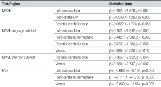
sig-niicant. MMSE total scores correlated positively with volumes of the left temporal lobe, right cerebellum, and posterior cerebellar lobe. he MMSE language sub-test correlated positively with the left temporal lobe, right cerebellar hemisphere, posterior cerebellar lobe, and vermis. Attention correlated positively with the pos-terior cerebellar lobe and vermis volumes. FAQ scores correlated negatively with the left temporal lobe, right cerebellum, and vermis volumes. For more details see Table 2.
Of the 26 subjects without dementia at baseline, 10 subjects (1 with CDR=0 and 4 with CDR=0.5) devel-oped dementia after two years. here were no difer-ences in intracranial volume, cerebral volume, temporal lobes, and right cerebellum between the two groups at baseline. In a logistic regression analysis, reduced left temporal lobe (OR=2.002, C.I.: 1.102-3.123, p=0.058), posterior cerebellar lobe (OR=1.402, C.I.: 1.004-2.903, p=0.068), and vermal volume (OR=1.480, C.I.: 1.024-2.803, p=0.042) were related to future dementia onset.
Table 1. Intracranial volume, cerebral and cerebellar volumes in cm3 for each group.32
CDR
0 Mean (SD)
0.5 Mean (SD)
1 Mean (SD)
2 Mean (SD)
3
Mean (SD) c2/F p
Duncan test (5%)
Intracranial volume 1722.19 (47.80) 1750.64 (99.12) 1728.96 (86.32) 1729.64 (78.39) 1747.19 (93.64) 0.334 0.854 0=0.5=1=2=3
Brain 596.09 (37.01); 1008.67 (95.30)
576.23 (42.24); 1034.39 (129.73)
536.44 (45.38); 1007.44 (114.49)
449.38 (87.39); 1019.38 (131.69)
418.91 (51.24); 1056.33 (66.36)
27.636 < 0.001 0 >0.5 >1 >2=3
Left temporal lobe 71.86 (5.45), 120.41(8.12)
70.23 (4.67); 120.65 (8.95)
58.46 (4.32); 98.18 (5.67)
51.45 (4.48); 88.62 (4.58)
47.96 (3.79); 83.92 (4.23)
124.041 < 0.001 0 >0.5 >1 >2 >3
Right temporal lobe 70.89 (5.22); 117.77 (8.56)
70.77 (5.55); 117.44 (6.78)
55.03 (4.46); 94.96 (7.19)
51.06 (4.65); 87.99 (7.68)
49.22 (5.67); 83.45 (7.41)
19.389 < 0.001 0=0.5 >1 >2=3
Left cerebellum 30.40 (2.47); 53.21 (5.17)
28.86 (2.38); 49.70 (4.22)
26.90 (2.16); 46.43 (3.43)
24.04 (1.58); 41.54 (2.76)
20.69 (1.63); 36.04 (1.83)
53.976 < 0.001 0 >0.5 >1 >2 >3
Right cerebellum 30.15 (2.10); 51.95 (4.31)
31.40 (2.44); 55.00 (5.78)
24.90 (2.27); 44.41 (3.22)
23.46 (1.58); 40.54 (2.76)
20.12 (1.60); 35.04 (1.84)
80.858 < 0.001 0=0.5 >1 >2 >3
Vermis 3.24 (0.48); 5.45 (0.82)
2.05 (0.31); 3.89 (0.54)
1.46 (1.84); 2.51 (0.33)
1.37 (0.29); 2.37 (0.50)
0.99 (0.27); 1.74 (0.50)
103.069 < 0.001 0 >0.5 >1=2 >3
Cerebellar anterior lobe 9.08 (1.49); 15.25 (4.84)
9.06 (1.64); 15.75 (2.38)
8.11 (1.93); 13.84 (1.38)
8.15 (2.46); 13.99 (3.23)
6.56 (2.31); 10.57 (3.11)
2.097 0.088 0=0.5 >1=2 >3
Cerebellar posterior lobe 53.85 (3.82); 88.42 (5.23)
51.22 (2.36); 84.35 (3.44)
42.53 (2.18); 76.55 (4.21)
39.95 (2.95); 69.72 (3.33)
34.85 (1.88); 60.35 (2.12)
17.563 < 0.001 0 >0.5 >1 >2 >3*
First volume: corrected by Intracranial volume. Second volume: Absolute volume.
Table 2. Significant correlations between all cerebellar region volumes and scores on MMSE and FAQ*.
Test/Region Statistical data
MMSE Left temporal lobe β=0.492, t=1.876, p=0.064
Right cerebellum β=0.0543, t=1.263, p<0.080 Posterior cerebellar lobe β=0.0627, t=1.774, p=0.058 MMSE language sub-test Left temporal lobe β=0.452, t=1.642, p=0.052
Right cerebellar hemisphere β=0.442, t=9.542, p < 0.020 Posterior cerebellar lobe β=0.422, t=1.284, p=0.062
Vermis β=0.380, t=4.520, p<0.018
MMSE attention sub-test Posterior cerebellar lobe β=0.365, t=2.252, p=0.044
Vermis β=0.260, t=2.747, p=0.007
FAQ Left temporal lobe β= –0.666, t= –3.196, p=0.002
Right cerebellar hemisphere β= –0.111, t= –1.736, p=0.086
Vermis β= –0.809, t= –2.884, p=0.005
Cerebellum in posttraumatic stress disorder. here were no age (t= –1.74, p=0.08) or gender (c2=1.42, df=1,
p=0.34) diferences between the PTSD and resilient control groups. he PTSD group presented signiicantly higher scores for history of early traumatic life events (t=2.49, p<0.01) and all clinical variables as follows: re-experiencing symptoms (t=6.22, p<0.01), avoidance and numbing symptoms (t=7.12, p<0.01), hyperarousal symptoms (t=6.445, p<0.01), total CAPS score (t=7.86, p<0.01), anxiety (t=4.65, p<0.01), and depressive symp-toms (t=4.46, p<0.01).
Of the PTSD patients, 81% (34 subjects; c2=42.60,
p<0.01) presented comorbid major depressive disorder (MDD), 9.5% (4 subjects) presented panic disorder (PD), and 2.4% (1 subject) presented alcohol abuse disorder (AAD). Of the resilient controls, 78.6% (33 subjects) did not fulill criteria for a psychiatric disorder, but 9.5% (4 subjects) presented MDD, 7.1% (3 subjects) PD, and 2.4% (1 subject) fulilled the criteria for ADD. he PTSD sample included a heterogeneous range of traumatic ex-periences as follows: assault (38.1%), sexual and
physi-cal abuse (28.6%), sudden death of a loved one (19%), kidnapping (7.1%) and others (7.2%). he average du-ration of symptoms was 43 months (from 1 month to 18 years). he controls were also primarily victims of assault (35.7%), sexual and physical abuse (26.2%), sud-den death of a loved one (16.7%), kidnapping (7.1%) and others (14.3%). Brain volume was smaller in the PTSD group (F=4.50, p=0.01). PTSD patients also had reduced left cerebellar hemisphere (F=2.55, p=0.04) and vermal volumes (F=13.49, p<0.01) compared to resil-ient controls. For more details see Table 3.
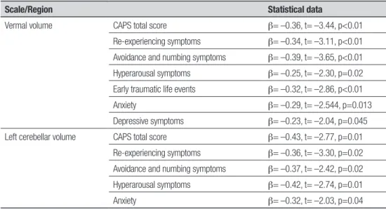
In the PTSD group (but not in the control group), a signiicant negative correlation was observed between vermal volume and CAPS total score, re-experiencing symptoms, avoidance and numbing symptoms, hyper-arousal symptoms, early traumatic life events, anxiety, and depressive symptoms. A negative correlation be-tween left cerebellum volume and CAPS total score, re-experiencing symptoms, avoidance and numbing symp-toms, hyperarousal sympsymp-toms, and anxiety was also observed . For more details see Table 4.
Table 3. Brain measurement in PTSD patients and resilient controls.35
Volumes*
PTSD (n=42) Controls (n=42)
F p
Mean 95% C.I.** Mean 95% C.I.b
Brain+ 1096.27 1069.05–1123.48 1161.03 1133.82–1188.24 4.50 0.01
Total cerebellum++ 101.93 98.97–104.88 105.52 102.55–108.46 1.45 0.22
Left cerebellar hemisphere++ 50.55 48.89–52.20 53.61 51.95–55.26 2.55 0.04
Right cerebellar hemisphere++ 51.38 49.89–52.88 51.89 50.40–53.39 1.01 0.41
Vermis++ 7.04 6.70–7.39 8.74 8.34–9.08 13.49 <0.01
*Volumes are in cm3; **Confidence Interval; +Adjusted for age, gender and comorbidity; ++Adjusted for brain volume, age, gender and comorbidity.
Table 4. Significant correlations between all cerebellar region volumes and clinical scale scores (only in PTSD group).
Scale/Region Statistical data
Vermal volume CAPS total score β= –0.36, t= –3.44, p<0.01
Re-experiencing symptoms β= –0.34, t= –3.11, p<0.01 Avoidance and numbing symptoms β= –0.39, t= –3.65, p<0.01 Hyperarousal symptoms β= –0.25, t= –2.30, p=0.02 Early traumatic life events β= –0.32, t= –2.86, p<0.01
Anxiety β= –0.29, t= –2.544, p=0.013
Depressive symptoms β= –0.23, t= –2.04, p=0.045 Left cerebellar volume CAPS total score β= –0.43, t= –2.77, p=0.01
Re-experiencing symptoms β= –0.36, t= –3.30, p=0.02 Avoidance and numbing symptoms β= –0.37, t= –2.42, p=0.02 Hyperarousal symptoms β= –0.42, t= –2.74, p=0.01
CAPS total score correlated positively with ETI – ear-ly trauma (β= –0.32, t= –3.02, p<0.01). Moreover, when both variables were controlled in linear regression anal-ysis, vermal volume continued to correlate negatively with CAPS total score (β= –0.34, t= –3.11, p<0.01) and ETI (β= –0.25, t= –2.14, p=0.04).
Cerebellum in bipolar disorder. here was no age, gender or schooling diference among patients with BD and healthy controls. he mean number of suicide attempts was 1.6±0.8. here was no diference between patients with BD who exhibited suicidal behavior and those who did not in terms of age (F=0.72, p=0.49), gender (c2=3.05
, p=0.22), and years of schooling (F=0.26,
p=0.77). BIS scores were higher in BD subjects with a history of suicide attempts (67.35±14.80) as compared to both those without attempts (58.34±8.64) and con-trol subjects (58.52±9.05) (p=0.05). here were no dif-ferences in intracranial and brain volumes between the BD group and the healthy control group. he left cerebellum (t=5.58, p=0.02), right cerebellum (t=5.43, p=0.02) and vermis (t=20.20, p<0.01) were smaller in the BD group. For more details see Table 5.
here was no correlation between vermal and cer-ebellar hemisphere volumes and BIS, number of suicide attempts, or number of mood episodes (manic, depres-sive or mixed episodes).
DISCUSSION
he evidence of structural alterations in cerebellum vol-ume in neuropsychiatric disorders is not novel. Howev-er, few studies have assessed cerebellar morphology in well-characterized samples and related this to psycho-pathology. Moreover, there was no previous research that related early trauma to cerebellar volume reduction in any psychiatric disorders.
In past studies, the cerebellum has only been
impli-cated in movement, gait, posture, and balance.2 However,
recent studies suggest signiicant interconnections between the cerebellum and prefrontal cortex subdivi-sions related to executive functioning (working mem-ory, attention, inhibition of behaviors, and decision making), verbal memory and language.3,4,52,53 Aferent
projections from parietal, temporal, and occipital corti-ces, and the limbic system implicated in the integration of sensitive and sensory information, visuospatial or-ganization, visual memory, and control of behavior and motivation have also been proposed.2,54,55 More recently,
a timekeeping or “clock” function has been postulated for the cerebellar cortex and the inferior olive (the sole source of climbing iber inputs to Purkinje cells) based on their unique microstructure and intrinsic rhythmic oscillatory properties.55-57 Xu (2006) proposed that the
primary role of the inferior olive and the climbing iber system in timing is to mediate the encoding of temporal information independent of motor behavior.56
he notion that the cerebellum is involved in the ex-perience and regulation of emotions was posited more than half a century earlier,14,58 and intimate aferent
and eferent connections to the brainstem and limbic system have provided neuroanatomical support.14,58
he cerebellum has monosynaptic projections not only to the hypothalamus, septum, hippocampus, amygda-la, and basal ganglia, but also to the brainstem nuclei, where the cerebellar projections stimulate dopamine and noradrenaline release by innervating the substantia nigra and locus coeruleus.14,58
One of the irst reports to relate the cerebellum to emotional experience involved a patient who reported unpleasant feelings after electrical stimulation of the dentate nucleus and superior peduncle.58
Further-more, electrophysiological responses in several limbic structures, including the hippocampus, amygdala, and septum, were recorded following electrical stimulation
Table 5. Intracranial, brain and cerebellar volumes (cm3) in control, bipolar without and with suicide attempts groups.36
Variable
Control n=22
Bipolar disorder n=40
Bipolar disorder without history of
suicide attempt (n=20)
Bipolar disorder with history of suicide attempt
(n=20) t / F p
Intracranial volume* 1429.70±157.32 1399.37±153.19 1380.91±138.82 1417.82±167.87 0.55**, 0.55+ 0.46**, 0.58+
Brain volume* 834.24±56.85 820.71±36.78 816.83±35.17 824.59±38.82 1.29**, 0.79+ 0.26**, 0.46+
Left cerebellum* 44.31±7.51 40.18±6.03 40.85±5.53 39.50±6.57 5.58**, 2.97+ 0.02**, 0.06+
Right cerebellum* 45.40±7.48 41.15±6.51 41.80±5.29 40.50±7.62 5.43**, 2.86+ 0.02**, 0.06+
Vermis 5.62±0.81 4.76±0.66 4.97±0.71 4.56±0.55 20.20**, 12.19+ <0.01**,+
of the fastigial portion of the deep cerebellar nuclei in mammals.58 Additional support for the connection
be-tween the cerebellum and emotions in humans is pro-vided by reports of an emotionally disturbed patient who received electrical stimulation in the fastigial nucle-us.58 It was found that electrical discharges induced by
electric stimulation correlated with the patient’s experi-ence of anger and tension. Moreover, there is evidexperi-ence that chronic stimulation of the vermis using implanted subdural electrodes can normalize behavior in severely emotionally (severe anxiety or depression) dysregulated patients.14,58
One result from these studies is highly important: cerebellar volume reduction related to early trauma. Although previous studies have demonstrated struc-tural49,59,60 and functional61,62 cerebellar
abnormali-ties in PTSD subjects, none have found a relationship between early-life traumatic experiences and cerebel-lar alterations in adulthood. he correlations between early-life trauma and CAPS with cerebellar volumes found in the current study suggest that both traumatic events and PTSD symptoms have an efect on cerebellar structure. However, it is not yet clear whether reduced brain regions (e.g., the cerebellum) represent anteced-ent vulnerability for developing PTSD upon exposure to a traumatic event or a consequence of PTSD symp-toms. Spinelli et al. (2009)63 conducted a study to
iden-tify structural abnormalities that may predict increased risk of stress-related neuropsychiatric disorders. In this study, mother-reared Rhesus monkeys were compared to peer-reared ofspring. An enlarged vermis, dorsome-dial prefrontal cortex, and dorsal anterior cingulated cortex were found in peer-reared monkeys; however, there were no diferences in the corpus callosum or the hippocampus.63 Comparing the present results with
those from previous studies, we speculate that cerebel-lar hyperactivity is present during the irst few months after the stress factor, and cerebellar volume reduction is a consequence of this chronic hyperactivity that ap-pears later. Adverse childhood factors may lead to an increased risk for later PTSD.64 Evidence has suggested
that the developing cerebellum is vulnerable to environ-mental insults, including those of a physical and psycho-logical nature.27,58,65 Environmental insults encountered
during childhood, such as exposure to toxic levels of lead,66 chronic irradiation,67 low birth weight,68 and
neo-natal exposure to dexamethasone,69 preferentially
dam-age cerebellar structures. he dysregulation of the lim-bic-hypothalamic-pituitary-adrenal (LHPA) axis with elevated levels of corticotrophin releasing hormone (CRH) has been consistently reported in traumatized
individuals.27,70 Adults with PTSD, maltreated children
with symptoms of mood and anxiety disorders, children with PTSD secondary to maltreatment, and infant pri-mate mothers all show this dysregulation.27,70-72
One hypothesis to explain PTSD is that childhood abuse acts as a severe stressor that unleashes a cascade of events that afect brain development.27 Adult
ani-mals submitted to a single prolonged episode of early maternal deprivation show stress-induced corticoste-rone responses.73 Maternal deprivation induces
neu-ronal degeneration and astroglial abnormalities in the hippocampus and cerebellar cortex of neonatal rats. Maternal separation may impair learning and memory in adult males by altering normal developmental fea-tures in glucocorticoid receptor expression.73 hus,
chil-dren exposed to trauma (early trauma) may experience chronically elevated CRH during pituitary development. Elevated CRH may lead to pituitary hypertrophy, which may be most pronounced during puberty, due to trophic factors.27 Chronic exposure to CRH may result in the
downregulation of pituitary CRH receptors over time. his downregulation may be an adaptive mechanism that regulates pituitary hypertrophy, as the resultant high cortisol levels would otherwise result in medical ill-ness, and damage to the brain27 and cerebellum.74
here are several limitations of this study that should be considered. First, although the overall sample size of patients was reasonable for a study using neuroimaging data, the examined subgroups in multivariate analysis was relatively small. hus, there was insuicient statisti-cal power to examine all aspects of psychopathologistatisti-cal phenomena in patients groups. Second, although our indings suggest that psychiatric disorders indicates more severe brain structural anomalies, our indings do not necessarily indicate that these indings are exclusive to dementia, PTSD and BD. hird, the cross-sectional design precluded the opportunity to examine cause and efect. Further, three studies had well characterized samples and two studies were conducted in a commu-nity sample; therefore, these results may not be general-izable to other care service settings.
disorders. Abnormalities in cerebellar structure and its functions have been reported in some psychiatric disor-ders. In subjects with cognitive impairment, it was ob-served that cerebellar volume is reduced and correlated with the loss of attention and language, even before the development of Alzheimer’s dementia. hese data re-late to previous studies that showed cerebellar abnor-malities and Alzheimer’s, but also extend the indings to early-stage disease and characterize more precisely the cognitive impairments. he post-traumatic stress disor-der was found to reduce the volume of the left cerebellar hemisphere, associated with symptoms of re-experienc-ing, avoidance, hyperactivity and anxiety. Although le-sions in hemispheres and associated emotional changes have previously been described, there is no support in the literature to elucidate the reason for this lateraliza-tion. A reduction in the volume of the cerebellar vermis associated with the same symptoms, and depressive symptoms and trauma in early life was also observed. his inding corroborates the literature, which observed the relationship of posterior-inferior regions of the cer-ebellum (more precisely the vermis) with the control of emotions. Further, the data may extend to the relation-ship between early life events in the genesis of mental disorders, a theory much discussed in the literature, yet diicult to prove. In bipolar disorder patients, a reduced total volume of the cerebellum, the hemispheres and
vermis was found compared with controls. A volume reduction in patients with the disease and a history of suicide was also observed, but, contrary to expectations based on literature data, no associations with symp-toms of impulsivity, number of mood episodes or dura-tion of disease were identiied. In the latter disorder this raises the question: Does the reduced volume of the cer-ebellum relect a relationship directly accompanying the course of the disease or constitute an early change that could predispose to disease? Future research is needed to investigate the importance of structural abnormali-ties in cerebellar patients with psychiatric disorders in more depth. his calls for research with larger samples, more detailed neuropsychological testing and neuroim-aging combining structural and functional techniques (such as the volume of interest - VOI). Also, it is impor-tant to observe diferent groups in the same disorder, based on diferent forms of activation of the cerebellum during the same task. he 21st Century holds much to be
discovered. Perhaps a new molecule, a new cellular pro-cess, unique signal processing in neuronal networks or an as yet unknown principle of control may change cur-rent concepts and further understanding on how this structure regulates and complements the other nervous functions. he cerebellar system appears to be the key to such discoveries, especially to clarify the operation of alternative circuits and circuits of reinement.
REFERENCES
1. Meneses MS. Neuroanatomia Aplicada. 3a ed. Rio de Janeiro:
Guana-bara Koogan; 2011.
2. Baldaçara L, Borgio JGF, Lacerda ALT, Jackowski AP. Cerebellum and psychiatric disorders. Rev Bras Psiquiatr 2008;30:281-289.
3. Baillieux H, De Smet HJ, Dobbeleir A, Paquier PF, De Deyn PP, Marien P. Cognitive and affective disturbances following focal cerebellar damage in adults: a neuropsychological and SPECT study. Cortex 2010;46:869-879. 4. Marien P, Baillieux H, De Smet HJ, et al. Cognitive, linguistic and affec-tive disturbances following a right superior cerebellar artery infarction: a case study. Cortex 2009;45:527-536.
5. Stoodley CJ, Schmahmann JD. Functional topography in the human cerebellum: a meta-analysis of neuroimaging studies. NeuroImage 2009;44:489-501.
6. Picard H, Amado I, Mouchet-Mages S, Olie JP, Krebs MO. The role of the cerebellum in schizophrenia: an update of clinical, cognitive, and functional evidences. Schizophr bull 2008;34:155-172.
7. Monkul ES, Hatch JP, Sassi RB, et al. MRI study of the cerebellum in young bipolar patients. Prog Neuropsychopharmacol Biol Psychiatry 2008;32:613-619.
8. Cheron G, Servais L, Dan B. Cerebellar network plasticity: From genes to fast oscillation. Neuroscience 2008;153:1-19.
9. De Bellis MD, Kuchibhatla M. Cerebellar volumes in pediatric maltreat-ment-related posttraumatic stress disorder. Biol Psychiatry 2006;60: 697-703.
10. Mackie S, Shaw P, Lenroot R, et al. Cerebellar development and clini-cal outcome in attention deficit hyperactivity disorder. Am J Psychiatry 2007;164:647-655.
11. Wegiel J, Wisniewski HM, Dziewiatkowski J, et al. Cerebellar atrophy in Alzheimer’s disease-clinicopathological correlations. Brain Res 1999; 818:41-50.
12. Sjobeck M, Englund E. Alzheimer’s disease and the cerebellum: a mor-phologic study on neuronal and glial changes. Dement Geriatr Cogn Disorders 2001;12:211-218.
13. Thomann PA, Schlafer C, Seidl U, Santos VD, Essig M, Schroder J. The cerebellum in mild cognitive impairment and Alzheimer’s disease - a structural MRI study. J Psychiatric Res 2008;42:1198-1202.
14. Schutter DJ, van Honk J. The cerebellum in emotion regulation: a repet-itive transcranial magnetic stimulation study. Cerebellum 2009;8:28-34. 15. Nashold BS, Jr., Slaughter DG. Effects of stimulating or destroying the
deep cerebellar regions in man. J Neurosurg 1969;31:172-86. 16. Lippmann S, Manshadi M, Baldwin H, Drasin G, Rice J, Alrajeh S.
Cerebellar vermis dimensions on computerized tomographic scans of schizophrenic and bipolar patients. Am J Psychiatry 1982;139:667-668. 17. Nasrallah HA, McCalley-Whitters M, Jacoby CG. Cortical atrophy in
schizophrenia and mania: a comparative CT study. J Clin Psychiatry 1982;43:439-441.
18. Nasrallah HA, Jacoby CG, McCalley-Whitters M. Cerebellar atrophy in schizophrenia and mania. Lancet 1981;8229:1102.
19. Weinberger DR, DeLisi LE, Perman GP, Targum S, Wyatt RJ. Computed tomography in schizophreniform disorder and other acute psychiatric disorders. Arch Gen Psychi 1982;39:778-783.
20. Moorhead TW, McKirdy J, Sussmann JE, Hall J, Lawrie SM, Johnstone EC, McIntosh AM. Progressive gray matter loss in patients with bipolar disorder. Biol Psychiatry 2007;62:894-900.
21. Mills NP, Delbello MP, Adler CM, Strakowski SM. MRI analysis of cer-ebellar vermal abnormalities in bipolar disorder. Am J Psychiatry 2005; 162:1530-1532.
22. DelBello MP, Strakowski SM, Zimmerman ME, Hawkins JM, Sax KW. MRI analysis of the cerebellum in bipolar disorder: a pilot study. Neuro-psychopharmacol 1999;21:63-68.
Post RM. Regional cerebral blood flow correlated with flashback inten-sity in patients with posttraumatic stress disorder. Biol Psychiatry 2001; 50:246-253.
24. Anderson CM, Teicher MH, Polcari A, Renshaw PF. Abnormal T2 re-laxation time in the cerebellar vermis of adults sexually abused in child-hood: potential role of the vermis in stress-enhanced risk for drug abuse. Psychoneuroendocrinology 2002;27:231-244.
25. Pissiota A, Frans O, Fernandez M, von Knorring L, Fischer H, Fredrikson M. Neurofunctional correlates of posttraumatic stress disorder: a PET symptom provocation study. Eur Arch Psychiatry Clin Neurosci 2002; 252:68-75.
26. De Bellis MD, Keshavan MS, Frustaci K, et al. Superior temporal gyrus volumes in maltreated children and adolescents with PTSD. Biol Psy-chiatry 2002;51:544-552.
27. Thomas LA, De Bellis MD. Pituitary volumes in pediatric maltreatment-related postraumatic stress disorder. Biol Psychiatry 2004;55:752-758. 28. d’Orsi E, Xavier AJ, Ramos LR. Work, social support and leisure protect
the elderly from functional loss: EPIDOSO study. Rev Saude Publica 2011;45:685-692.
29. Montaño MBMM, Ramos LR. Validade da versão em português da Clinical Dementia Rating. Rev Saúde Pública 2005;39:912-917. 30. Petersen RC, Smith GE, Waring SC, Ivnik RJ, Tangalos EG, Kokmen E.
Mild cognitive impairment: clinical characterization and outcome. Arch Neurol 1999;56:303-308.
31. Dubois B, Feldman HH, Jacova C, et al. Research criteria for the di-agnosis of Alzheimer’s disease: revising the NINCDS-ADRDA criteria. Lancet Neurol 2007;6:734-746.
32. Baldaçara L, Borgio JGF, Moraes WAS, et al. Cerebellar volume in pa-tients with dementia. Rev Bras Psiquiatr 2011;33:122-129.
33. Andreoli SB, Ribeiro WS, Quintana MI, et al. Violence and post-traumat-ic stress disorder in Sao Paulo and Rio de Janeiro, Brazil: the protocol for an epidemiological and genetic survey. BMC Psychiatry 2009;9:34. 34. Bressan RA, Quarantini LC, Andreoli SB, et al. The posttraumatic stress
disorder project in Brazil: neuropsychological, structural and molecu-lar neuroimaging studies in victims of urban violence. BMC Psychiatry 2009;9:30.
35. Baldacara L, Jackowski AP, Schoedl A, et al. Reduced cerebellar left hemisphere and vermal volume in adults with PTSD from a community sample. J Psychiatric Res 2011;45:1627-1633.
36. Baldacara L, Nery-Fernandes F, Rocha M, et al. Is cerebellar volume related to bipolar disorder? J Affect Disorder 2011;135:305-309. 37. Spitzer RL, Williams JB, Gibbon M, First MB. The Structured Clinical
In-terview for DSM-III-R (SCID). I: History, rationale, and description. Arch Gen Psychiatry 1992;49:624-629.
38. Williams JB, Gibbon M, First MB, et al. The Structured Clinical Interview for DSM-III-R (SCID). II. Multisite test-retest reliability. Arch Gen Psychia-try 1992;49:630-636.
39. Folstein MF, Folstein SE, McHugh PR. Mini-mental state: a practical method for grading the cognitive state of patients for the clinican. J Psychiatric Res 1975;12:189-198.
40. Pfeffer RI, Kurosaki TT, Harrah CH, Jr., Chance JM, Filos S. Measure-ment of functional activities in older adults in the community. J Gerontol 1982;37:323-329.
41. Pupo MC, Jorge MR, Schoedl AF, et al. The accuracy of the Clinician-Administered PTSD Scale (CAPS) to identify PTSD cases in victims of urban violence. Psychiatry Res 2011;185:157-160.
42. Beck AT, Epstein N, Brown G, Steer RA. An inventory for measuring clinical anxiety: psychometric properties. J Consult Clin Psychol 1988; 56:893-897.
43. Beck AT, Rial WY, Rickels K. Short form of depression inventory: cross-validation. Psychol Reports 1974;34:1184-1186.
44. Mello MF, Schoedl AF, Pupo MC, Souza AA, Andreoli SB, Bressan RA, Mari JJ. Early Trauma Inventory (ETI): cross-cultural adaptation and in-ternal consistency. Cad Saude Publica 2010;26:713-724.
45. Hamilton M. A rating scale for depression. J Neurol Neurosurg Psychia-try 1960;23:56-62.
46. Young RC, Biggs JT, Ziegler VE, Meyer DA. A rating scale for mania: reli-ability, validity and sensitivity. Br J Psychiatry 1978;133:429-435. 47. Bayle FJ, Bourdel MC, Caci H, et al. Factor analysis of french
transla-tion of the Barratt impulsivity scale (BIS-10). Can J Psychiatry 2000; 45:156-165.
48. Ashburner J, Friston KJ. Unified segmentation. Neuroimage 2005; 26: 839-851.
49. De Bellis MD, Kuchibhatla M. Cerebellar Volumes in Pediatric Maltreat-ment-Related Posttraumatic Stress Disorder. Biol Psychiatry 2006; 60:697-703.
50. Luft AR, Skalej M, Schulz JB, et al. Patterns of age-related shrinkage
in cerebellum and brainstem observed in vivo using three-dimensional MRI volumetry. Cereb Cortex 1999;9:712-721.
51. Pierson R, Corson PW, Sears LL, et al. Manual and semiautomated measurement of cerebellar subregions on MR images. Neuroimage 2002;17:61-76.
52. Walter H, Vasic N, Hose A, Spitzer M, Wolf RC. Working memory dys-function in schizophrenia compared to healthy controls and patients with depression: evidence from event-related fMRI. Neuroimage 2007; 35:1551-1561.
53. Timmann D, Daum I. Cerebellar contributions to cognitive functions: a progress report after two decades of research. Cerebellum 2007;6: 159-162.
54. Schmahmann JD, Weilburg JB, Sherman JC. The neuropsychiatry of the cerebellum - insights from the clinic. Cerebellum 2007;6:254-267. 55. Schmahmann JD. The cerebellum and cognition. Volume 41. San
Di-ego: Academic Press; 1997.
56. Xu D, Liu T, Ashe J, Bushara KO. Role of the olivo-cerebellar system in timing. J Neurosci 2006;26:5990-5995.
57. Ito M. Cerebellar circuitry as a neuronal machine. Prog Neurobiol. 2006; 78:272-303.
58. Schutter DJ, van Honk J. The cerebellum on the rise in human emotion. Cerebellum 2005;4:290-294.
59. Carrion VG, Weems CF, Watson C, Eliez S, Menon V, Reiss AL. Con-verging evidence for abnormalities of the prefrontal cortex and evalua-tion of midsagittal structures in pediatric posttraumatic stress disorder: an MRI study. Psychiatry Res 2009;172:226-234.
60. Levitt JJ, Chen QC, May FS, Gilbertson MW, Shenton ME, Pitman RK. Volume of cerebellar vermis in monozygotic twins discordant for combat exposure: lack of relationship to post-traumatic stress disorder. Psychiatry Res 2006;148:143-149.
61. Bonne O, Gilboa A, Louzoun Y, Brandes D, Yona I, Lester H. Resting regional cerebral perfusion in recent posttraumatic stress disorder. Biol Psychiatry 2003;54:1077-1086.
62. Fernandez M, Pissiota A, Frans O, von Knorring L, Fischer H, Fredrikson M. Brain function in a patient with torture related post-traumatic stress disorder before and after fluoxetine treatment: A positron emission to-mography provocation study. Neurosci Lett 2001;297:101-104. 63. Spinelli S, Chefer S, Suomi SJ, Higley JD, Barr CS, Stein E. Early-life
stress induces long-term morphologic changes in primate brain. Arch Gen Psychiatry 2009;66:658-665.
64. LeardMann CA, Smith B, Ryan MA. Do adverse childhood experiences increase the risk of postdeployment posttraumatic stress disorder in US Marines? BMC Public Health 2010;10:437.
65. Teicher MH, Tomoda A, Andersen SL. Neurobiological consequences of early stress and childhood maltreatment: Are results from human and animal studies comparable? Ann NY Acad Sci 2006;1071:313-323. 66. Sanders T, Liu Y, Buchner V, Tchounwou PB. Neurotoxic effects and
bio-markers of lead exposure: a review. Rev Environ Health 2009;24:15-45. 67. Altman J. Morphological and behavioral markers of environmentally
induced retardation of brain development: an animal model. Environ Health Perspect 1987;74:153-168.
68. Martinussen M, Flanders DW, Fischl B, et al. Segmental brain volumes and cognitive and perceptual correlates in 15-year-old adolescents with low birth weight. J Pediatr 2009;155:848-853.
69. Ferguson SA, Holson RR. Neonatal dexamethasone on day 7 in rats causes mild hyperactivity and cerebellar stunting. Neurotoxicol Teratol 1999;21:71-76.
70. Mello MF, Faria AA, Mello AF, Carpenter LL, Tyrka AR, Price LH. [Child-hood maltreatment and adult psychopathology: pathways to hypotha-lamic-pituitary-adrenal axis dysfunction]. Rev Bras Psiquiatr 2009;31 Suppl 2:S41-S48.
71. Coplan JD, Smith EL, Altemus M, et al. Maternal-infant response to variable foraging demand in nonhuman primates: effects of timing of stressor on cerebrospinal fluid corticotropin-releasing factor and cir-culating glucocorticoid concentrations. Ann N Y Acad Sci 2006;1071: 525-533.
72. De Bellis MD. Developmental Traumatology: The psychobiological de-velopment of maltreated children and its implications for research, treat-ment, and policy. Dev Psychopathol 2001;13:537-561.
73. Llorente R, Gallardo ML, Berzal AL, Prada C, Garcia-Segura LM, Vive-ros MP. Early maternal deprivation in rats induces gender-dependent effects on developing hippocampal and cerebellar cells. Int J Dev Neu-rosci 2009;3:233-241.