DOI: 10.1590/2317-1782/20162015057
CoDAS 2016;28(2):193-196
Brief Communication
Comunicação Breve
Jaw movement in people with Parkinson’s
Disease
Características do percurso da movimentação
mandibular dos diferentes tipos de Doença de
Parkinson
Lucas Carvalho Aragão Albuquerque1
Hilton Justino da Silva1
Keywords
Parkinson
Jaw Motion
Chewing
Elders
Descritores
Parkinson
Mandíbula
Movimento Mastigação
Idosos
Correspondence address: Lucas Carvalho Aragão Albuquerque Av. Conselheiro Aguiar, 3321, apto. 305, Recife (PE), Brazil, CEP: 50021-020.
E-mail: [email protected]
Received: December 17, 2014
Accepted: June 14, 2015
Study carried out at Universidade Federal de Pernambuco – UFPE, Ambulatório de Neurologia and Departamento de Fonoaudiologia of Hospital das Clínicas, Recife (PE), Brazil.
1 Universidade Federal de Pernambuco – UFPE - Recife (PE), Brazil.
Financial support: nothing to declare.
Conlict of interests: nothing to declare.
ABSTRACT
This study aimed to characterize the amplitude and speed of isolated jaw movements and chewing using electrognathography in a volunteer and to compare these data with those of two other Parkinson Disease (PD) subjects, differentiated by the motor characteristics. Method: The 3 participants were divided into three categories: one with 1 non-PD volunteer, a second category with 1 volunteer characterized by Parkinson’s hypokinesia, and a third with 1 volunteer characterized by Parkinson’s tremor. Results: There were differences among the
three groups; however the most signiicant was between the non-PD and the PD-rigidity, in the amplitude and
speed when performing the jaw movements and chewing. Factors related to the adaptive and compensatory processes derived from rigidity process seemed to better explain the observed changes among the PD groups.
RESUMO
O objetivo deste trabalho foi caracterizar a amplitude e a velocidade dos movimentos mandibulares isolados e
mastigatórios avaliados por eletrognatograia de dois indivíduos com Doença de Parkinson (DP) e confrontar esses
CoDAS 2016;28(2):193-196
Albuquerque LCA, Silva HJ 194
INTRODUCTION
Movement disorders caused by Parkinson’s disease (PD) can affect the whole musculoskeletal system, including the muscles of the stomatognathic system. Sometimes, subjects with the disease may predominantly have one of the three symptoms
of the pathology, namely: rigidity, tremor, and slowness(1-4).
Jaw movements (JMs), chewing, speech, and swallowing are among the functions of the stomatognathic system most affected by PD. The precise and objective assessment of the functions of the stomatognathic system can help the speech-language pathologist to adjust the therapeutic goals according to the patient needs(5-7).
Consequently, this study aimed at identifying the jaw motion path, range of motion, and velocity during isolated JMs and chewing in the subjects with and without PD.
METHODS
This was a cross-sectional study conducted at the Neurology Clinic, approved by the Research Ethics Committee of the Federal University of Pernambuco (Process No. 353.911). The study was a characterization project with two phases: screening and millimetric scanning of the range of motion, velocity, and motion path of JMs.
The study sample comprised three subjects, matched for gender and age and differentiated according to gross motor characteristics. Participants included an otherwise healthy individual without PD, a patient with PD and predominant rigidity (PD-rigidity), and a patient with PD and predominant tremor (PD-tremor).
After classifying participants according to their motor condition, screening was performed based on previous studies that dynamically and statically described the structures that are
directly and indirectly involved in JMs(8).
After anamnesis and clinical evaluation of the oral health status, the electrognathography (EGN) was performed. For that purpose, volunteers were comfortably seated in a chair, with a
90-degree angle of lexion at the hip, knees, and ankles, hands
placed on their thighs, head erect, and gaze directed forward. Afterward, volunteers received all the guidelines on how the examination would be performed.
The equipment used was a JT-3D™ electrognathograph,
manufactured by BioRESEARCH, and the software used to
read the data collected during the EGN. For the exam, a small magnet was attached to the labial surface of the lower incisors, corresponding to the midline level. Subsequently, the head support was adjusted symmetrically.
Following the protocol suggested by the equipment manufacturer, during the JMs assessment, the volunteers were asked to open their mouths to the maximum, return to the closed position, make lateral movements to the right and to the left to the maximum, and always return to the initial position after each excursion, with a closed mouth. Subsequently, JMs range of motion and velocity were measured in the three orthogonal spatial planes, which can be represented in the three-dimensional
envelope of motion in millimeters(9).
Masticatory cycle was assessed in a different projection, in which 25 g of bread was offered to subjects to analyze the preferred chewing side.
JMs and the consequent movement of the magnetic sensor were captured by the electrognathograph and transmitted to a laptop. The laptop was used to observe the graphic movements of mandibular symmetry as well as range of motion and velocity of the masticatory cycle. Data related to the symmetry of jaw opening, the lateral movements to the right and the left, and the
preferred chewing side were veriied and stored on the computer
for later analysis as well as registered in the EGN chart. All the information was organized in a spreadsheet using
Excel for Windows, 2007 version, in which the statistical–
descriptive parameters mean, median, and standard deviation were analyzed. In view of the small sample size, it was not possible to collect analytical statistics.
RESULTS
We analyzed three areas related to JMs and chewing (range of motion, velocity, and motion path), comparing the range and path of the healthy volunteer with those of the patient with PD-tremor. The velocity of the healthy volunteer was compared with the patient with PD-rigidity.
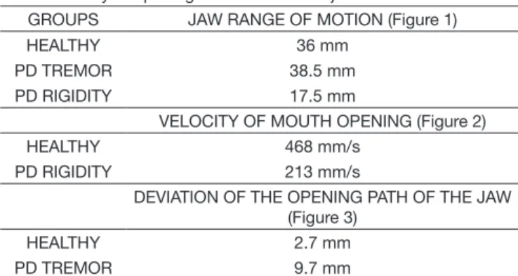
The analyses of results and JMs characteristics during the opening of the mouth are described in Table 1.
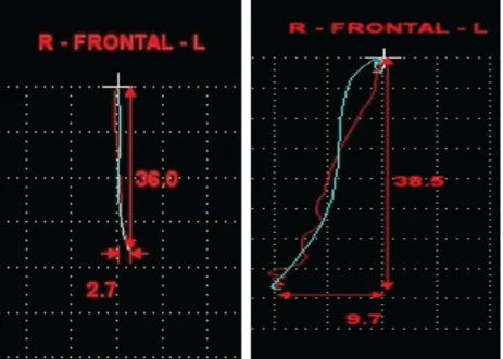
With regard to the range of the motion of mouth opening during JMs and chewing, we observed that there was a difference between the patient with predominant PD-rigidity and the other two non rigid subjects, the volunteer and the patient with PD-tremor (Figure 1).
Regarding velocity during the mouth-opening movement in the JMs and chewing, we observed that there was a noticeable delay in the displacement of the jaw during the requested task when comparing the healthy volunteer with the patient with PD-rigidity (Figure 2).
Analyzing jaw path millimetrically during masticatory movements and JMs, we observed that there were considerable eccentric deviations from the midline in the patient with PD-tremor compared with the volunteer without PD (Figure 3).
Table 1. Study of opening movement of the jaw
GROUPS JAW RANGE OF MOTION (Figure 1)
HEALTHY 36 mm
PD TREMOR 38.5 mm
PD RIGIDITY 17.5 mm
VELOCITY OF MOUTH OPENING (Figure 2)
HEALTHY 468 mm/s
PD RIGIDITY 213 mm/s
DEVIATION OF THE OPENING PATH OF THE JAW (Figure 3)
HEALTHY 2.7 mm
PD TREMOR 9.7 mm
CoDAS 2016;28(2):193-196
Jaw movement in people with Parkinson’s disease 195
Caption: R = Right; L = Left
Figure 1. Charts of the jaw range of motion
Caption: C = Closing; O = Opening
Figure 2. Charts of the velocity of mouth opening
CoDAS 2016;28(2):193-196
Albuquerque LCA, Silva HJ 196
DISCUSSION
Studies on the velocity, the range in motion, and the path of
body, and JMs have been performed in other research ields such
as neurology, odontology, physiotherapy, and speech-language pathology.
A prior systematic review of the literature conducted by the
team found reports similar to the indings in this brief description
of JMs in patients with PD(10-14).
JMs characteristics and their correlation to parameters that
are predominant, which are classiied and shown in the Hoehn
and Yahr scale(15) for patients with PD, are indings of paramount
importance related to speech-language pathology that are still scarcely reported in the literature.
Patients with PD-tremor may present deviations in JMs without apparent harm to their range of motion or velocity, in contrast with subjects with PD-rigidity, who may develop changes in velocity and range of motion during JMs and chewing while maintaining the mandibular path without deviation.
The main indings in this study and in the literature were
directly related to the dopamine levels found in the brain stem, considering that all the requested movements, despite being voluntary, were repetitive and partly unconscious; thus, they were controlled by subcortical structures.
CONCLUSIONS
The results showed that PD creates changes in the stomatognathic system, which are parafunctions represented by alterations in the range of motion, path, and velocity of JMs.
Therefore, we can conclude that patients with PD-tremor may not have limitations on range of motion of jaw and masticatory movements, but they may present alterations in the path. However, patients with PD-rigidity have limitations on the range of motion and velocity of JMs and chewing, but without harming the JMs path.
This led us to believe that, with regard to the functions of the stomatognathic system, the differentiation in treatments offered to patients with PD is necessary, while adapting and improving the techniques used in therapy, assessment, and diagnosis in speech-language pathology for different groups of patients with PD. Conducting further studies with a larger number of patients and methodological criteria adapted to the sample characteristics is equally necessary.
REFERENCES
1. Hornykiewicz O. Basic research on dopamine in Parkinson’s disease and the discovery of the nigrostriatal dopamine pathway: the view of an eyewitness. Neurodegener Dis. 2008;5(3-4):114-7. http://dx.doi.org/10.1159/000113678. PMid:18322366.
2. Leopold NA, Kagel MC. Prepharyngeal dysphagia in Parkinson’s disease. Dysphagia. 1996;11(1):14-22. http://dx.doi.org/10.1007/BF00385794. PMid:8556872.
3. Troche MS, Sapienza CM, Rosenbek JC. Effects of bolus con- sistency on timing and safety of swallow in patients with Par- kinson’s disease. Dysphagia. 2008;23(1):26-32. http://dx.doi.org/10.1007/s00455-007-9090-7. PMid:17551793.
4. Ali GN, Wallace KL, Schwartz R, DeCarle DJ, Zagami AS, Cook IJ. Mechanisms of oral-pharyngeal dysphagia in patients with Parkinson’s disease. Gastroenterology. 1996;110(2):383-92. http://dx.doi.org/10.1053/ gast.1996.v110.pm8566584. PMid:8566584.
5. Nagaya M, Kachi T, Yamada T, Igata A. Videofluorographic study of swallowing in Parkinson’s disease. Dysphagia. 1998;13(2):95-100. http:// dx.doi.org/10.1007/PL00009562. PMid:9513304.
6. Ertekin C, Tarlaci S, Aydogdu I, Kiylioglu N, Yuceyar N, Turman AB, et al. Electrophysiological evaluation of pharyngeal phase of swallowing in patients with Parkinson’s disease. Mov Disord. 2002;17(5):942-9. http:// dx.doi.org/10.1002/mds.10240. PMid:12360543.
7. Sarr MM, Pinto S, Jankowski L, Teston B, Purson A, Ghio A, et al. Contribution de la mesure de la pression intra-orale pour la compréhension des troubles de la coordination pneumophoniquedans la dysarthrieparkinsonienne. Rev Neurol (Paris). 2009;165(12):1055-61. http://dx.doi.org/10.1016/j. neurol.2009.03.012. PMid:19406446.
8. Bataglion SAN. Aplicação do questionário TMJ scale e eletrognatografia em indivíduos disfuncionados temporamandibulares com e sem tratamento ortodôntico e tratamento odontológico restaurador de dentística: estudo comparativo [dissertação]. Ribeirão Preto (SP): Universidade de São Paulo; 2001.
9. Walsh B, Smith A. Basic parameters of articulatory movements and acoustics in individuals with Parkinson’s disease. Mov Disord. 2012;27(7):843-50. http://dx.doi.org/10.1002/mds.24888. PMid:22729986.
10. Mazzone P, Padua L, Falisi G, Insola A, Florio TM, Scarnati E. Unilateral deep brain stimulation of the pedunculopontine tegmental nucleus improves oromotor movements in Parkinson’s disease. Brain Stimulat. 2012;5(4):634-41. http://dx.doi.org/10.1016/j.brs.2012.01.002. PMid:22410474.
11. Robertson LT, St George RJ, Carlson-Kuhta P, Hogarth P, Burchiel KJ, Horak FB. Site of deep brain stimulation and jaw velocity in Parkinson’s disease. J Neurosurg. 2011;115(5):985-94. http://dx.doi.org/10.3171/2011.7.JNS102173. PMid:21838506.
12. Umemoto G, Tsuboi Y, Kitashima A, Furuya H, Kikuta T. Impaired food transportation in Parkinson’s disease related to lingual bradykinesia. Dysphagia. 2011;26(3):250-5. http://dx.doi.org/10.1007/s00455-010-9296-y. PMid:20803220.
13. Bakke M, Larsen SL, Lautrup C, Karlsborg M. Orofacial function and oral health in patients with Parkinson’s disease. Eur J Oral Sci. 2011;119(1):27-32. http://dx.doi.org/10.1111/j.1600-0722.2010.00802.x. PMid:21244508.
14. Robertson LT, Horak FB, Anderson VC, Burchiel KJ, Hammerstad JP. Assessments of axial motor control during deep brain stimulation in parkinsonian patients. Neurosurgery. 2001;48(3):544-51, discussion 551-2. http://dx.doi.org/10.1097/00006123-200103000-00017. PMid:11270544.
15. Hoehn MM, Yahr MD. Parkinsonism: onset, progression, and mortality. Neurology. 1967;17(5):427-42. http://dx.doi.org/10.1212/WNL.17.5.427. PMid:6067254.
Author contributions