Department of Vascular Surgery, São Paulo University Medical School -São Paulo/SP, Brazil.
Email: [email protected]
Received for publication on January 01, 2006. Accepted for publication on March 17, 2006.
ORIGINAL RESEARCH
OBJECTIVE EVALUATION OF UPPER LIMB
CLAUDICATION: USE OF ISOKINETIC
DYNAMOMETRY
Lívio Nakano, Nelson Wolosker, Ruben Ayzin Rosoki, Baptista Muraco Netto, and Pedro Puech-Leão
Nakano L, Wolosker N, Rosoki RA, Muraco Netto B, Puech-Leão P. Objective evaluation of upper limb claudication: use of isokinetic dynamometry. Clinics. 2006;61(3):189-96.
OBJECTIVE: The objective of this work is to present an objective, practical, and reproducible method for evaluating the functional limitation caused by occlusive arterial disease in upper limbs: a stress test using an isokinetic dynamometer.
METHODS: Twenty-three patients with unilateral subclavian artery occlusive disease were included in the study, forming group 1. Seven patients of similar age, with atherosclerotic or Takayasu’s disease in the aorto-iliac segment, without subclavian artery occlusive disease, were included as a control group (group 2). For all tests, we utilized the CYBEX© 6000 isokinetic dynamometer. The elbow was tested using a series of 30 repetitions of extension and flexion of the arm, performed up to a maximum of 270 repetitions (9 series) or until the limit of the tested limb was reached.
RESULTS: We initially compared all the limbs without arterial disease of both groups to analyze whether they presented similar functional performance. No significant difference was found for any of the parameters studied. Considering that upper limbs without arterial disease have a similar response to exercise in these analyzed parameters, we compared the upper limbs in group 1 (with and without subclavian artery occlusive disease). For all parameters, the limbs with subclavian artery occlusive disease presented significantly lower values than the control limbs (P < 0.05), which can be objectively attributed to the ischemia (there
were different responses in different individuals, which allows the quantification of the limitation caused by subclavian artery occlusive disease).
CONCLUSION: In this study, we present a new stress test for impairment in patients with subclavian artery occlusive disease that might facilitate the classification of patients according to their functional impairment, and thus result in a better choice of therapeutic approach for each case as well as reporting of objective parameters that allow comparisons of the results of different treatments, including for long-term follow-up.
KEYWORDS: Ischemia. Upper extremity/blood supply. Exercise test/instrumentation. Intermittent claudication. Blood pressure. Subclavian steal syndrome.
INTRODUCTION
With the evolution of diagnosis and intervention tech-niques, increasing numbers of patients with occlusions in supra-aortic trunk arteries have been identified. The num-bers of procedures that such patients undergo have also
been increasing.1,2,3
The clinical status of subclavian artery occlusive dis-ease (SAOD) varies according to the degree of stenosis/oc-clusion4,5,6 and the quantity of collateral circulation.7,8,9,10 The presence of trophic lesions or ischemic pain is uncom-mon, but claudication of the upper limbs occurs with no-table frequency.11
measurements of segmental pressure differences, Doppler-calibrated pulse wave, and plethysmography.13,14,15
Although these tests are well established, the functional limitation caused by SAOD still cannot be objectively measured, and it continues to be subjectively estimated on the basis of the patient’s history, from the patient’s percep-tion of his own physical limitapercep-tions. Hence, it has not been possible to objectively evaluate the results from the vari-ous types of treatments available nowadays. This is a very important clinical problem.
The isokinetic dynamometer is today one of the more precise available methods for analysis of muscular perform-ance.16 The objective of this work was to present an objec-tive, practical, and reproducible method for evaluating the functional limitation caused by SAOD: ie, a stress test for upper limbs using an isokinetic dynamometer.
SAMPLE
Twenty-three patients with unilateral SAOD were in-cluded in the study, forming group 1. Seven patients of similar age, with atherosclerotic or Takayasu’s disease in the aortoiliac segment but without SAOD, were included as a control group (group 2). We selected the control group patients so as to match the group 1 patients with regard to sex, age, risk factors, and their sedentary lifestyles.
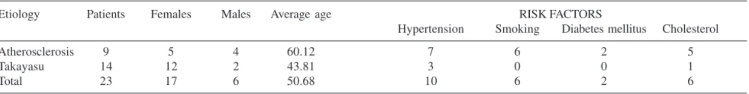
All the patients in group 1 were followed up regularly for at least 6 months, and they presented stable symptoms at the time of the test. All these patients were questioned about their symptoms; these ranged from mild and nonspe-cific symptoms to significant limitations on performing any daily activities (Table 1). Their ages ranged from 19 to 70 years (average of 50.6 years), and 17 patients were women (74%). Takayasu disease occurred in 14 patients, and atherosclerosis occurred in the other 9 (Table 2). The left limb (nondominant) was affected in 17 patients, and the right (dominant) was affected in 6 cases. The diagnosis and location of the arterial disease in group 1 were confirmed by arteriography or magnetic angioresonance.
Patients were not included in the study if they presented the following: an orthopedic disease or condition in the upper limbs that limited their physical performance; neu-rological disease that interfered with the perception of pain or led to motor deficiency or deformity of the upper limbs; active inflammatory disease progressing with limitation of mobilization or movement amplitude; or finally, functional limitation of physical activity for reasons other than SAOD, namely, acute myocardial infarct or angina, congestive heart failure of NYHA Class III (shortness of breath upon less-than-ordinary exertion), or states of dementia. Nor were patients with bilateral SAOD included.
The patients in group 2 were selected from among pa-tients with atherosclerosis or Takayasu’s disease but with-out SAOD who were attended at the vascular surgery with- out-patient service. Their ages ranged from 24 to 68 years (av-erage of 53 years), and there were 5 women. The absence of vascular lesions in the upper limbs was confirmed by physical examination, segmental pressure measurements, and Duplex scan.
Segmental pressure was recorded in both upper limbs for all patients. Those in group 1 presented a systolic arte-rial pressure gradient between their arms of at least 20 mm Hg. The patients in group 2 did not present pressure dif-ferences between their arms of more than 10 mm Hg.
METHOD
All patients underwent the isokinetic test using a CYBEXâ 6000 isokinetic dynamometer (Cybex Division of Lumex, Inc., 2100 Smithtown Ave, Ronkonkoma, NY 11779). All tests were monitored personally by the research-ers. All patients received continuous cardiac monitoring via cardioscopy, and 2 measurements of systolic blood pressure
Table 1 - Functional limitation imposed by patients’ symptoms
Number Symptomatic side Dominance Symptoms
1 left right Grade 2
2 right right Grade 3
3 left right Grade 2
4 left right Grade 2
5 left right Grade 2
6 left right Grade 1
7 left right Grade 1
8 right right Grade 1
9 left right Grade 1
10 right right Grade 2
11 left right Grade 1
12 left right Grade 2
13 left right Grade 2
14 left right Grade 1
15 right right Grade 2
16 right right Grade 1
17 right right Grade 2
18 left right Grade 1
19 left right Grade 1
20 left right Grade 3
21 left right Grade 2
22 left right Grade 1
23 left right Grade 2
Grade 0: No limitation of physical activity (Ordinary physical activity does not cause symptoms)
Grade 1: Slight limitation of physical activity (Ordinary physical activity does cause symptoms)
Grade 2: Moderate limitation of activity (Patient is comfortable at rest, but less than ordinary activities cause symptoms)
using a Doppler ultrassound were made in the tested limb immediately before and after the test, in order to observe all possible changes caused by the stress test.
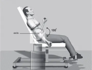
The patients were positioned on a chair that forms part of the equipment (UBXT module). The chest was secured to the bench, the contralateral limb was supported on a bar, and the arm to be tested was secured to the chest, so that interference from the trunk and shoulder musculature would be avoided during the test. The axis of movement was set up in the elbow immediately distal to the lateral epicondyle (Figure 1).
The elbow was tested using flexion/extension movement at an angular velocity of 180º per second. For this, an ad-aptation of the elbow flexion/extension protocol (DAP0115) recommended by the equipment manufacturer was utilized (equipment manual).
The amplitude of the flexion/extension movement was established across the range from zero (total extension) to 100o (total flexion), considering anatomical zero to be the extension of the elbow (Figure 1). Several series of 30 rep-etitions of the flexion/extension movement were performed with an interval of 5 seconds between each one, up to a maximum of 270 repetitions (9 series).
The patients underwent the test without any restriction on eating before the test. They only needed to observe that they were to avoid extensive physical activity using the up-per limbs during the 48 hours preceding the examination.
Before the test, patients were advised to concentrate only on their task, attempting to achieve as many repetitions as possible until the pain triggered by the exercise impeded them from continuing with the test. Patients were also re-quested to indicate the exact moment when pain began.
For the patients in group 1, the first limb tested was the
one without arterial disease, while for the patients in group 2, it was the dominant arm. This was followed by a period of rest until the heart rate, arterial pressure, and breathing rate returned to the levels measured before the test, and until the patient was ready to do the test on the other arm. At that time, the contralateral limb was also submitted to the standardized stress test.
All patients who interrupted the test before reaching 270 repetitions had to do so because of pain in the arm muscles that limited their test performance. Although all the patients presented occlusion of the proximal section of the subcla-vian artery before the emergence of the vertebral artery, no patient presented symptoms of subclavian steal syndrome, incapacitating dyspnea, or chest pain triggered by the test.
The following variables were studied: maximum number of repetitions; systolic arterial pressure before and immediately after the end of the exercise; heart rate moni-tored throughout the test and recorded after every 5 rep-etitions; measurements of muscle performance gauged by the isokinetic dynamometer; and maximum torque and to-tal work.
STATISTICAL ANALYSIS
Initially, the parameters for the limb without SAOD of group 1 were compared with those of group 2 (control). Student’s t test for independent samples was used for
study-ing the systolic arterial pressure (normal distribution). Other variables with nonparametric distribution (maximum number of repetitions, total work, and maximum torque) were analyzed using the Mann-Whitney test for independ-ent samples.
Similarly, the comparison between the limbs with and without SAOD among the patients in group 1 was done via Student’s t test for paired samples for the pressure variables
and via the Wilcoxon test for other variables.
The significance level of 0.05 (α = 5%) was adopted,
and probability levels (P) that were lower than this value
were considered to be significant and were marked by an asterick (*).
RESULTS
Considering the variables defined by our protocol, we chose an angular velocity of 180 degrees per second, be-cause this velocity is intermediate between a slow move-ment requiring great strength and a high velocity that re-quires sports reflex. Thus, the test could be performed by any individual without specific prior training. This veloc-ity allowed a larger number of repetitions and more pre-cise differentiation between the different degrees of
tional limitation. Within this protocol, 68% of the limbs without SAOD accomplished the test to conclusion, with 86% achieving 210 or more repetitions. The diseased limbs attained varying proportions of the number of repetitions possible, thus allowing objective quantification of their limitation. Only 17% of the limbs with SAOD achieved the 270 repetitions proposed, and 65% of these limbs attained less than 210 repetitions. The movement amplitude of 100 degrees was chosen because this is the normal articulation amplitude within which the best biomechanical perform-ance of muscle leverage by the flexors and extensors of the elbow takes place.
The dynamometer also registers muscular performance parameters that enable quantification of the limitation caused by SAOD according to the torque, ie, the force ap-plied through the distance traversed. This was analyzed in terms of peak torque, ie, the maximum torque value ob-tained for each series of 30 repetitions and thus represent-ing the best movement among all the repetitions. The work done by the movement was the product of the torque over the whole movement arc. This was analyzed as total work, ie, the sum of the work done through each movement.
We initially compared all the limbs without arterial dis-ease in group 1 versus all the limbs in group 2 to evaluate whether they presented similar functional performance. No statistical difference was found between groups 1 and 2 re-garding to the number of repetitions (P = 0.117), total work
throughout the test (P = 0.604), average work per series
of 30 repetitions (P = 0.916), average work per repetition
(P = 0.916), peak torque (P = 0.204), or the ratio between
the systolic arterial pressure measurements (P = 0.449).
The only parameter that presented a significant differ-ence between the two groups was the measurement of systolic arterial pressure before (P = 0.037) and after the
test (P = 0.031), which was greater in group 1 (unilateral
SAOD) than in group 2 (control).
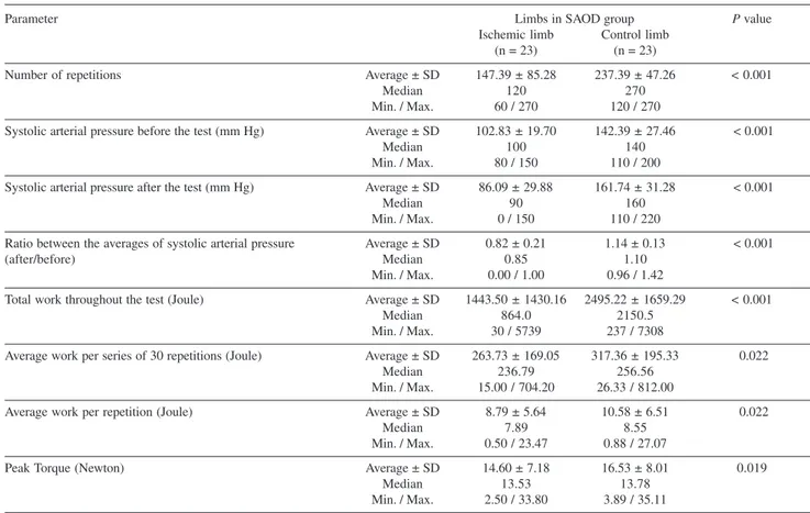
Considering that the parameters analyzed showed that upper limbs without arterial disease had similar responses to exercise, we decided to make comparisons between the upper limbs of each patient in group 1 (P with and without
SAOD). The parameters analyzed are shown in Table 3. Significant differences were found between the two groups in relation to number of repetitions (P < 0.001); total
work throughout the test (P < 0.001); average work per
se-ries of 30 repetitions (P = 0.022); average work per
rep-etition (P = 0.022); peak torque (P = 0.019); systolic
arte-rial pressure before the test (P < 0.001); systolic pressure
after the test (P < 0.001); and the ratio between the systolic
pressures (P < 0.001). For all these parameters, the limbs
with SAOD presented significantly lower values than the limbs without SAOD.
The maximum number of repetitions was registered as a first estimate of the functional limitation of the member submitted to the test.
The differences in systolic arterial blood pressure were used as a first estimate of the degree of limitation to perfusion caused by the arterial obstruction and the changes caused by the test. Although these measurements were con-sidered valuable, they are limited in many clinical situa-tions. On the basis of previous experience with lower limbs, absolute Doppler measurements alone do not necessarily correlate with clinical limitations and are not raised when patients increase their maximum walking distance.17
DISCUSSION
Treadmill test protocols are being used in the objective evaluation of the functional limitations of lower limbs due to ischemia (intermittent claudication).18,19,20 Subjective pa-rameters for estimating maximum walking distances are no longer used, and scientifically, they cannot be accepted anymore. When the ischemic functional limitation occurs in upper limbs, there is not a reliable method for objec-tively determining this limitation.
In 1986, Gerdle et al observed in lower limbs that meas-urements of work performed by patients with intermittent claudication that were obtained via an isokinetic dynamometer had a direct relationship with the maximum walking distance obtained in a treadmill test.21 Hedberg, in 1988, analyzed the results from the treatment of 2 groups of patients with intermittent claudication; the first group underwent surgical treatment with bypass surgery (8 pa-tients), and the second group underwent clinical treatment with physical exercises (9 patients), with the parameters obtained from isokinetic dynamometer prior to and after the treatment compared. Both groups increased the
maxi-Table 2 - Subclavian artery occlusive disease group - Patients’ characteristics
Etiology Patients Females Males Average age RISK FACTORS
Hypertension Smoking Diabetes mellitus Cholesterol
Atherosclerosis 9 5 4 60.12 7 6 2 5
Takayasu 14 12 2 43.81 3 0 0 1
mum walking distance, but only the first group achieved a significant increase for all parameters studied; the authors concluded that surgery had the best results for increasing muscular performance (maximum performance, electrical efficacy, and fatiguability level) for patients with intermit-tent claudication.22
However, there are clear differences between the mus-culature of the upper and lower limbs. Over the course of human evolution, the upper limbs have become specialized in performing movements that are finer and more precise than those of the lower limbs, thereby coming to present lesser but more specialized muscle mass that has lower blood support needs. The rich network of collateral circu-lation and lower demand for blood support make the symp-toms of obstructive arterial disease less evident and more heterogeneous.7
Isokinetic dynamometers are electromechanical devices coupled to microcomputers that generate a resistance equal and opposite to the force exerted by the patient’s muscu-lature when the movement is made at a predetermined con-stant angular velocity. These devices allow objective and dynamic evaluation of muscle performance in specific mus-cle groups.16
To facilitate study and analysis, we decided to select only patients with SAOD. The choice was made via a pro-tocol that allowed specific study to be made of the desired parameters of the muscle activity of the flexor and exten-sor groups of the elbow.
The obstruction of the subclavian artery often takes place at its origin, as was observed in all of our cases. Thus, one concern resulting from performing exercises of the upper limb would be the triggering of subclavian steal symptoms.23 This was not, however, observed in any of our cases.
The presence of SAOD is considered to be a risk fac-tor for heart disease,24,25,26 and debilitating physical exertion may cause severe consequences. In evaluating heart rate by continuous monitoring of all the patients, we observed that none of them reached rates close to their maximum cardiovascular capacity, and no alteration in the electrical activity of the heart was seen.
Because the test is done selectively for a specific ar-ticular movement, it has low cardiovascular impact.21 This allows us to conclude that the test is safe from the point of view of heart exertion. No cases of test interruption be-cause of clinical or electrocardiographic cardiac alteration were recorded in this series.
Table 3 - Parameters analyzed comparing the ischemic and control limbs in Subclavian artery occlusive disease (SAOD) group
Parameter Limbs in SAOD group P value
Ischemic limb Control limb
(n = 23) (n = 23)
Number of repetitions Average ± SD 147.39 ± 85.28 237.39 ± 47.26 < 0.001
Median 120 270
Min. / Max. 60 / 270 120 / 270
Systolic arterial pressure before the test (mm Hg) Average ± SD 102.83 ± 19.70 142.39 ± 27.46 < 0.001
Median 100 140
Min. / Max. 80 / 150 110 / 200
Systolic arterial pressure after the test (mm Hg) Average ± SD 86.09 ± 29.88 161.74 ± 31.28 < 0.001
Median 90 160
Min. / Max. 0 / 150 110 / 220
Ratio between the averages of systolic arterial pressure Average ± SD 0.82 ± 0.21 1.14 ± 0.13 < 0.001
(after/before) Median 0.85 1.10
Min. / Max. 0.00 / 1.00 0.96 / 1.42
Total work throughout the test (Joule) Average ± SD 1443.50 ± 1430.16 2495.22 ± 1659.29 < 0.001
Median 864.0 2150.5
Min. / Max. 30 / 5739 237 / 7308
Average work per series of 30 repetitions (Joule) Average ± SD 263.73 ± 169.05 317.36 ± 195.33 0.022
Median 236.79 256.56
Min. / Max. 15.00 / 704.20 26.33 / 812.00
Average work per repetition (Joule) Average ± SD 8.79 ± 5.64 10.58 ± 6.51 0.022
Median 7.89 8.55
Min. / Max. 0.50 / 23.47 0.88 / 27.07
Peak Torque (Newton) Average ± SD 14.60 ± 7.18 16.53 ± 8.01 0.019
Median 13.53 13.78
In addition, this type of approach offers control of the movement in relation to the angular velocity. It allows re-cording of the torque produced during the whole amplitude of the movement, as well as identification of regions of strength or weakness within the amplitude of the move-ment. The tests can be repeated indefinitely with the im-mediate obtaining of results. The data can be stored on magnetic media for later use, especially for studies of pa-tients’ sequential evolution.
We initially sought to compare the limbs without arte-rial disease in the two groups to determine whether arms with SAOD(group 1) interfered in the functional perform-ance of the contralateral limb (without arterial disease). We verified that there was no significant difference in the mus-cle performance between the normal limbs. Therefore, we could move on to analyze the test results between the mem-bers of group 1.
Both upper limbs were tested until they reached their maximum limit, ie, the moment when the degree of pain or fatigue started to impede the continuation of the test, or when the maximum number of repetitions (270) was reached. This maximum number was set via a prior pilot study and had been found to be the most appropriate limit for the analysis of muscle function in the manner desired. This number of repetitions proved to be optimal, because the healthy limbs completed the test, while the diseased ones remained below this number. If the number of repeti-tions had been fewer, it would have been insufficient to pro-voke symptoms in cases with lower ischemic limitations.
The analysis of the maximum number of repetitions al-lowed the identification of a significant difference between the limbs with and without SAOD. There was a correla-tion with the degree of limitacorrela-tion reported by the patient, although we observed a significant variation in the number of repetitions among the limbs with SAOD.
The behavior of the systolic arterial pressure in each upper limb before and after the test was similar to what has been observed in lower limbs submitted to physical ex-ercise.27 We observed that the systolic pressure ratio tended to fall in limbs with SAOD and rise in normal limbs in a similar manner to what occurs in lower limbs with inter-mittent claudication. Although we consider this to be im-portant data for clinical diagnosis, neither arterial pressure recorded while resting, nor the degree of fall in systolic arterial pressure had a direct relationship with the restric-tion in the number of repetirestric-tions or work achieved by the limb. In lower limbs with intermittent claudication under-going conservative treatment with physical activity, we have often observed an increase in the maximum walking distances without any increase in the absolute arterial pres-sure of the limb or alteration in the ankle brachial index.
The differences in systolic arterial blood pressure were used as a first estimate of the degree of limitation to perfusion caused by arterial obstruction as well as the changes caused by the test. Although these measurements were considered valuable, they are limited in many clini-cal situations. On the basis of previous experience with lower limbs, absolute Doppler measurements alone do not necessarily correlate with clinical limitations and are not raised when patients increase their maximum walking dis-tance.28
Analysis of the maximum number of repetitions allowed identification of a significant difference between the limbs with and without SAOD .
While Isokinetic dynamometry is commonly used to evaluate strength, it also can be utilized as a resistance test.29-33
Peak torque is considered to be the most important isokinetic parameter for evaluating muscular strength.32 It can be used for finding any early impairment in muscular performance of the tested limbs as well as for measurement of the level of strength achieved for both limbs,
Total work was analyzed as a complementary param-eter for the muscular strength, offering data about work done by the muscular group during the whole test, and in this case, showing different levels of the patients’ exercise resistance between the two groups. Gerdle, in 1987, con-sidered it the most sensitive parameter for evaluating mus-cular fatigue.34
Data recorded by the dynamometer also showed lower values in peak torque and total work for the ischemic limb. For these parameters, the lower performance of limbs with SAOD in comparison to normal limbs can be objectively at-tributed to ischemia. These data allowed the degree of mus-cle performance during the test to be detailed. Some patients began the test with greater effort that subsequently dimin-ished, while others maintained a constant level throughout the test. Patients probably develop different mechanisms to overcome the limitation caused by their SAOD.
In practical terms related to patients with conservative management, we will be able to follow up the long-term results of drug or exercise therapy as well as to propose interventions in the event of a poor response to therapy, decrease in muscle performance, or occurrence of a sig-nificant limitation. Regarding patients undergoing
revascularization, we will be able to estimate the increase in muscle performance and follow up the clinical success. If thrombosis of the angioplasty or graft were to occur, we would be able to estimate the degree of worsening in terms of functional limitation, and thus decide more accurately on the necessity for further intervention.
RESUMO
Nakano L, Wolosker N, Rosoki RA, Muraco Netto B, Puech-Leão P. Avaliação objetiva da isquemia de membros superiores: uso do dinamômetro isocinético. Clinics. 2006;61(3):189-96.
OBJETIVO: O objetivo deste trabalho é apresentar um método para a avaliação da limitação funcional causada por doença arterial oclusiva de artéria subclávia: o teste de es-forço utilizando o dinamômetro isocinético.
MÉTODO: Pacientes com trombose unilateral de artéria subclávia foram selecionados, reunindo 23 pacientes no Gru-po com doença arterial oclusiva de artéria subclávia. Sete pa-cientes com idade semelhante, sem doença arterial em mem-bros superiores foram incluídos, formando o grupo controle. Para a realização do teste, utilizou-se o dinamômetro isocinético CYBEX® 6000. O cotovelo foi testado em séries consecutivas de 30 repetições do movimento de extensão e flexão, até que se atingisse o máximo de 270 repetições (9 séries), ou até que se alcançasse o limite do membro testado.
RESULTADO: Inicialmente comparou-se todos os mem-bros sem doença arterial dos dois grupos, para analisar se apresentavam desempenho semelhante. Não houve
diferen-ça estatística entre os grupos em relação a todos os parâmetros estudados. Comparou-se então, os dois mem-bros de cada paciente do Grupo doença arterial oclusiva de artéria subclávia. Em todos os parâmetros analisados, os membros com doença arterial oclusiva de artéria subclávia apresentaram diferença estatística (p < 0.05) em relação aos membros controle, o que foi objetivamente atri-buído à isquemia. (Foram registrados diferentes graus de limitação entre os paciente, o que permite estimar objeti-vamente o grau de limitação causado pela isquemia cau-sada pela oclusão da subclávia)
CONCLUSÃO: Este teste de esforço permite que pacien-tes com isquemia de membros superiores sejam avaliados e estratificados, conforme o grau de sua limitação funcio-nal, o que facilitará a escolha da melhor terapêutica para cada caso e a obtenção de parâmetros para comparação do resultado de diferentes tratamentos e para o seguimento clí-nico em longo prazo.
UNITERMOS: Isquemia. Extremidade Superior/irrigação sangüínea. Teste de esforço/instrumentação. Claudicação intermitente. Pressão arterial. Síndrome do roubo subclávio.
REFERENCES
1. Berguer R, Morasch MD, Kline RA, Kazmers A, Friedland MS. Cervical reconstruction of the supra-aortic trunks: a 16-year experience. J Vasc Surg. 1999;29:239-46.
2. Mesh CL, McCarthy WJ, Pearce WH, Flinn WR, Shireman PK, Yao JS. Upper extremity bypass grafting. A 15-year experience. Arch Surg. 1993;128:795-801.
3. Queral LA, Criado FJ. The treatment of focal aortic arch branch lesions with Palmaz stents. J Vasc Surg. 1996;23:368-75.
4. Fields WS, Lemak NA. Joint Study of Extracranial Arterial Occlusion. VII. Subclavian steal – a review of 168 cases. JAMA. 1972;222:1139-43.
5. Johnston KW. Neurovascular conditions involving the upper extremity. In: Rutherford RB, editor. Vascular Surgery. 4th ed. Philadelphia, USA: WB Saunders; 1995. p. 913-7.
6. Rapp JH, Reilly LM, Goldstone J, Krupsky WC, Ehremfeld WK, Stoney RJ. Ischemia of the upper extremity: Significance of proximal arterial disease. Am J Surg. 1986;152:122-6.
7. Williams SJ 2nd. Chronic upper extremity ischemia: current concepts in management. Surg Clin North Am. 1986;66:355-75.
9. Gosselin C, Walker PM. Subclavian Steal Syndrome: Existence, Clinical Features, Diagnosis and Management. Sem Vasc Surg. 1996;9:93-7. 10. Ackermann H, Diener HC, Seboldt H, Huth C. Ultrasonographic
follow-up of subclavian stenosis and occlusion: Natural history and surgical treatment. Stroke. 1988;19:431-5.
11. Welling RE, Cranley JJ, Krause RJ, Hafner CD. Obliterative arterial disease of the upper extremity. Arch Surg. 1981;116:1593-96. 12. Ouriel K. Noninvasive diagnosis of upper extremity vascular disease.
Sem Vasc Surg .1998;11(2):54-9.
13. Baxter BT, Blackburn D, Payne K, Pearce WH, Yao JS. Noninvasive evaluation of the upper extremity. Surg Clin North Am. 1990;70:87-97. 14. Edwards JM, Porter JM. Upper extremity arterial disease: etiological considerations and differential diagnosis. Sem Vasc Surg. 1998;11:60-6. 15. Edwards WH. Vertebral artery reconstruction: indications and
techniques. Sem Vasc Surg. 1996;9:105-10.
16. Gardner AW, Skinner JS, Smith LK. Reliability of testing the knee extensors and flexors in healthy adult women using a Cybex II isokinetic Dynamometer. J Orthop Sports Phys Ther. 1991;14:37-47.
17. Wolosker N, Rosoky RA, Nakano L, Basyches M, Puech-Leao P. Predictive value of the ankle-brachial index in the evaluation of intermittent claudication. Rev Hosp Clin Fac Med Sao Paulo. 2000;55:61-4.
18. Gardner AW, Skinner JS, Smith LK. Effects of handrail support on claudication and hemodynamic responses to single-stage and progressive treadmill protocols in peripheral vascular occlusive disease. Am J Cardiol. 1991;68:99-105.
19. Hiatt WR, Hirsch AT, Regensteiner JG, Brass EP. Clinical trials for claudication. Assessment of exercise performance, functional status and clinical end points. Circulation. 1995;92:614-21.
20. Wolosker N, Nakano L, Rosoky RA, Puech-Leao P.Evaluation of walking capacity over time in 500 patients with intermittent claudication who underwent clinical treatment. Arch Intern Med. 2003;163:2296-300.
21. Gerdle B, Hedberg B, Angquist KA, Fugl-Meyer AR. Isokinetic strength and endurance in peripheral arterial insufficiency with intermittent claudication. Scand J Rehabil Med. 1986;18:9-15.
22. Hedberg B, Langstrom M, Angquist KA, Fugl-Meyer AR. Isokinetic plantar flexor performance and fatiguability in peripheral arterial insufficiency. Effects of training vs. vascular surgery. Acta Chir Scand. 1988;154:363-9.
23. Webster MW, Downs L, Yonas H, Makaroun MS, Steed DL. The effect of arm exercise on regional cerebral blood flow in the subclavian steal syndrome. Am J Surg. 1994;168:91-3.
24. Vogt DP, Hertzer NR, O’Hara PJ, Beven EG. Brachiocephalic artery reconstruction. Ann Surg. 1982;196:541-52.
25. Brewster DC, Moncure AC, Darling RC, Ambrosino JJ, Abbott WM. Innominate artery lesions: problems encountered and lessons learned. J Vasc Surg 1985;2(1):99-112.
26. Evans WE, Williams TE, Hayes JP. Aortobrachiocephalic reconstruction. Am J Surg. 1988;156:100-2.
27. Carter SA: Response of ankle systolic pressure to leg exercise in mild or questionable arterial disease. N Engl J Med. 1972;287:578-82. 28. Jonason T, Ringqvist I. Changes in peripheral blood pressures after five
years of follow up in nonoperated patients with intermittent claudication. Acta Med Scand. 1986;220:127-32.
29. Patton RW, Hinson MM, Arnold BR, Lessard B. Fatigue curves of isokinetic contractions. Arch Phys Med Rehabil. 1979;59:507-9. 30. Burdett RG, Swearinger JV. Reliability of isokinetic muscle endurance
tests. J Orthop Sports Phys Ther. 1987;8:484-8.
31. Montgomery LC, Douglas LW, Deuster PA. Reliability of an isokinetic test of muscle strength and endurance. J Orthop Sports Phys Ther. 1989;11:315-22
32. Kannus P. Isokinetic evaluation of muscular performance: implications for muscle testing and rehabilitation. Int J Sports Med. 1994;15 Suppl 1:S11-8.
33. Thorstensson A, Karlsson J. Fatiguability and fibre composition of human skeletal muscle. Acta Physiol Scand. 1976;98:318-22. 34. Gerdle B, Langström M. Repeated isokinetic plantar flexions at different