In vitro
cytotoxicity of self-curing acrylic resins
of different colors
Luciana Borges Retamoso1, Taís de Morais Alves da Cunha2, Matheus Melo Pithon3, Rogério Lacerda dos Santos3,
Fernanda Otaviano Martins4, Maria Teresa Villela Romanos5, Orlando Motohiro Tanaka6
Objective: The aim of this study was to assess the in vitro cytotoxicity of acrylic resins of different colors over time. Methods: Specimens were divided into 4 groups (n = 6) according to the color of the acrylic resin (Orto Class, Clássico, Campinas, São Paulo, Brazil): Group 1: clear acrylic resin; group 2: pink acrylic resin; group 3: blue acrylic resin and group 4: green acrylic resin. All specimens were fabricated according to the mass manipulation technique and submitted to mechanical polishing protocol. The control was performed with an amalgam specimen (C+), a glass specimen (C-) and cell control (CC). Specimens were immersed in Minimum Eagle’s Medium (MEM) and incubated for 24 h at 37o C. The extracts from the experimental
material were filtered and mixed with L929 fibroblast. Cytotoxicity was evaluated at 4 different times, 24, 48, 72 and 168 h. After contact, cells were incubated for 24 h and added to 100 µ of 0.01% neutral red dye. The cells were incubated for 3 h for pigment incorporation and fixed. Cells viability was determined by a spectroscopic (BioTek, Winooski, Vermont, USA) with a 492-nm wavelength λ=492 nm). Results: There were no statistical differences between the experimental groups and the CC and C- groups. Conclusion: Clear, pink, blue and green self-curing acrylic resins fabricated by means of the mass manipula-tion technique and mechanically polished are not cytotoxic. Neither the pigment added to the self-curing acrylic resin nor the factor of time influenced the cytotoxicity of the material.
Keywords:Acrylic resins. Cell culture techniques. Cytotoxins.
How to cite this article: Retamoso LB, Cunha TMA, Pithon MM, San-tos RL, Martins FO, Romanos MTV, Tanaka OM. In vitro cytotoxicity of self-curing acrylic resins of different colors. Dental Press J Orthod. 2014 July-Aug;19(4):66-70. DOI: http://dx.doi.org/10.1590/2176-9451.19.4.066-070.oar
» The authors report no commercial, proprietary or financial interest in the products or companies described in this article.
Contact address: Orlando Motohiro Tanaka
R. Imaculada Conceição, 1155 ‒ CEP: 80215-901 ‒ Curitiba/PR — Brazil E-mail: [email protected]
1 PhD resident in Dental Material, Catholic University of Rio Grande do Sul
(PUC-RS).
2 MSc in Orthodontics, Catholic University of Paraná (PUC-PR).
3 PhD resident in Orthodontics, Federal University of Rio de Janeiro (UFRJ).
4 MSc in Immunobiological Technology, Oswaldo Cruz Foundation
(FIOCRUZ).
5 Adjunct Professor, UFRJ.
6 Full Professor, Department of Orthodontics, –PUCPR.
Submitted: May 18, 2009 - Revised and accepted: April 12, 2010
DOI: http://dx.doi.org/10.1590/2176-9451.19.4.066-070.oar
Objetivo: avaliar, in vitro, a citotoxicidade de resinas acrílicas autopolimerizáveis, de diferentes cores, ao longo do tempo. Métodos: os corpos de prova foram divididos em quatro grupos (n = 3), de acordo com a cor da resina acrílica utilizada (Orto Class, Clássico, São Paulo/SP), sendo: grupo 1, acrílica incolor; grupo 2, acrílica rosa; grupo 3, acrílica azul; e, grupo 4, acrílico verde. Todos os corpos de prova foram confeccionados pela técnica de massa e polidos mecanicamente. Um corpo de prova de amálgama, um de vidro e célula constituíram o controle positivo (C+), controle negativo (C-), e controle de célula (CC), respectivamente. Em seguida, esses foram imersos em meio mínimo essencial de Eagle (MEM) por 24h, quando se removeu o sobrenadante e colocou-os em contato com fibroblastos L929. Avaliou-se a citotoxicidade em quatro períodos: 24, 48, 72 e 168h. Após o contato com o meio, as células foram incubadas por 24h e adicionou-se 100µ do corante vermelho neutro a 0,01%. Posteriormente, as células foram incubadas por 3h, para incorporação do co-rante, e fixadas. A contagem das células viáveis foi realizada em espectrofotômetro (BioTek, Winooski, EUA), com um comprimento de onda de 492nm (λ = 492nm). Resultados: não houve diferença estatística entre os grupos experimen-tais e os grupos CC e C-. Conclusões: as resinas acrílicas autopolimerizáveis incolor, rosa, azul e verde, manipuladas pela técnica de massa e polidas mecanicamente não são citotóxicas. O corante utilizado em resinas autopolimerizáveis e tempo não influenciam na citotoxocidade do material.
INTRODUCTION
Chemically activated acrylic resins are widely used in the fabrication of ixed, removable and reten-tion orthodontic appliances.4 Resin is sold in two
vi-als: one containing the powder, the polymer, and the other containing the liquid, the monomer. The mono-mer is a clear, lammable and volatile liquid at room temperature.1 It is considered cytotoxic and possibly
genotoxic.9,10,22,23,24 The polymer usually has the
pig-ment that gives color to the resin.
Adding the monomer (methyl methacrylate, MMA) to the polymer causes a resin polymerization reaction that occurs without the formation of by-products. Nevertheless, conversion of monomer into polymer is generally not complete,4 for this reason,
some amount of monomer, known as residual,2
re-mains. According to some studies, residual monomer remains in the manufactured orthodontic applianc-es, which indicates that varying amounts of residual monomer may be released into the oral cavity during the use of these appliances.4,10
The residual monomer of methyl methacrylate not only changes the inal physical properties of resins,3,12
but also induces the onset of systemic and local sue reactions when in contact with saliva and sot tis-sues,8,9,11,17 thus causing hypersensitivity, lip swelling,
chronic urticaria and sialorrhea.5,6 Furthermore, the
pigment added to the powder may be another causative factor of hypersensitivity.
Although the in vitro cytotoxicity of MMA has al-ready been demonstrated, no studies were conducted to assess the inluence of pigments present in colored acrylics on cell viability. Thus, the aim of this study was to evaluate the in vitro cytotoxicity of acrylic resin at dif-ferent periods and compare the cytotoxicity of acrylic resins of diferent colors.
MATERIAL AND METHODS
Preparation of specimens
For preparation of specimens, a metal matrix (10 mm X 5 mm X 2 mm) was molded with addition silicone (Express®, 3M/ESPE, St. Paul, USA) and the mold illed
with self-curing acrylic resin (Ortho Class®, Classic,
Campinas, São Paulo, Brazil). Powder-liquid ratio was obtained according to the manufacturer’s instructions.
Each acrylic resin used for preparation of specimens was manipulated by means of the mass technique in
a dappen dish with a lid where the monomer was in-serted immediately before the polymer was poured until its saturation. Subsequently, the dish was covered with the lid, which allowed the resin to go through a sandy and ibrillar phase until it reached its plastic phase during which it was inserted into the mold. The acrylic resin was processed in a resin polymerizer M-1000®
(EDG Equipment and Control Ltda.) at 20°C and pres-sure of 25 psi (1.75 kg/cm²) for a period of 15 minutes.
Ater polymerization, mechanical polishing was car-ried out in a vise using a bristle brush with a mixture pumice and water for 1 minute, followed by felt with white paste of Spain used for 1 minute.
Groups
Four self-curing acrylic resins of diferent col-ors were divided into four groups as follows (n = 3): Group 1 (clear), 2 (pink), 3 (blue) and 4 (green).
Control
To assess cellular response against extremes, other three groups (n = 3) were included: Group CC (cell control), cells which were not exposed to any ma-terial. This group was used to monitor normal cell growth. Group C+ (positive control) consisting of specimen made of amalgam. Silver amalgam was used because of its well known cytotoxic ability.18
Speci-mens of 10 mm x 5 mm x 2 mm were manufactured in amalgamator (SDI®, Bayswater, Australia) and polished
with abrasive rubber tips. Group C- (negative control) consisting of glass specimen. Glass was the material of choice for not triggering cytotoxicity efect.19
Cell culture
Cell lineage used was L929 obtained from the Ameri-can Type Culture Collection (ATCC, Rockville, MD) (mouse ibroblasts) grown in Eagle’s minimum essen-tial medium (MEM) (Cultilab, Campinas, São Paulo, Brazil), supplemented with 2 mM L-glutamine (Sigma, St. Louis, Missouri, USA) 50 µg/mL gentamycin (Scher-ing Plough, Kenilworth, New Jersey, USA), 2.5 µg/mL fungizone (Bristol-Myers-Squibb, New York, USA), 0.25 mL sodium bicarbonate solution (Merck, Darm-stadt, Germany), 10 mM HEPES (Sigma, St. Louis, Mis-souri, USA) and 10 % fetal bovine serum (FBS) (Culti-lab, Campinas, São Paulo, Brazil). It was kept at 37oC in
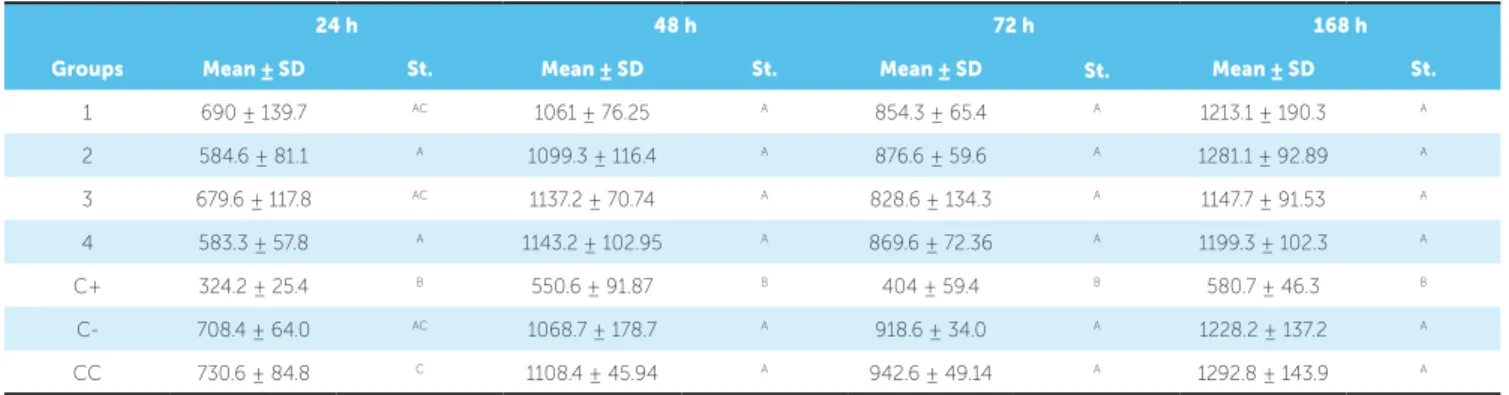
Table 1 - Mean and standard deviation of the amount of viable cells and statistical analysis of evaluated groups.
Mean: mean values of the amount of viable cells; SD: Standard deviation;
St: Statistics. Same letters account for the absence of statistical difference for Tukey’s test (p < 0.05).
24 h 48 h 72 h 168 h
Groups Mean ± SD St. Mean ± SD St. Mean ± SD St. Mean ± SD St.
1 690 ± 139.7 AC 1061 ± 76.25 A 854.3 ± 65.4 A 1213.1 ± 190.3 A
2 584.6 ± 81.1 A 1099.3 ± 116.4 A 876.6 ± 59.6 A 1281.1 ± 92.89 A
3 679.6 ± 117.8 AC 1137.2 ± 70.74 A 828.6 ± 134.3 A 1147.7 ± 91.53 A
4 583.3 ± 57.8 A 1143.2 ± 102.95 A 869.6 ± 72.36 A 1199.3 ± 102.3 A
C+ 324.2 ± 25.4 B 550.6 ± 91.87 B 404 ± 59.4 B 580.7 ± 46.3 B
C- 708.4 ± 64.0 AC 1068.7 ± 178.7 A 918.6 ± 34.0 A 1228.2 ± 137.2 A
CC 730.6 ± 84.8 C 1108.4 ± 45.94 A 942.6 ± 49.14 A 1292.8 ± 143.9 A
Cytotoxicity assay
Acrylic resin, silver amalgam and glass specimens were sterilized by exposure to UV light (Labconco, Kansas, Missouri, USA) for 1 hour.19 Then, three samples of each
material were placed in 24-well plates containing culture medium (MEM) (Cultilab, Campinas, São Paulo, Bra-zil). Supernatants were collected according to the time of evaluation, 24, 48, 72 and 168 hours (7 days), being the culture medium renewed every 24 hours.
Supernatants were placed, in triplicate, in 96-well plates containing conluent monolayer of L929 cells and incubated for 24 hours at 37oC in an environment
con-taining 5 % CO2. Ater incubation, the efect on cell vi-ability was determined by means of the dye-uptake tech-nique, as described by Neyndorf et al,16 but with minor
modiications. The technique consists in adding 100 µL of 0.01 % neutral red (Sigma, St. Louis, Missouri, USA) into culture medium and incubation at 37°C for 3 hours for penetration of the dye in living cells.
Ater this period, the dye was discarded and the cells ixed for 5 minutes by adding 100 µL of formaldehyde solution (Reagen, Rio de Janeiro, Rio de Janeiro, Bra-zil) to 4 % in PBS (130 mM NaCl, 2 mM KCl; 6 mM Na2HPO4.2H2O, 1 mM K2HPO4, pH 7.2). Subse-quently, the dye was extracted by adding 100 µL of a solution of 1 % acetic acid (Vetec, Rio de Janeiro, Rio de Janeiro, Brazil) and 50 % methanol (Reagen, Rio de Janeiro, Rio de Janeiro, Brazil). Ater 20 minutes, a spectrophotometer (BioTek, Winooski, Vermont, USA) at a wavelength of 492 ηm (λ = 492 ηm) was used to read the data.
Statistical analysis
Statistical analysis was performed with the SPSS 13.0 sotware (SPSS Inc., Chicago, Illinois, USA). Initially, data were submitted to Kolmogorov Smirnov and Lev-ene’s test to determine normality and homogeneity, re-spectively. The values of the amount of viable cells were subjected to analysis of variance (ANOVA), with two factors (color and time) to determine whether there were statistical diferences between groups, and subsequently to Tukey’s test (Table 1). Signiicance level was set at 5 %.
RESULTS
Results revealed increased cell viability from 24 to 48 h, a reduction in cell viability ater 72 h and, an in-crease in cell viability ater 168 h; however, with no sta-tistically signiicant diferences (P≥ 0.05).
The color of resin proved not to inluence material cytotoxicity, since there were no diferences between groups 1, 2, 3 and 4 at all times (P≥ 0.05).
DISCUSSION
Acrylic resins are widely used in Dentistry; however, some studies have demonstrated that this material can cause allergic reactions.13,14,15 Nevertheless, most
re-searches assessed material used for prosthetic purposes, most of which are heat-curing
Self-curing acrylic resins are the most frequently used in Orthodontics. According to Hensten-Pettersen and Wictorin,7 polymerization inluences cytotoxicity.
Their studies revealed lower cell growth in self-curing resins in comparison to heat-curing ones, and for both, cell growth was lower than in the control group.
Baker et al2 found that residual monomer
concentra-tion was four times higher in saliva adjacent to the pala-tal surface of appliances manufactured with acrylic resin in comparison to total saliva, thus indicating the impor-tance of assessing cytotoxicity of this material, as well as the efects produced by the pigment and by the time of exposure on material cytotoxic potential.
Acrylic resin color proved not to afect cell viabil-ity, thus suggesting that the pigment does not inlu-ence cytotoxicity levels. However, when specimens were made of pink and green acrylic resin, normal cell growth was modiied, as shown by experimental groups 2 and 4 which difered from CC. Thus, toxic reaction seems not to be associated with neither pig-ment nor the other substances that constitute the poly-mer, but with increase in the level of residual mono-mer present in the material.6
In this study, groups 1, 2, 3 and 4 showed no statis-tically signiicant diferences over time (P ≤ 0.05), thus indicating that cytotoxicity was not afected within the times tested. This inding is in disagreement with Gonçalves et al6 who assessed cytotoxicity of acrylic resins
for orthodontic purposes within 24 and 48 hours. Their results showed that there was less cell viability ater 24 hours. This diference can be explained by the cell type used, given that Gonçalves et al6 used epithelial cells
and not ibroblasts. Polishing may also be considered as,
according to Rocha Filho et al20 and Gonçalves et al,4 it
alters the level of residual monomer present in the acrylic resins. Release of residual monomer is responsible for the reduction in cell viability, and this release is more intense within the irst 24 h.4 However, mechanical polishing
de-creases the levels of residual monomer6. For this reason,
it is suggested that the polishing procedure performed in this study was key not to trigger toxic reaction.
Although there was no signiicant diference be-tween the times of assessment, in all experimen-tal groups, from 48 to 72 hours there was a decrease in the number of viable cells. The fact that release of residual monomer is considered crucial in de-termining cell viability corroborates the results by Rocha Filho et al.20 In the graphs of this study, the
au-thors demonstrate increased concentration of residual monomer from 2 to 5 days.
It is recommended that acrylic resin used to manu-facture orthodontic appliances be properly propor-tioned and manipulated, following the manufactur-er’s recommendations to ensure safety for patients’ health. Some measures may be taken to reduce the amount of residual monomer, such as: polymerization in water or under pressure, the use of correct mono-mer : polymono-mer proportion, and storage in water for 72 hours ater polymerization.2,17,21 Moreover, the
difer-ent colors of resin tested can be used without causing any damage to the biocompatibility of the material.
CONCLUSION
According to the methodology used and the condi-tions established in this research, it can be concluded that: 1) Acrylic resin manufactured by means of the mass technique, polymerized under pressure and mechanically polished does not alter cell viability.
2) Color of acrylic resin has no efect on cell vi-ability.
1. Aydin O, Attila G, Dogan A, Aydin MV, Canacankatan N, Kanik A. The efects of methyl methacrylate on nasal cavity, lung, and antioxidant system (an experimental inhalation study). Toxicol Pathol. 2002;30(3):350-6. 2. Baker S, Brooks SC, Walker DM. The release of residual monomeric methyl
methacrylate from acrylic appliances in the human mouth: an assay for monomer in saliva. J Dent Res. 1988;67(10):1295-9.
3. Del Bel Cury AA, Rached RN, Ganzarolli SM. Microwave-cured acrylic resins and silicone-gypsum moulding technique. J Oral Rehabil. 2001;28(5):433-8. 4. Gonçalves TS, Menezes LM, Silva LE. Residual monomer of autopolymerized
acrylic resin according to diferent manipulation and polishing methods. Angle Orthod. 2008;78(4):722-7.
5. Gonçalves TS, Morganti MA, Campos LC, Rizzatto SM, Menezes LM. Allergy to auto-polymerized acrylic resin in an orthodontic patient. Am J Orthod Dentofacial Orthop. 2006;129(3):431-5.
6. Siqueira Gonçalves T, Minghelli Schmitt V, Thomas M, Lopes de Souza MA, Macedo de Menezes L. Cytotoxicity of two autopolymerized acrylic resins used in orthodontics. Angle Orthod. 2008;78(5):926-30.
7. Hensten-Pettersen A, Wictorin L. The cytotoxic efect of denture base polymers. Acta Odontol Scand. 1981;39(2):101-6.
8. Hochman N, Zalkind M. Hypersensitivity to methyl methacrylate: mode of treatment. J Prosthet Dent. 1997;77(1):93-6.
9. Jorge JH, Giampaolo ET, Machado AL, Vergani CE. Cytotoxicity of denture base acrylic resins: a literature review. J Prosthet Dent. 2003;90(2):190-3.
10. Kedjarune U, Charoenworaluk N, Koontongkaew S. Release of methyl methacrylate from heat-cured and autopolymerized resins: cytotoxicity testing related to residual monomer. Aust Dent J. 1999;44(1):25-30.
11. Lassila LVJ, Vallittu PK. Denture base polymer Alldent Sinomer®: mechanical properties, water sorption and realease of residual compounds. J Oral Rehabil. 2001;28(7):607-13.
12. Lee SY, Lai YL, Hsu TS. Inluence of polymerization conditions on monomer elution and microhardness of autopolymerized polymelhyl methacrylate resin. Eur J Oral Sci. 2002;110(2):179-83.
13. Lunder T, Rogl-Butina M. Chronic urticaria from an acrylic dental prosthesis. Contact Dermatitis. 2000;43(4):232-3.
REFERENCES
14. McCabe JF, Basker RM. Tissue sensitivity to acrylic resin. A method of measuring the residual monomer content and its clinical application. Br Dent J. 1976;140(10):347-50.
15. Nealey ET, Del Rio CE. Stomatitis venenata: reaction of a patient to acrylic resin. J Prosthet Dent. 1969;21(5):480-4.
16. Neyndorf HC, Bartel DL, Tufaro F, Levy JG. Development of a model to demonstrate photosensitizer-mediated viral inactivation in blood. Transfusion. 1990;30(6):485-90.
17. Nunes de Mello JA, Braun KO, Rached RN, Del Bel Cury AA. Reducing the negative efects of chemical polishing in acrylic resins by use of an additional cycle of polymerization. J Prosthet Dent. 2003;89(6):598-602.
18. Pithon MM, Santos RL, Ruellas ACO, Sant’Anna EF, Romanos MTV, Silva-Mendes G. Avaliação in vitro da citotoxicidade de elásticos ortodônticos intermaxilares. Rev Odonto Ciênc. 2008;23(3):287-90.
19. Pithon MM, Santos RL, Martins FO, Romanos MTV. Avaliação in vitro da citotoxicidade de parafusos expansores palatinos. Rev Odonto Ciênc. 2009;24(2):168-72.
20. Rocha Filho R, Paula LV, Costa VC, Seraidarian PI. Avaliação de monômero residual em resinas acrílicas de uso ortodôntico e protético: análise por espectroscopia. Rev Dental Press Ortod Ortop Facial. 2007;12(2):96-104.
21. Rose EC, Bumann J, Jonas IE, Kappert HF. Contribution to the biological assessment of orthodontic acrylic materials. Measurement of their residual monomer output and cytotoxicity. J Orofac Orthop. 2000;61(4):246-57. 22. Schweikl H, Schmalz G, Spruss T. The induction of micronuclei in vitro by
unpolymerized resin monomers. J Dent Res. 2001;80(7):1615-20. 23. Sheridan PJ, Koka S, Ewoldsen NO, Lefebvre CA, Lavin MT. Cytotoxicity of
denture base resins. Int J Prosthodont. 1997;10(1):73-7.