Juliana Fernandes de Morais1, Marcos Roberto de Freitas2, Karina Maria Salvatore de Freitas3, Guilherme Janson4,
Nuria Cabral Castello Branco5, Marcelo Zanda6
Maxillary incisors mesiodistal angulation changes in patients with
orthodontically treated anterior superior diastemas
original article
Objective: The aims of this study were to describe the patterns of maxillary incisor angulation in patients with up-per interincisive diastemas, to evaluate angulation changes with treatment and posttreatment up-period, and to assess whether there are association between incisor angulation and interincisive diastema relapse.
Methods: The sample comprised 30 Class I or Class II patients with at least one pretreatment anterior diastema of 0.77 mm or greater after eruption of maxillary permanent canines. Data were obtained from panoramic radiographs at pretreatment, posttreatment and at least 2 years post-retention.
Results: Incisors presented a mesial tipping tendency after treatment, but only lateral incisors showed significant changes between pre and posttreatment stages.
Conclusion: Regarding post-retention period, no changes were found. Finally, no relation was found between dia-stema relapse and maxillary incisor axial angulation.
Keywords: Angulation. Diastema. Stability. Panoramic radiograph. Orthodontics.
How to cite this article: Morais JF, Freitas MR, Freitas KMS, Janson G, Branco NCC, Zanda M. Maxillary incisors mesiodistal angulation changes in patients with orthodontically treated anterior superior diastemas. Dental Press J Orthod. 2012 July-Aug;17(4):65-71.
Submitted: April 29, 2009 - Revised and accepted: October 22, 2009
» Patients displayed in this article previously approved the use of their facial and in-traoral photographs.
» The authors report no commercial, proprietary or financial interest in the products or companies described in this article.
Contact address: Juliana Fernandes de Morais
Alameda Dr. Octávio Pinheiro Brisolla, 9-75 – CEP 17012-901 – Bauru/SP –Brazil E-mail: [email protected]
1 MSc and PhD in Orthodontics, FOB-USP.
2 Head Professor of Orthodontics, Head of the Graduate Course in
Orthodontics, FOB-USP.
3 Associate Profesor of Orthodontics, UNINGÁ-Maringá.
4 Head Professor FOB-USP, Head of the Department of Pedodontics, Orthodontics
and Public Health, Head of the Masters Course in Orthodontics, FOB-USP.
5 MSc and PhD student in Orthodontics, FOB-USP.
Juliana Fernandes de Morais1, Marcos Roberto de Freitas2, Karina Maria Salvatore de Freitas3, Guilherme Janson4,
Nuria Cabral Castello Branco5, Marcelo Zanda6
Alteração das angulações mesiodistais dos incisivos superiores em
pacientes com diastemas anterossuperiores tratados ortodonticamente
Objetivo: esse trabalho teve por objetivo descrever o padrão de angulações mesiodistais dos incisivos superiores em pacientes com diastemas nessa área e observar as alterações dessas angulações durante e após o tratamento ortodôntico. Também objetivou analisar se existe associação entre as angulações mesiodistais desses dentes nos estágios inicial, final de tratamento e pós-contenção e a recidiva dos diastemas anterossuperiores.
Métodos: a amostra consistiu de 30 pacientes que foram avaliados antes do tratamento, logo após e passados 7 anos da remoção do aparelho ortodôntico. Os indivíduos apresentavam má oclusão de Classe I ou Classe II e, pelo menos, um diastema interincisivos com largura mínima de 0,77mm antes do tratamento. Além disso, os caninos superiores permanentes apresentavam a metade da coroa na cavidade bucal, no mínimo.
Resultados: as angulações dos incisivos apresentaram tendência à mesialização com o tratamento ortodôntico, sen-do que houve diferença significativa apenas para as angulações sen-dos incisivos laterais. No períosen-do pós-contenção, as angulações de todos os incisivos permaneceram estáveis. Não foi observada associação entre a recidiva dos diastemas e as angulações dos incisivos superiores.
Conclusão: as angulações mesiodistais dos incisivos de pacientes com diastemas nessa área sofreram poucas modifi-cações com o tratamento, havendo uma maior tendência de mesialização das coroas dos incisivos laterais. Os resulta-dos obtiresulta-dos com o tratamento permaneceram estáveis. Não houve associação entre a recidiva resulta-dos diastemas anteros-superiores e as posições mesiodistais dos dentes analisados.
Palavras-chave: Diastema. Radiografia panorâmica. Incisivo. Ortodontia. Recidiva.
Como citar este artigo: Morais JF, Freitas MR, Freitas KMS, Janson G, Branco NCC, Zanda M. Maxillary incisors mesiodistal angulation changes in patients with orthodontically treated anterior superior diastemas. Dental Press J Orthod. 2012 July-Aug;17(4):65-71.
Enviado em: 29 de abril de 2009 - Revisado e aceito: 22 de outubro de 2009
» Os pacientes que aparecem no presente artigo autorizaram previamente a publica-ção de suas fotografias faciais e intrabucais.
» Os autores declaram não ter interesses associativos, comerciais, de propriedade ou financeiros, que representem conflito de interesse nos produtos e companhias des-critos nesse artigo.
Endereço para correspondência: Juliana Fernandes de Morais Alameda Dr. Octávio Pinheiro Brisolla, 9-75 – CEP 17012-901 – Bauru/SP E-mail: [email protected]
1 Mestre e Doutoranda em Ortodontia, FOB-USP.
2 Professor Titular de Ortodontia, FOB-USP. Coordenador do Programa de
Pós-Graduação em Ortodontia, FOB-USP.
3 Professora Adjunta de Ortodontia, UNINGÁ.
4 Chefe do Departamento de Odontopediatria, Ortodontia e Odontologia em Saúde
Coletiva, FOB-USP. Coordenador do curso de Mestrado, FOB-USP.
5 Mestre e Doutoranda em Ortodontia, FOB-USP.
Maxillary incisors mesiodistal angulation changes in patients with orthodontically treated anterior superior diastemas
original article
INTRODUCTION
Mesiodistal angulation of teeth may influence the
harmony of the smile and the posterior occlusion.4
Furthermore, orthodontic finishing with parallel roots promotes correct occlusal force distribution
and contributes for treatment stability.9
Depending on the amount of mesiodistal tipping, the space occupied by a tooth can be altered, modify-ing the arch length or fillmodify-ing previous spacmodify-ing. This association is more perceptible in the maxillary an-terior segment due to the longer crown shapes com-pared to the buccal crown shapes.
The ideal root mesiodistal positioning is still dubious in the literature. Some authors defend that finishing with parallel roots reduces the risk of space reopening after space closure mechan-ics.9,10,20 Andrews4 determined average values for
each tooth angulation based on normal occlusion patients and asserted that those values should be aimed when treating orthodontic patients. On the other hand, it was cited that positioning the maxil-lary central incisors with more divergent roots
im-prove median diastema closure stability.17
It is well-known that during the development of occlusion the maxillary incisors exhibit divergent crowns and generalized spacing. These spaces are normally closed spontaneously and the incisors
be-come more mesially tipped.8 However, a prevalence
ranging from 1.7 to 38% of patients remains with
ante-rior spacing during permanent dentition.14,16,23,25,29
One of the most common treatment approach of maxillary anterior spacing is closing the spaces by
orthodontic movement.2,5,12,13,24 Yet there is evidence
that orthodontic closure of median diastema is not
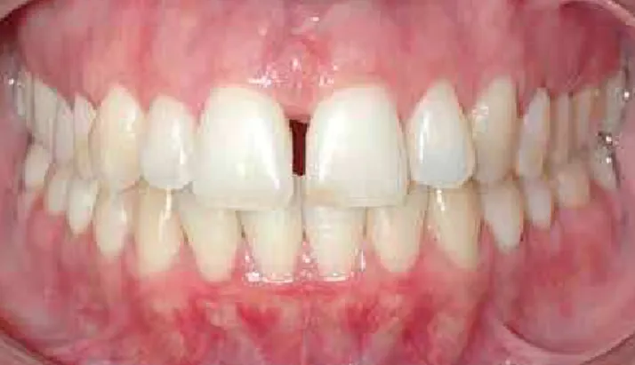
stable out of retention10,24,26 (Fig 1). Among the
pos-sible contributing factors for diastema relapse is the incisor root mesiodistal positioning.
Few studies evaluated changes in teeth
angula-tions during and after orthodontic treatment.1,7 But
changes in incisor angulations specifically in patients with maxillary anterior spacing was still not assessed.
Therefore, the objective of this study was to describe the mesiodistal angulation pattern of the maxillary incisors in patients with anterior spac-ing and evaluate changes in the incisor angulations during treatment and after retention. Additional-ly, we aim to analyze whether there is association
between incisor angulations at pretreatment, post-treatment and post-retention stages, and maxillary anterior spacing relapse.
OBJECTIVE
The aim of this study was to describe the mesio-distal angulation pattern of the upper incisors in patients with diastema in this area and observe the alteration of this angulation during the orthodontic treatment and after removing the upper retention. Also, verify whether there is an association between the mesiodistal angulation of these teeth in the ini-tial and final stages of treatment and the post-reten-tion and the anterior superior diastema relapse.
MATERIAL AND METHODS Material
The subjects evaluated in this study were select-ed from the files of the Orthodontic Department at the University of São Paulo, which comprised 4334 patients. Inclusion criteria comprised maxillary in-terincisor diastema equal to or greater than 0.77 mm, after partial eruption of the maxillary canines. Sub-jects with missing anterior teeth, periodontal disease with bone loss, generalized microdontia, maxillary pathologies, mesiodens, diastema closure by a non-orthodontic method and post-non-orthodontic restora-tion of maxillary anterior teeth resulting in increase in mesiodistal width were excluded.
Ninety panoramic radiographs of 30 subjects (13
male and 17 female) taken at pre-treatment (T1),
post-treatment (T2) and post-retention (T3) stages
A
B were selected. All patients were treated nonextrac-tion. Eighteen subjects showed a Class I and 12 of them showed a Class II malocclusion. Maxillary ca-nines were completely erupted in 20 patients. The average initial age was 12.94 years (SD 1.27) and the treatment time was 2.38 years (SD 1.06). The reten-tion time and the period between the appliance re-moval and the last follow-up appointment were 1.44 (SD 0.48) and 7.05 (SD 3.60) years, respectively.
Methods
Measurements of spaces between maxillary cen-tral and lateral incisors were taken on dental casts
taken at T1, T2, and T3 with a 0.01 mm precision digital
caliper (Mitutoyo, Japan). Widths were considered to be the smallest distance between adjacent teeth at the gingival level (Fig 2). The sum of the maxillary
in-terincisor widths at post-treatment (T2) and
post-re-tention (T3) stages were obtained. The space relapse
was calculated by subtracting the sum of the spaces
at T3 minus the sum of the spaces at T2. According to
this result, the sample was divided into two groups stable and unstable. Unstable cases occurred when the result was greater than zero.
Panoramic radiographs of 24 patients were tak-en with a Rotograph Plus unit (Villa Sistemi Medi-cali, Italy) which shows a mean linear magnifica-tion of 25%. While six patients had their pretreat-ment and post-treatpretreat-ment panoramic projections taken with as Orthophos CD unit (Siemens Medi-cal Systems, Germany), which presents a mean lin-ear magnification of 27%.
The panoramic radiographs were traced by the same examiner using a 0.5 mm pencil on a sheet of ac-etate paper (14 x 21 cm) placed over the radiographic film. The inferior outline of the orbits, the inferior outlines of the zygomatic processes of the max-illa and the contours of the maxmax-illary incisors were traced. The horizontal reference lines used were interorbital (IO), passing through the most inferior point of the right and left orbits, and the interzygo-matic processes of the maxilla (IZP), passing through the most inferior point of the right and left zygomatic processes of the maxilla. Each incisor long-axis was traced determined by its pulp canal long-axis (Fig 3).
The analyzed variables were: RLI (angle be-tween the long axis of the right lateral maxillary
incisor and the IZP line), RCI (angle between the long axis of the right central maxillary incisor and the IZP line), LCI (angle between the long axis of the left central maxillary incisor and the IZP line), and LLI (angle between the long axis of the left lat-eral maxillary incisor and the IZP line).
In this study, the IZP line was used because only twenty patients presented both orbital inferior con-tours in the panoramic radiographs of the initial, fi-nal and follow-up stages. The angle IO.IZP between the IO and the IZP reference lines was evaluated in
Figure 2 - A) Digital caliper Mitutoyo used to measure interincisor
spac-ing. B) Diastema measurement method.
Figure 3 - Schematic design of the radiographic tracings performed showing the orbital contours, zygomatic processes of maxilla, contour of maxillary incisors, as well as the reference lines interorbitary (IO), inter-zygomatic processes (IZP), and teeth long axes.
Ror
RIZP RLI RCI LCI LLI LIZP
Maxillary incisors mesiodistal angulation changes in patients with orthodontically treated anterior superior diastemas
original article
twenty patients where the orbital contours could be traced. When the lines converged at the right side the values were written positive, while when the convergence occurred at the left side the values were written negative.
Statistical analyses
Thirty panoramic radiographs were retraced and remeasured by the same examiner after a month in-terval. The casual error was calculated according to
Dahlberg’s formula.2 The systematic errors were
an-alyzed with a t test16 at p<0.05.
Mean values and standard deviations of each maxillary incisor angulations were calculated at the
initial (T1), final (T2), and post-retention (T3) stages
and were compared by repeated measures ANOVA, followed by Tukey post hoc test, in order to evaluate
the changes occurred during treatment (T1 and T2), to
analyze the stability of treatment outcomes (T2 and
T3) as well as to verify if the angulations after
reten-tion and before treatment were similar.
Since the patients evaluated are in growing phase, repeated measures ANOVA, followed by Tukey tests, were used to assess the changes in the parallelism be-tween IO and IZP lines.
Student’s t tests were used to compare
mesio-distal angulations between the stable and unstable groups at T1, T2 and T3.
RESULTS
Method precision
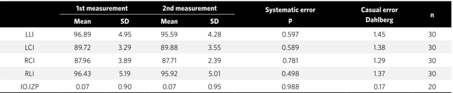
The casual error was found to be small and ac-ceptable for all measurements, while the system-atic error was not statistically significant for any measurement (Tab 1).
The angle between the IZP and IO lines with a maximum divergence of 2° at all stages (Tab 2)
demonstrates an acceptable parallelism between these lines.
Incisor angulations showed a tendency for me-sial inclination during orthodontic treatment but the changes were significant only for lateral incisor angu-lations (Tab 3). On the other hand, all incisor angula-tions remained stable during post-retention period.
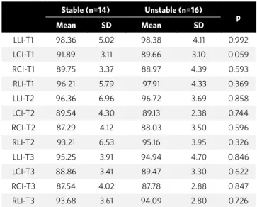
No differences were found between stable and unstable groups (Tab 4). This suggests that there was no association between space relapse and me-siodistal angulations.
ANOVA and Tukey test results
Generally, the angulation of the incisors pre-sented a tedency to mesialization with orthodontic treatment, and there was a significant difference only for the lateral incisors angulation (Tab 3). In the postretention stage, the angulation of all inci-sors remained stable.
Angulation comparison between stable and non-stable groups
No significant difference was verified between groups (Tab 4). This fact suggests that there was no relation between diastemas relapse and mesio-distal angulations.
DISCUSSION
Since the development and improvement of the
panoramic radiograph technique by Paatero,18,19 it has
been recognized as an essential diagnosis method be-cause it is possible to show on the same film the whole dentition, alveolar bone, temporomandibular joint and adjacent structures. Additionally, panoramic ra-diographs help the evaluation of mesiodistal teeth
angulations before and after treatment1,11,15,30 and root
parallelism near extraction areas.11,15
1st measurement 2nd measurement Systematic error
p
Casual error
Dahlberg n
Mean SD Mean SD
LLI 96.89 4.95 95.59 4.28 0.597 1.45 30
LCI 89.72 3.29 89.88 3.55 0.589 1.38 30
RCI 87.96 3.89 87.71 2.39 0.781 1.29 30
RLI 96.43 5.19 95.92 5.01 0.498 1.37 30
IO.IZP 0.07 0.90 0.07 0.95 0.988 0.17 20
It is known that panoramic radiographs as well as any extraoral radiograph exhibit magnification of
the real image.3,6 But in panoramic radiographs the
amount of magnification varies in different sites of the film due to the method of image acquisition by moving the X-ray tube. The linear measurements, especially horizontal ones, demonstrate signifi-cant magnification and are not reliable. In the other hand, angular measurements exhibit smaller
dis-tortions6,11,15,22, specially on the anterior region,3,21
confirming that panoramic radiographs can be a re-liable method to evaluate angular measurements on maxillary anterior teeth.
It is traditionally advocated that appropriate
root parallelism improves treatment stability,9,10,20
especially in the areas of space closure after
extrac-tions.9,20 In cases with anterior spacing, the incisor
mesiodistal positions has been mentioned to
in-fluence stability,10,17 even though there is no study
evaluating the mesiodistal angulations of maxillary anterior teeth in cases with interincisor diastema.
Some studies1,7,28,30 have evaluated teeth
angula-tions with panoramic radiographs, using the inter-orbital line (IO) as reference. Nevertheless, in our study, ten patients did not show both orbital infe-rior contours in the panoramic radiographs of the initial, final and follow-up stages. Therefore, the zy-gomatic processes of the maxilla were traced so that interzygomatic processes of the maxilla (IZP) line,
similar to IO line, was used. The results showed an acceptable parallelism (Tab 2) and a maximum divergence of 2° between these lines. This demon-strates that the IZP line can be as reliable as the in-terorbital line and the values obtained in this study
can be compared to those proposed by Ursi30
ob-tained from patients with normal occlusion.
According to repeated measures analysis of vari-ance (Tab 3), the changes in the maxillary central
in-cisors angulations were not significant during (T1-T2)
Total (3 stages) N=20
Initial (T1)
N=20
Final (T2)
n=20
Post-retention (T3) n=20
ANOVA p
Mean SD Min Max Mean SD Mean SD Mean SD
IO.IZP 0.07 0.90 -2.0 2.0 0.08 0.95 0.03 0.64 0.13 1.10 0.94
Table 2 - Descriptive statistics and repeated measures analysis of variance comparing the angle between interorbitary and inter-zigomatic processes of maxilla in initial, final, and postretention stages.
LLI LCI RCI RLI
Mean SD Mean SD Mean SD Mean SD
Initial (T1) 98.37A 4.47 90.70 3.26 89.33 3.90 97.12A 5.05
Final (T2) 96.55AB 5.36 89.32 3.36 87.68 3.76 94.25B 5.30
Post-retention (T3) 95.08
B 4.28 89.18 3.31 87.67 3.40 93.90B 3.16
p (ANOVA) 0.031* 0.151 0.139 0.015*
Table 3 - Results of repeated measures analysis of variance, followed by Tukey tests comparing mesiodistal angulations of each incisor at all stages T1, T2 and T3.
* statistically significant for p<0,05.
Stable (n=14) Unstable (n=16)
p
Mean SD Mean SD
LLI-T1 98.36 5.02 98.38 4.11 0.992
LCI-T1 91.89 3.11 89.66 3.10 0.059
RCI-T1 89.75 3.37 88.97 4.39 0.593
RLI-T1 96.21 5.79 97.91 4.33 0.369
LLI-T2 96.36 6.96 96.72 3.69 0.858
LCI-T2 89.54 4.30 89.13 2.38 0.744
RCI-T2 87.29 4.12 88.03 3.50 0.596
RLI-T2 93.21 6.53 95.16 3.95 0.326
LLI-T3 95.25 3.91 94.94 4.70 0.846
LCI-T3 88.86 3.41 89.47 3.30 0.622
RCI-T3 87.54 4.02 87.78 2.88 0.847
RLI-T3 93.68 3.61 94.09 2.80 0.726
Table 4 - Results of Student t tests comparing the incisor angulations in
Maxillary incisors mesiodistal angulation changes in patients with orthodontically treated anterior superior diastemas
original article
and after treatment (T2-T3), except for the lateral
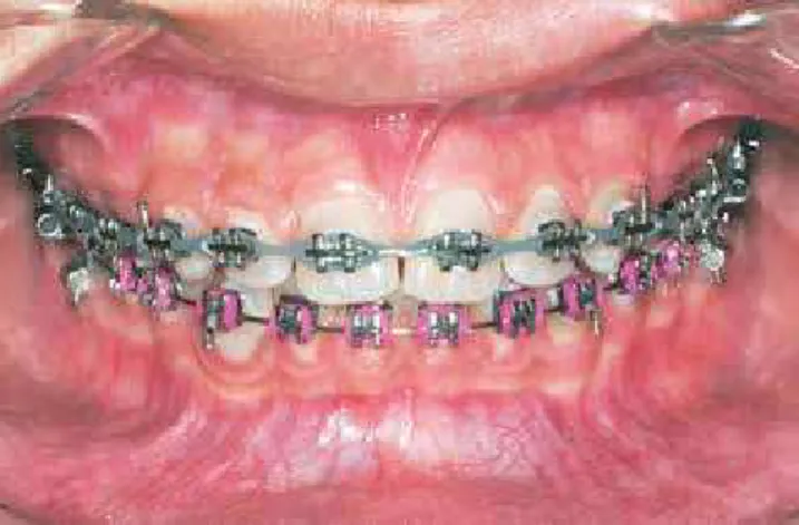
in-cisor angulations, which showed significant changes in both periods. There was a tendency for crown me-sialization, which may be explained by the mechanics used, which included an association of preadjusted appliances with elastomeric chains. The high forces produced might cause great crown mesialization if used on light wires (Fig 4).
The mesiodistal angulations achieved with treatment remained stable (Tab 3). These findings shows that finishing with ideal angulations should be a relevant treatment objective, since the pa-tient’s smile will show the positions obtained, even after a long period out of retention.
Diastema relapse is quite often on maxillary
anterior area.10,24,26,27 Some authors reported that
space reopening might be associated with an inap-propriate root positioning at the end of treatment. However our results showed no difference between stable and unstable groups in central and lateral incisor angulations when compared in each ini-tial, final, post-retention stages. In conclusion, this demonstrates that no association between space re-opening and mesiodistal angulations was found.
CONCLUSIONS
Based on the method and the results we conclude: 1) Incisor mesiodistal angulations in patients with
anterior spacing showed slight changes during treatment with greater crown mesialization on the lateral incisors, which remained stable posttreatment.
2) There was no association between anterior spacing relapse and mesiodistal angulations of the maxillary incisors.
1. Almeida-Pedrin RR, Pinzan A, Almeida RR, Ursi W, de Almeida MR. Panoramic evaluation of mesiodistal axial inclinations of maxillary anterior teeth in orthodontically treated subjects. Am J Orthod Dentofacial Orthop. 2006 Jul;130(1):56-60; discussion 60-1.
2. Almeida RR, Garib DG, Almeida-Pedrin RR, Almeida MR, Pinzan A, Junqueira MHZ. Diastema interincisivos centrais superiores: quando e como intervir? R Dental Press Ortodon Ortop Facial. 2004 Maio-Jun;9(3):137-56.
3. Almeida SM, Bóscolo FNE, Montebello Filho A. Estudo das distorções da imagem radiográfica produzida em aparelhos panorâmicos que se utilizam dos princípios ortopantomográficos e elipsopantomográficos. Rev Odont USP. 1995 Abr-Jun;9(2):91-9.
4. Andrews LF. The six keys to normal occlusion. Am J Orthod. 1972 Sep;62(3):296- 309.
5. Bishara SE. Management of diastemas in orthodontics. Am J Orthod. 1972 Jan;61(1):55-63.
6. Blackman S. Mass dental radiography. Radiography. 1956 Feb;22(254):21-5. 7. Brandão AG. Estudo ortopantomográfico longitudinal das inclinações axiais mesiodistais em pacientes tratados ortodonticamente com extrações dos quatro primeiros pré-molares [Dissertação]. Bauru (SP): Universidade de São Paulo, Faculdade de Odontologia de Bauru; 2002.
8. Cuoghi OA, Bertoz FA, de Mendonça MR, Santos EC, Li AT. Labiolingual and mesiodistal positioning of maxillary permanent incisors during the eruption process. J Clin Pediatr Dent. 2000 Fall;25(1):13-21.
9. Edwards JG. The prevention of relapse in extraction cases. Am J Orthod. 1971 Aug;60(2):128-44.
10. Edwards JG. The diastema, the frenum, the frenectomy: a clinical study. Am J Orthod. 1977 May;71(5):489-508.
11. Frykholm A, Malmgren O, Sämfors KA, Welander U. Angular measurements in orthopantomography. Dentomaxillofac Radiol. 1977;6(2):77-81.
12. Gardiner JH. Midline spaces. Dent Pract Dent Rec. 1967 Apr;17(8):287-97. 13. Janson GRP, Silva, CCA, Henriques JFC, Freitas MR, Martins DR. Fechamento
ortodôntico de diastema entre os incisivos centrais superiores durante a dentadura mista: relato de um caso clínico. Rev Dental Press Ortodon Ortop Facial. 1998 Jul-Ago;3(4):72-8.
14. Lavelle CL. Crowding and spacing within the human dental arch of different racial groups. Arch Oral Biol. 1970 Nov;15(11):1101-3.
REFERENCES
15. McKee IW, Williamson PC, Lam EW, Heo G, Glover KE, Major PW. The accuracy of 4 panoramic units in the projection of mesiodistal tooth angulations. Am J Orthod Dentofacial Orthop. 2002 Feb;121(2):166-75; quiz 192.
16. McVay TJ, Latta GH Jr. Incidence of the maxillary midline diastema in adults. J Prosthet Dent. 1984 Dec;52(6):809-11.
17. Mulligan TF. Diastema closure and long-term stability. J Clin Orthod. 2003 Oct;37(10):560-74.
18. Paatero YV. A new tomographical method for radiographing curved outer surfaces. Acta Radiol. 1949 Sep 30;32(2-3):177-84, illust.
19. Paatero YV. Pantomography and orthopantomography. Oral Surg Oral Med Oral Pathol. 1961 Aug;14:947-53.
20. Parker GR. Transseptal fibers and relapse following bodily retraction of teeth: a histologic study. Am J Orthod. 1972 Apr;61(4):331-44.
21. Peck JL, Sameshima GT, Miller A, Worth P, Hatcher DC. Mesiodistal root angulation using panoramic and cone beam CT. Angle Orthod. 2007 Mar;77(2):206-13. 22. Philipp RG, Hurst RV. The cant of the occlusal plane and distortion in the panoramic
radiograph. Angle Orthod. 1978 Oct;48(4):317-23.
23. Richardson ER, Malhotra SK, Henry M, Little RG, Coleman HT. Biracial study of the maxillary midline diastema. Angle Orthod. 1973 Oct;43(4):438-43.
24. Shashua D, Artun J. Relapse after orthodontic correction of maxillary median diastema: a follow-up evaluation of consecutive cases. Angle Orthod. 1999 Jun;69(3):257-63.
25. Steigman S, Gershkovitz E, Harari D. Characteristics and stability of spaced dentition. Angle Orthod. 1985 Oct;55(4):321-8.
26. Sullivan TC, Turpin DL, Artun J. A postretention study of patients presenting with a maxillary median diastema. Angle Orthod. 1996;66(2):131-8.
27. Surbeck BT, Artun J, Hawkins NR, Leroux B. Associations between initial, posttreatment, and postretention alignment of maxillary anterior teeth. Am J Orthod Dentofacial Orthop. 1998 Feb;113(2):186-95.
28. Tavano O, Ursi WJSE, Almeida RR. Determinação das linhas de referência para medições angulares em radiografias ortopantomográficas. Odontol Mod. 1989;16(9):22-5.
29. Taylor JE. Clinical observations relating to the normal and abnormal frenum labii superians. Am J Orthod and Oral Surg. 1939 Jul;25(7):646-60.