J of Evolution of Med and Dent Sci/ eISSN- 2278-4802, pISSN- 2278-4748/ Vol. 4/ Issue 24/ Mar 23, 2015 Page 4157
ROLE OF COMPUTED TOMOGRAPHY AND 3D RECONSTRUCTIONS IN
PELVIC RIM AND ACETABULAR FRACTURES
Somasekhar R1, A. V. K. Adithya2, Kalra V. B3HOW TO CITE THIS ARTICLE:
Somasekhar R, A. V. K. Adithya, Kalra V. B. Role of Computed Tomography and 3D Reconstructions in Pelvic RIM and Acetabular Fractures . Journal of Evolution of Medical and Dental Sciences 2015; Vol. 4, Issue 24, March 23; Page: 4157-4164, DOI: 10.14260/jemds/2015/598
ABSTRACT: To determine the role of computed tomography and 3D Reconstructions in classification of pelvic rim and acetabular fractures and assessing possible changes in fracture classification. We collected retrospective information in a period of 18 months in our institution, of patients with pelvic injuries considering---demographic data, radiological examination performed and the moment when it was performed, fracture classification and management. In 12 cases (54%) there were isolated pelvic rim fractures and 7 cases of isolated acetabular fractures (32%) and 3 cases (14%) involving both. After the CT scan was obtained, the initial classification was changed in five cases (22.7%). Tridimensional CT based modeling is very helpful in the classification of pelvic fractures and is a complement of the plain X-ray.
KEYWORDS: Computed tomography, 3d reconstructions, pelvic rim, acetabular fractures.
INTRODUCTION:
Since long, fractures of the pelvis have been posing problems in terms of diagnosis, treatment, and prognosis. According to recently published series nearly 15% of multi trauma patients have pelvic fractures. There will be need for urgent intervention regarding hemodynamic stability in these persons with pelvic fractures, so the physicians approach must be clear and quick in following the diagnostic protocol.1,2 For all of this, emphasis should be made on an
accurate diagnosis of the pelvic injury and its extent.
With the new technological advances, both in helical Computed Tomography and 3D multi-slice devices like volumetric processing software for images, we are able to guarantee better evaluation of injuries.
Conventional axial tomography is a very useful modality in the evaluation of posterior elements, mainly the sacroiliac joint. In fact, axial views are still the ones that define sacral fractures better,3 as has happened in some cases picked up in this work.
When it comes to the 3D reconstruction of the osteo-muscular system, the pelvis turns out to be one of the most complex modeling elements. The volumetric representation of the pelvic cavity, as well as the individualized subtraction of elements, allows for a thorough study of injuries in this region.4
Visualization of 3D models allows for a global view of the entire pelvic rim, as well as a detailed view of each element damaged.5
MATERIAL AND METHODS:
We collected retrospective information in a period of 18 months in our institution, of patients with pelvic injuries considering---demographic data, type of accident, radiological examination performed and the moment when it was performed, fracture classification and management.
J of Evolution of Med and Dent Sci/ eISSN- 2278-4802, pISSN- 2278-4748/ Vol. 4/ Issue 24/ Mar 23, 2015 Page 4158
Before surgery, all patients underwent a 3D reconstruction performed with a 3D (helical volumetric) 16 channel multi-slice CT scanner (Philips).
The classification of the fracture was performed first after the simple X-ray, being reviewed on two occasions: once with CT scan and then with 3D reconstruction.
Tiles classification was used to classify pelvic rim fractures and the AO classification was used for socket fractures.
22 patients were studied without any significant medical-surgical background. The sample was made up of 14 men and 8 women with an average age of 46. 4 years (20-73).
When patients were seen in the ER a CT scan examination was performed in 59% of them and remaining did not undergo an emergency radiological examination because the patient’s clinical condition was unstable. All patients had an emergency simple AP X-ray of the pelvis and 17% had 3 view X-rays. But after hemodynamic stability and before planning surgical management all the patients were seen to undergo CT scan and 3D reconstructions.
Tile classification of Pelvic fractures6
AO CLASSIFICATION OF ACETABULAR FRACTURES7:
Type A Partial articular, involving only one of the two columns: A1: Posterior wall fracture.
A2: Posterior column fracture. A3: Anterior wall or column fracture.
Type B Partial articular, involving a transverse component: B1: Pure transverse fractures.
B2: T-Shaped fractures.
B3: Anterior column and posterior hemi transverse.
Type C Complete articular fractures, both columns: C1: High variety, extending to the iliac crest.
J of Evolution of Med and Dent Sci/ eISSN- 2278-4802, pISSN- 2278-4748/ Vol. 4/ Issue 24/ Mar 23, 2015 Page 4159
OBSERVATIONS AND DISCUSSION:
In 12 cases (54%) there were isolated pelvic rim fractures and 7 cases of isolated acetabular fractures (32%) and 3 cases (14%) involving both. In the cases of type A3 and B1 pelvic fractures the classification did not change when performing the tomography or the 3D reconstruction. Difficulty emerged when evaluating posterior elements and when differentiating types B and C.
TOTAL NUM OF CASES
ISOLATED PELVIC RIM FRACTURES
ISOLATED ACETABULAR
FRACTURES
BOTH
22 12 7 3
There are two cases (9%) where initially A2 (Tile classification) given on x rays as iliopubic and ischiopubic rami are fractured but with CT and 3D images involvement of sacrum noted and thus classification changed to B2.
PELVIC RIM FRACTURES PLAIN XRAY CT, 3D RECON NO. OF CASES
GRADING A3 B2 2
CASE 1
Fig. 1 Fig. 2
J of Evolution of Med and Dent Sci/ eISSN- 2278-4802, pISSN- 2278-4748/ Vol. 4/ Issue 24/ Mar 23, 2015 Page 4160
CASE 1: X-ray frontal view shows fracture involving right inferior pubic ramus. CT axial sections and 3d reconstruction imaging show fractures involving iliac wing, anterior acetabular wall and inferior pubic ramus on the right side.
With regards to acetabular fractures, in two cases (9%) there were fractures involving anterior column and posterior hemi transverse region of acetabulum on CT and 3D reconstructions of which only anterior column fracture was identified on X-ray pelvis, by which the classification changed from A1 to B1 (AO Classification) and thus altered the management. And in another case (4%) classification was changed from A3 (A3: Anterior wall or column fracture) to B2 (B2: T-Shaped fractures). Thus accounting for alteration of classification of acetabular fractures in 13. 6% of cases in this study (4 cases out of 22). Fractures of the acetabulum mostly caused the pelvis to shatter into a wide array of complex configurations which were difficult to fully delineate with plain radiography8 as evidenced in this study.
ACETABULAR FRACTURES PLAIN XRAY CT, 3D RECON NO OF CASES
GRADING A3 B3 2
A3 B2 1
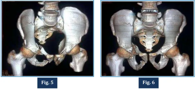
CASE 2
J of Evolution of Med and Dent Sci/ eISSN- 2278-4802, pISSN- 2278-4748/ Vol. 4/ Issue 24/ Mar 23, 2015 Page 4161
Fig. 7 Fig. 8
Fig. 9 Fig. 10
J of Evolution of Med and Dent Sci/ eISSN- 2278-4802, pISSN- 2278-4748/ Vol. 4/ Issue 24/ Mar 23, 2015 Page 4162
CASE 2: X-ray frontal view shows fracture involving right pubic bone and left ischiopubic ramus and fracture involving left iliac wing extending into the left acetabulum.
CT axial sections and 3d reconstruction show fractures involving right pubic bone and left ischiopubic ramus and fracture involving left iliac wing extending into left acetabulum. Additionally fracture involving left posterior acetabular wall noted.
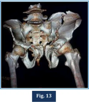
Fig. 13
Fig. 14 Fig. 15
J of Evolution of Med and Dent Sci/ eISSN- 2278-4802, pISSN- 2278-4748/ Vol. 4/ Issue 24/ Mar 23, 2015 Page 4163
CASE 3: X-ray frontal view shows posterior dislocation of right femoral head and pubic diastasis and displacement noted at right sacroiliac joint CT axial views and 3d reconstruction show fractures involving right ischiopubic ramus, right actabulum and left ischiopubic ramus with displacement of right sacroiliac joint and posterior dislocation of right femoral head.
In few cases though fracture classifications were not altered, the 3D CT images have shown clear advantages in saving time and in understanding the fractures from a mechanical and anatomical perspective.9
The 3D model reproduces the findings that the surgeon will find intra-operatively. However, care should be taken in usage of proper technique and avoiding the errors made by the software when interpreting attenuation coefficients.
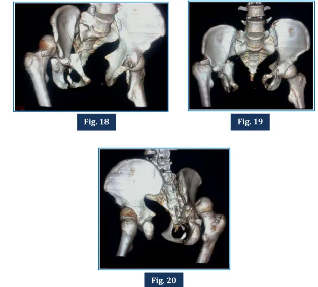
3D reconstruction images are a very good addition to the investigation list and help a lot in the presurgical planning of pelvic fractures. Spiral CT datasets coupled with real time 3D-VR allows visualization of the entire pelvis through almost any plane or perspective.10
Fig. 18 Fig. 19
J of Evolution of Med and Dent Sci/ eISSN- 2278-4802, pISSN- 2278-4748/ Vol. 4/ Issue 24/ Mar 23, 2015 Page 4164
REFERENCES:
1. Turen CH, Dube MA, LeCroy MA: The polytraumatized patient with musculoskeletal injuries. JAAOS 1999; 7: 154-65.
2. Tile M, Rubenstein J: Fractures of the pelvis and acetabulum. Baltimore: Williams & Wilkins 1995: 1221.
3. Falchi M, Rollandi GA: CT of pelvic fractures. Eur J Radiol 2004; 50 (1): 96-105.
4. Brown GA, Firoozbakhsh K, Gehlert RJ: Three-dimensional CT modeling versus traditional radiology techniques in treatment of acetabular fractures. Orthop J 2001; 21: 20-4.
5. Mitton D, Deschenes S, Laporte S, Godbout B, Bertrand S, de Guise JA, Skalli W: 3D reconstruction of the pelvis from biplanar radiography. Comput Methods Biomech Biomed Engin 2006; 9 (1): 1-5.
6. Int. braz j urol. vol. 37 no. 3 Rio de Janeiro May/June 2011.
7. Keith Mayo, Michel Oransky, Pol Rommens, Carlos Sancineto. AO Foundation [Internet].Grisons, Switzerland: AO Foundation; c2011 [cited 2015 Feb 10]; [about 9 screens]. Available from: https://www2.aofoundation.org/wps/portal/surgery?showPage=diagnosis&bone=Pelvis&seg ment=Acetabulum.
8. True three dimensional stereographic display of 3D reconstructed CT scans of the pelvis and acetabulum. Gautsch TL1, Johnson EE, Seeger LL. PMID: 8050223.
9. Gautier E, Bachler R, Heini PF, Nolte LP: Accuracy of computer-guided screw fixation of the sacroiliac joint. Clin Orthop 2001; 393: 310-7.
10.Stroszczynski C, Schedel H, Stockle U, et al. Clinical application of multiplanar and 3D reconstruction of spiral CT in diagnosis of acetabulum fractures (German) Aktuelle Radiol 1996; 6 (2): 91-95.
1.
AUTHORS:
1. Somasekhar R. 2. A. V. K. Adithya 3. Kalra V. B.
PARTICULARS OF CONTRIBUTORS:
1. Post Graduate, Department of Radio-diagnosis,Konaseema Institute of Medical Sciences, Amalapuram, A. P.
2. Post Graduate, Department of Radio-diagnosis,Konaseema Institute of Medical Sciences, Amalapuram, A. P.
FINANCIAL OR OTHER
COMPETING INTERESTS: None
3. HOD, Department of Radio-diagnosis, Konaseema Institute of Medical Sciences, Amalapuram, A. P.
NAME ADDRESS EMAIL ID OF THE CORRESPONDING AUTHOR:
Dr. Somasekhar R, D. No. 19-7-15, Opp. P. A. Quarters, Kanukurthi Vari Street, Vizianagaram-535002.
E-mail: [email protected]
< <,