06 artigo 462
original article
1 – Orthopedist in the Hip Surgery Team, Hospital São Bento Cardioclínica S/A, Belo Horizonte, MG, Brazil.
2 – MSc and PhD in Orthopedics and Traumatology from Unifesp; Head of the Residence Service, Hospital São Bento Cardioclínica S/A, Belo Horizonte, MG, Brazil. 3 – Resident Physician in Orthopedics and Traumatology, Hospital São Bento Cardioclínica S/A, Belo Horizonte, MG, Brazil.
Work performed at Hospital São Bento Cardioclínica S/A, Belo Horizonte, MG.
Correspondence: Rua Crucis 50, Santa Lucia, 30360-290 Belo Horizonte, MG. E-mail: [email protected] Work received for publication: December 22, 2010; accepted for publication: June 16, 2011.
PROSPECTIVE STUDY ON SEVENTY-SIX CASES OF FRACTURED
ACETABULUM WITH SURGICAL TREATMENT
André Gaudêncio Ignácio de Almeida1, Carlos Antônio Garrido2, Leonardo Eustáquio Vaz Amaral3, Luiz Fernando Lindenberg Vargas3
aBstract
Objective: To conduct a prospective study on unstable and incongruent fractures of the acetabulum, in comparison with the literature, covering the type of access, fixation materials, degree of reduction, type of fracture and results after surgery. Methods: The authors evaluated 76 patients with unstable and incongruent fracture of the acetabulum between January 1999 and December 2009. The Marvin Tile classification was used, and all cases were treated surgically using the technique recommended by the AO-ASIF group. Results: The radiological evaluation was done in accordance with the
The authors declare that there was no conflict of interest in conducting this work
this article is available online in portuguese and english at the websites: www.rbo.org.br and www.scielo.br/rbort introdUction
The first surgical treatment for a fracture of the acetabulum was performed by Levine, in 1943(1).
In 1951, Cauchoix produced a study on the surgical approach to the acetabulum(2). A major change took
place starting in 1955, thanks to Letournel and Judet(3,4), who greatly influenced the knowledge and
systematization of this surgical treatment. These last authors introduced a didactic classification of fractures of the acetabulum that aids in dealing with them. They improved the radiological assessments by instituting specific radiographic views and publicized the ilioinguinal, Kocher-Langenbeck and iliofemoral access routes. Their studies showed good results from the surgical approach in 70 to 80% of their cases, thus leading to good clinical results.
More recently, Marvin Tile created a classification system for fractures of the acetabulum in an attempt to achieve better standardization of the approaches and treatments(5,6).
It has become essential to use computed tomogra-phy, in order to achieve better preparation and com-prehension of the fracture(7).
The aim of this study was to make a prospective evaluation in comparison with the literature, covering the type of access, fixation material, degree of reduc-tion, type of fracture and results after surgery.
methods
Seventy-six patients with unstable and incongruent fractures of the acetabulum were evaluated between January 1999 and December 2009. The Marvin Tile Ruesch criteria, and good, excellent or perfect results were obtained for 64 patients (84.2%). The clinical evaluation was done in accordance with the Harris criteria, and good or excellent results were obtained for 62 patients (81.6%). The complications were assessed and, in comparison with the literature, were shown to be compatible with previous reports even after the statistical analysis. Conclusion: It was concluded that anatomical reduction and stable fixation are important for good results.
classification was used and all the patients were treated surgically in accordance with the technique recommended by the AO-ASIF group. The mean length of follow-up was 4.9 years, with a minimum of four years and a maximum of nine years. The patients’ mean age was 38.4 years (minimum of 17 years and maximum of 76 years), with a standard deviation of 11.4 years. Regarding sex, 64 were male (84.2%) and 12 were females (15.8%). Regarding color, 65 were dark-skinned (86%), seven were intermediate (9.3%) and four were white-skinned (4.7%). Regarding the type of trauma, there were 51 cases of car and motorcycle accidents (67.1%), nine cases of being run over (11.8%), 11 cases of falls from a height (14.5%) and another five indeterminate cases (6.6%).
Regarding the side affected, 54 fractures (71%) were on the left side and 22 (29%) were on the right side. Both sides were affected in three patients, but only one side was treated surgically and the other side was excluded (Table 1).
All the patients underwent radiographic evaluation in anteroposterior (AP) view of the pelvis and Judet 45° oblique view (wing and obturator), and 53 pa-tients also underwent tomography. The criterion used for indicating surgery was the presence of an unstable and/or incongruent hip. The criterion for instability was hip misalignment associated with displacement of the wall or the anterior or posterior column. The cri-terion for incongruence was a deviation greater than 3 mm in fractures of the acetabular roof, transverse fractures, “T” fractures or fractures of the two col-umns. To evaluate the displacement, the Matta criteria were also taken into consideration in measuring the arch of the acetabular roof in the three Judet views(8,9).
The fracture distribution according to the Marvin Tile classification(6) is shown in Table 2.
Regarding the surgical approach, the access used for two patients (2.6%) was anterior iliofemoral; for 11 (14.5%), an anterior ilioinguinal access was used; for 17 (22.4%), a combined access was used (ilioin-guinal and Kocher-Langenbeck); and for 46 (60.5%), a posterior Kocher-Langenbeck access was used.
All the patients underwent clinical and radiograph-ic evaluations. They were then taken to the surgradiograph-ical block, where transkeletal traction was established on the distal femur on the day of admission. Exposed fractures or associated dislocations were treated as
table 1 – Lesions associated with fractures of the acetabulum.
types of lesions frequency percentage
Other fractures 52 80.0%
Sciatic nerve lesions 7 10.8 %
Vascular lesions 4 6.1%
Urological lesions 2 3.1%
Total 65 100%
Source: Medical Archive Service (SAME), Hospital São Bento.
table 2 – Frequencies and percentages of acetabular fractures according to the Marvin Tile classification (AO).
type of fractures frequency percentage
A1: posterior wall 7 9.3%
A2: posterior column 12 15.8%
A3: anterior wall and/or posterior column 2 2.6%
B1: transverse 18 23.7%
B2: ¨T¨-shape 10 13.2%
B3: posterior hemitransverse plus
anterior column 2 2.6%
C1: both columns; high 14 18.4%
C2: both columns; low 9 11.8%
C3: both columns, involving sacroiliac
joint 2 2.6%
Total 76 100%
Source: SAME, Hospital São Bento.
emergencies. Treatment for the acetabular fractures was scheduled for a second occasion.
All the patients received a prophylactic regimen against deep vein thrombosis, consisting of sodium heparin at a dose of 5,000 UI subcutaneously, every eight hours, from the time of admission until discharge from hospital. They also received venous antibiotic therapy (first-generation cephalosporin) starting from the perioperative period, consisting of two grams intravenously at the time of induction of anesthesia and a further two grams every six hours after the operation, for another 24 hours.
Radiographic examinations were performed during the postoperative period, in Judet views, in or-der to verify the remaining degree of displacement. The Ruesch criteria were used: three points for absence of displacement (or if it was less than one millimeter), two points if the displacement was between one and three millimeters and one point if it was more than three and less than five millimeters(10). If the patient
Also during the postoperative period, the patients were assessed by means of a questionnaire in which they were classified according to the Harris scoring criteria, and this examination was repeated at all the return visits(11).
Evaluations were made on the 15th, 30th, 60th and 90th days after the operation, six months after the operation and annually thereafter. At all these return visits, clinical and radiological assessments were made using the criteria of Harris(11) and Ruesch et al(10). The
patients were released for passive movements under guidance on the first day after the operation, and the loading placed on the operated joint was limited over the first 90 days. No prophylaxis for heterotopic ossification was administered.
A satisfaction questionnaire was applied, which included asking whether the patient would undergo this surgical procedure again.
For each fracture, between one and four acetabular reconstruction plates were used.
resUlts
The patients were examined within the Harris criteria(11), receiving scores from 0 to 100. Their range
of motion, pain levels and functional abilities were evaluated, and these results are shown in Table 3.
Regarding the patients’ degree of satisfaction, it was observed that 14.5% were very satisfied, 67.1% were satisfied and 18.4 were dissatisfied but would still undergo the surgery again.
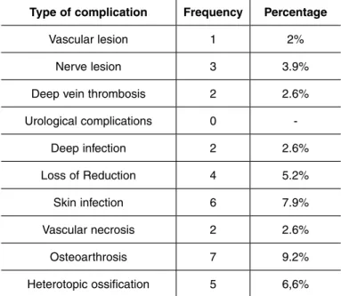
The complications most often found were nerve injuries (mostly neuropraxia), osteoarthrosis, infection, heterotopic ossification and loss of reduction, as shown in Table 4.
The mean duration of the operation was two hours (standard deviation of 10.92), with a range from one hour and 50 minutes to five hours.
There was one case of preoperative lesion of the superior gluteal vein that required ligature, but without further complications.
Out of the five cases of heterotopic ossification, two were grade I and three were grade III of Brooker et al(12).
It was observed that fractures of types B and C presented worse prognoses and correlated with a greater number of postoperative complications.
In the radiographic evaluations, it was seen that
there were some perfect reductions (Figures 1 and 2), excellent reductions (Figures 3 and 4) and good and poor reductions (Table 5).
The distribution of the types of access in relation to the types of fracture is shown in Table 6.
All the fractures consolidated and in a single case of deep infection, the plate was removed two months after the operation, without loss of the reduction. Nevertheless, this patient evolved with chronic osteomyelitis and osteoarthrosis.
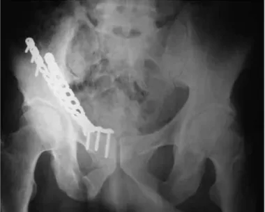
There were two cases of avascular necrosis of the femoral head. In these cases, a displacement of 2 mm remained after the surgical reduction. These patients underwent reduction of their coxofemoral dislocation on the day of the trauma. At the most recent assess-ment, one of these patients presented total collapse of the head (Figure 5).
table 3 – Evaluation of patients’ scores according to the Harris criteria.
score frequency percentage
Excellent (90 to 100 points) 11 14.5%
Good (80 to 89 points) 51 67.1%
Fair (70 to 79 points) 12 15.8%
Poor (less than 70 points) 2 2.6%
Total 76 100%
Source: SAME, Hospital São Bento.
table 4 – Postoperative complications of acetabular fractures
type of complication frequency percentage
Vascular lesion 1 2%
Nerve lesion 3 3.9%
Deep vein thrombosis 2 2.6%
Urological complications 0
-Deep infection 2 2.6%
Loss of Reduction 4 5.2%
Skin infection 6 7.9%
Vascular necrosis 2 2.6%
Osteoarthrosis 7 9.2%
Heterotopic ossification 5 6,6%
table 6 – Distribution of type of access in relation to type of fracture.
n° of patients type of fracture surgical access
19 A1 and A2 Kocher-Langenbeck
2 A3 Ilioinguinal
19 B1, B2 and B3 Kocher-Langenbeck
7 B1 Ilioinguinal
4 B2 Ilioinguinal
9 C1 Kocher-Langenbeck e
Ilioinguinal
5 C1 Kocher-Langenbeck
7 C2 Kocher-Langenbeck e
Ilioinguinal
2 C2 Iliofemoral
2 C3 Kocher-Langenbeck e
Ilioinguinal
figure 1 – AP view of case 32 with type B3 fracture.
Source: Archived photo from Hospital São Bento Cardioclínica, BH/MG
figure 2 – AP view of case 31 with type B2 fracture.
Source:Archived photo from Hospital São Bento Cardioclínica, BH/MG
figure 5 – Case 13, which evolved to necrosis of the femoral
head, six months after acetabular fracture of Tile type B1.
Source: Archived photo from Hospital São Bento Cardioclínica, BH/MG
table 5 – Scoring in radiographic evaluation in anteroposterior
and oblique views, in accordance with Ruesch et al.
score frequency percentage
9 points (perfect) 9 11.8%
8 points (excellent) 36 47.3%
7 points (good) 19 25.1%
Less than 7 points (poor) 12 15.8%
Source: SAME do Hospital São Bento.
figure 3 – Case 4, presenting fracture-dislocation of the right
acetabulum, of Tile type C2.
Source:Archived photo from Hospital São Bento Cardioclínica, BH/MG
figure 4 – After reduction and stabilization of the fracture using two acetabular reconstruction plates.
discUssion
As has been stated in several studies on fractures of the acetabulum, the treatment of choice for displaced fractures with significant joint involvement is open re-duction and anatomical internal fixation(13-17).
The number of excellent and good results (81.6%), according to the Harris criteria, coincided with the majority of published studies(18-21). These results were
compatible with the degree of satisfaction (p = 0.008), since the higher the score was, the greater the degree of satisfaction was.
The distribution of the fractures according to the Mar-vin Tile classification showed that the number of type B fractures (39.5%) was greater than the numbers of type C (32.9%) and type A (27.6%).
This distribution is not in agreement with what is found in the literature(20-24), in which type A fractures
predominate. It is believed that this difference is due to the greater severity of the accidents attended at our clinic. Moreover, exclusion of fractures of the posterior wall that were treated conservatively or by means of minimal fixation would also explain these differences.
Only in the cases of heterotopic ossification was there any significant association between the type of surgical access and the complications found, and this occurred only with the Kocher-Langenbeck access.
Heterotopic ossification was found in only 6.6% of the patients, even though no prophylaxis had been used. This incidence was much lower than in the literature, where rates of around 20% have been reported(23-27).
The statistical analysis on the possible associations between the duration of the operation, age, color, sex and type of fracture did not find any significant values. It was noted that heterotopic ossification only occurred in cases with a posterior approach, which was in line with the literature(28).
Two patients (2.6%) evolved with aseptic necrosis of the femoral head. Their fractures were of Marvin Tile type B3; their scores in the Harris classification were 72 and 70 points, and their radiological scores were poor in the Ruesch assessment. The incidence of aseptic necrosis of the femoral head found in the literature is approximately 5%(17,29-32). It is believed that the low incidence found in
the present study was due to early treatment.
Sciatic nerve injuries were seen in 10 patients: five with lesions affecting the fibular nerve portion and five with lesions affecting the entire nerve. Of these ten cases, seven already presented lesions before the surgery; the
other three presented neuropraxia, with full recovery at the last assessment. Only two patients with sciatic nerve injuries due to trauma did not recover. These data coincide with those of Machado and Araújo(29), who
reported that recovery of the sciatic nerve was achieved in 60% of the cases(13-17,30-33). Out of the patients with
Tile type C fractures, 23.7% underwent surgery with double access, and in 60% of these, there was a three-day interval between the two surgical procedures.
Seven patients who evolved with osteoarthrosis pre-sented radiographic scores of less than seven. Among these patients, one received a cemented total hip pros-thesis. All of these patients had scores of lower than 75 points in the Harris assessment. This demonstrates that there was a direct relationship between evolution to osteoarthrosis and poor reduction of the fracture, with consequent patient dissatisfaction(2).
In correlating the types of fracture with the final result, it was seen that the fractures of types C and B3 had a worse prognosis when evaluated as a function of the presence of complications and the Harris score (p < 0.005). Thus, early intervention, a correct ap-proach, good stabilization and reduction are important for minimizing the number of complications(10-12,18-31,33).
Other factors that influenced the prognosis for the fractures included deep infection (2.6%) and loss of reduction (5.2%). This was evident from the statisti-cally significant correlation that these two complica-tions showed in relation to the rate of unsatisfactory results (p = 0.00198).
It was observed that tomography was extremely important for scheduling and good progression of the surgery. It facilitates the approach and should be used routinely. Tomography was used in 53 cases, and it was observed that in the 23 cases (30.2%) in which it was not performed, the duration of the operation was longer (p = 0.00867).
conclUsions
references
1. Levine MA. A treatment of central fractures of the acetabulum. J Bone Joint Surg. 1943;25(4):902-6.
2. Cauchoix J, Truchet P. Les fratures articulaires de la hanche. In: XXVI Reunión annuelle de Societé Française d’Orthopedie et de Traumatologie, Paris; 1951. 3. Letournel E. Les fratures du cotyle. Étude d’une série de 75 cas [tesis]. Paris:
Arnette; 1961.
4. Judet R, Judet J, Letournel E. Fractures of the acetabulum: classification and surgical approaches for open reduction. preliminary report. J Bone Joint Surg Am. 1964;46:1615-46.
5. Tile M. Fractures of the acetabulum. Orthop Clin North Am. 1980;11(3):481-506. 6. Tile M, Kelleman JF, Joyce M. Fractures of the acetabulum: classification, management protocol and results of treatment. J Bone Joint Surg Br. 1985;67:324-5.
7. Brown GA, Willis MC, Firoozbakhsh K, Barmada A, Tessman CL, Montgomery A. Computed tomography image-guided surgery in complex acetabular fractures. Clin Orthop Relat Res. 2000;(370):219-26.
8. Matta JM, Anderson LM, Epstein HC, Hendricks P. Fractures of the acetabulum. A retrospective analysis. Clin Orthop Relat Res. 1986;(205):230-40. 9. Matta JM, Mehne DK, Roffi R. Fractures of the acetabulum. Early results of a
prospective study. Clin Orthop Relat Res. 1986;(205):241-50.
10. Ruesch PD, Holdener H, Ciaramitaro M, Mast JW. A prospective study of surgically treated acetabular fractures. Clin Orthop Relat Res. 1994;(305):38-46. 11. Harris WH. Traumatic arthritis of the hip after dislocation and acetabular
fractures: treatment by mold arthroplasty. An end-result study using a new method of result evaluation. J Bone Joint Surg Am. 1969;51(4):737-55. 12. Brooker AF, Bowerman JW, Robinson RA, Riley LH Jr. Ectopic ossification
following total hip replacement. Incidence and a method of classification. J Bone Joint Surg Am. 1973;55(8):1629-32.
13. Cole JD, Bolhofner BR. Acetabular fracture fixation via a modified Stoppa limited intrapelvic approach. Description of operative technique and preliminary treatment results. Clin Orthop Relat Res. 1994;(305):112-23.
14. Mayo KA. Open reduction and internal fixation of fractures of the acetabulum. Results in 163 fractures. Clin Orthop Relat Res. 1994;(305):31-7.
15. Mayo KA, Letournel E, Matta JM, Mast JW, Johnson EE, Martimbeau CL. Surgical revision of malreduced acetabular fractures. Clin Orthop Relat Res. 1994;(305):47-52.
16. Beaulé PE, Dorey FJ, Matta JM. Letournel classification for acetabular fractures. Assessment of interobserver and intraobserver reliability. J Bone Joint Surg Am. 2003;85(9):1704-9.
17. Issack PS, Toro JB, Buly RL, Helfet DL. Sciatic nerve release following fracture or reconstructive surgery of the acetabulum. J Bone Joint Surg Am. 2007;89(7):1432-7.
18. Johnson EE, Matta JM, Mast JW, Letournel E. Delayed reconstruction of acetabular fractures 21-120 days following injury. Clin Orthop Relat Res. 1994;(305):20-30.
19. Oliveira LP, Guimarães JAM., Magalhães TJS, Franzoisi TR. Fraturas do acetábulo: resultados do tratamento cirúrgico. Rev Bras Ortop. 1993;28(8):579-82. 20. Tornetta P 3rd. Displaced acetabular fractures: indications for operative and
nonoperative management. J Am Acad Orthop Surg. 2001;9(1):18-28. 21. de Ridder VA, de Lange S, Kingma L, Hogervorst M. Results of 75 consecutive
patients with an acetabular fracture. Clin Orthop Relat Res. 1994 Aug;(305):53-7. 22. Aristide RS, Honda E, Polesello G, Fernandez MS. Fratura em “T” do acetábulo:
análise de 45 casos. Rev Bras Ortop. 1996 31(11):919-24.
23. Köberle G, Miranda JB, Yamanaka E, Grava AL, Belangero WD. Fraturas do acetábulo: estudo dos resultados clínicos do tratamento cirúrgico precoce e tardio. Rev Bras Ortop. 1993;28:(6) 361-6.
24. Ghalambor N, Matta JM, Bernstein L. Heterotopic ossification following operative treatment of acetabular fracture. An analysis of risk factors. Clin Orthop Relat Res. 1994;(305):96-105.
25. Honda E, Aristide RS, Polesello G, Miashiro EH, Ferreira EA, Oliveira FR. Estudo da ossificação heterotópica no tratamento das fraturas acetabulares. Rev Bras Ortop. 1998;33(2):155-8.
26. Johnson EE, Kay RM, Dorey FJ. Heterotopic ossification prophylaxis following operative treatment of acetabular fracture. Clin Orthop Relat Res. 1994;(305):88-95.
27. Santin RA. Dupla abordagem no tratamento cirúrgico das fraturas complexas do acetábulo. Rev Bras Ortop. 1993;28(1/2):74-8.
28. Glas PY, Fessy MH, Carret JP, Béjui-Hugues J. [Surgical treatment of acetabular fractures: outcome in a series of 60 consecutive cases]. Rev Chir Orthop Reparatrice Appar Mot. 2001;87(6):529-38.
29. Machado F, Araújo FAS: Tratamento cirúrgico das fraturas de acetábulo. Rev Bras Ortop. 1990;25:323-7.
30. Moed BR, WillsonCarr SE, Watson JT. Results of operative treatment of fractures of the posterior wall of the acetabulum. J Bone Joint Surg Am. 2002;84(5):752-8.
31. Russell GV Jr, Nork SE, Chip Routt ML Jr. Perioperative complications associated with operative treatment of acetabular fractures. J Trauma. 2001;51(6):1098-103. 32. Guimarães RP, Kaleka CC, Cohen C, Ono NK. Análise do colapso segmentar
da cabeça femoral nas fraturas do acetábulo tratadas cirurgicamente. Rev Bras Ortop. 2009:44(5):427-31.