15 artigo 274
CAsE REPoRT
SPONTANEOuS RESOluTION OF PSEudOANEuRySm OF AN IlIAc
ARTERy bRANcH IN A mulTIPlE TRAumA PATIENT wITH
PElvIc FRAcTuRE: clINIcAl cASE
Cristina Maria Varino Sousa1, Luís Filipe Pires Silva1, Maria Elisa Rodrigues1, António Félix1, Bruno Alpoim1, Pedro Marques1, Joana Alexandra Gonçalves Oliveira2, Carlos Alves3, Maieiro Costa3, António Rodrigues4
The authors declare that they did not have any conflict of interests in producing this article
1 – Complementary Orthopedics and Traumatology Clinician at Local Healthcare Unit, Alto Minho, SA, Portugal. 2 – Complementary Neurosurgery Clinician at Hospital de São João, EPE
3 – Specialist Orthopedics and Traumatology Clinician at Local Healthcare Unit, Alto Minho, SA, Portugal.
4 – Specialist Physician and Director of Orthopedics and Traumatology Service, Local Healthcare Unit, Alto Minho, SA, Portugal. Work performed at the Orthopedics and Traumatology Service, Local Healthcare Unit, Hospital de Santa, Alto Minho, SA, Portugal. Correspondence:Rua da Praia 204m - 3º direito - Aguçadoura - 4495-031 - Póvoa de Varzim, Portugal. E-mail: [email protected] Work received for publication: December 21, 2009; accepted for publication: July 26, 2010.
Rev Bras Ortop. 2011;46(1):87-90 INTRODUCTION
Severe pelvic injuries have considerable incidence at emergency services in Portugal, particularly among the victims of high-energy trauma. They are more common among men, traffic accident victims and in-dividuals in their third decade of life. Pelvic fractures indicate that high energy was transferred, with severe trauma(1). It has been estimated that, for the pelvic ring
to be ruptured, a frontal collision at a speed of at least 48 km/h or a lateral collision at 24 km/h would be ne-cessary. This great dissipation of energy is responsible for the associated lesions that are frequently present(2).
Significant advances have been achieved over recent decades. On the one hand, there has been progress wi-thin surgery, with the use of external fixators to reduce unstable pelvic fractures, thereby making it possible to ABSTRACT
In patients who have been the victims of high-energy trau-ma, severe pelvic injury should always be suspected. Most of these fractures are stable and respond well to conserva-tive treatment. Pelvic fractures constitute 3% of all skeletal fractures and are associated with high-energy trauma. They are potentially serious injuries with significant mortality and large numbers of associated lesions. There are fun-damentally three sources of bleeding in pelvic fractures: arterial, venous and through the bone ends of the fracture. Arterial bleeding is more associated with hemodynamic
instability. In such cases, both early external fixation of the pelvic fracture and angiography with selective embo-lization of the bleeding vessels are effective methods for achieving hemostasis. Aneurysms of iliac artery branches are rare and are mostly pseudoaneurysm relating to the traumatic event. The natural history of pseudoaneurysms is unknown because of their rarity, but if they rupture, the mortality rate is high. We report a case of spontaneous thrombosis of a pseudoaneurysm of a branch of the right iliac artery.
Keywords – Hip Fractures; False Aneurysm; Iliac Artery
restore mobility to the patient. On the other hand, diffu-sion of advanced trauma life support (ATLS) theories has contributed towards diminishing mortality among patients with pelvic fractures(3).
The prognosis for pelvic fracture victims seems mostly to be related to the associated lesions, given that such fractures are often mild and do not give rise to great hemorrhage. However, in some cases, volu-minous retroperitoneal hemorrhage may occur, which may sometimes be lethal(2).
88
Rev Bras Ortop. 2011;46(1):87-90
replacement and progress in support measures, mor-tality persisted at close to 30% in the 1960s. Early ex-ternal fixation of pelvic fractures, in association with percutaneous angiography with selective embolization of the vessels with active bleeding, was responsible for changing the prognosis for these patients. Flint et al(4)
reported that the mortality rate was five times lower when angiography and embolization were used. Riemer et al(5) observed that there was a significant decrease in
mortality if early external fixation was implemented for complex pelvic fractures. Currently, the mortality rate ranges from 7 to 23%. Among multiple trauma victi-ms, hemorrhage is the main cause of death (39% of the cases), followed by associated cranioencephalic trauma (35%) and sepsis with multiple organ failure (25%).
At our service, the protocol for dealing with mul-tiple trauma victims with pelvic fractures includes an initial assessment in accordance with the ATLS pro-posals. After the initial radiological investigation, ab-dominal and pelvic assessments of hemodynamically stable patients are made using computed tomography.
Vascular lesions of the iliac arteries are more com-mon in cases of penetrating wounds(6). However, closed
pelvic trauma may lead to lesions in several branches of these arteries. Such lesions may result in fatal he-morrhage and should always be taken into considera-tion in cases of occult major hemorrhage. The vascular surgeon should be promptly consulted if arterial lesions are suspected or present. In cases of severe arterial hemorrhage, the initial treatment includes direct com-pression of the wound and blood volume resuscitation. Lesions of the iliac artery caused by pelvic fractures are not common. Hemorrhage in cases of pelvic frac-ture, resulting from lesions of the internal iliac artery and its branches, is more common in cases of posterior pelvic fracture. Fractures due to lateral compression more often cause hemorrhage of the pudendal and ob-turator vessels(7).
Vessels with lesions may be extremely difficult to identify, and attempting to do so on the operating table may result in abundant hemorrhage and fatal shock(6).
Arteriography is indicated for hemodynamically stable patients, both for diagnosing and for treating hemorrha-ge by means of embolization.
The therapeutic options have evolved over recent years and go from traditional surgery to approaches that are less invasive, and they include radiological pro-cedures such as echo-guided compression, echo-guided percutaneous thrombin injection and endovascular pro-cedures (embolization and placement of endoprostheses)(8).
Endovascular approaches are the gold standard for treating deep arterial hemorrhage(9,10).
Intervention radiology enables selective emboliza-tion of arteries that external fixaemboliza-tion is unable to plug. During angiography, signs of macrovascular lesions that would be sources of extravasation of contrast me-dium (false aneurysm), arteries with wall irregularities, arteries that are unfilled downstream, or stagnation of contrast medium in veins, are sought(11).
The time at which pseudoaneurysm is presented is variable and can be at any time from the original injury until years later(12).
Damage to the arterial wall and extravasation of blood to the periarterial fascial plane may result in
a pseudoaneurysm(13). Thus, pseudoaneurysms result
from transmural rupture of the arterial wall, extrava-sation of blood and formation of a hematoma that re-mains in communication with the arterial lumen. Un-like true aneurysms, pseudoaneurysms lack the three layers of the vessel wall (intima, media and adventitia) and are contained by perivascular tissue. Coagulation occurs on the periphery of the hematoma, which on ultrasound examination appears to be hyperechoic, while the center remains anechoic. During the intra--arterial high pressure of the systole, the blood flow is anterograde towards the pseudoaneurysm; during the diastole, the flow direction is retrograde. This pheno-menon causes swirling, which is the usual flow pattern seen on Doppler images(14).
Pseudoaneurysms of branches of the iliac arteries account for a tiny percentage of all false aneurysms(15).
They are usually asymptomatic, except when they rup-ture(16). Individuals with this disease usually have a
previous history of trauma, and this disease can also be diagnosed as a surgical finding(12).
Pseudoaneurysms may undergo spontaneous throm-bosis, or may evolve with development of complications such as infections, development of local compression over the neurovascular structures, or rupture.
Dilution coagulopathy may also be a reason for con-cern with regard to patients who receive large quantities of fluids. Thus, two to three units of frozen fresh plasma and seven to eight units of platelets should be prescribed for every five liters of volume replacement(1).
CLINICAL CASE
89
Figure 1 – Radiograph of the pelvis and axial computed tomo-graphy – images of pelvic fractures.
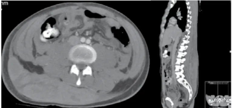
Figure 2 – Axial computed tomography showing increased den-sity and edema in the dorsal-lumbar tissue layers and the right buttock region, with a voluminous hematoma.
SPONTANEOUS RESOLUTION OF PSEUDOANEURYSM OF AN ILIAC ARTERY BRANCH IN A MULTIPLE TRAUMA PATIENT WITH PELVIC FRACTURE: CLINICAL CASE
Rev Bras Ortop. 2011;46(1):87-90 a four-wheel vehicle). Under observation in the
emer-gency service on admission, he was hemodynamically stable and presented Glasgow 15. He presented chest trauma, with bruising of the lungs, trauma to the pelvis with fracturing in the left wing of the sacrum, frac-turing of the right ileopubic branch and fracfrac-turing of the right acetabulum without displacement. Computed tomography on the abdomen and pelvis showed that the pelvis was fractured on the left side, with a frac-ture of the sacrum wing and the obturator ring, and on the right side, with a fracture of the anterior pillar of the acetabulum and ischiopubic branch. None of these fractures presented any significant displacement. There were also fractures of the L2, L3 and L4 right trans-verse apophyses, without signs of fracturing of the ver-tebral bodies or posterior elements of the lumbar spine (Figure 1). There were no other apparent abdominal or pelvic lesions. The patient was administered fluid therapy and a transfusion of red blood cell concentrate, and remained clinically stable. It was decided to use conservative treatment for the pelvic fractures, consist-ing of restconsist-ing in bed and skin traction on the right leg. Eight days later, the patient started to complain of increased discomfort in the right lumbar region, with palpable tumefaction, accompanied by a fall in hemo-globin levels and hemodynamic instability (hypotension and tachycardia). Transfusional support was needed.
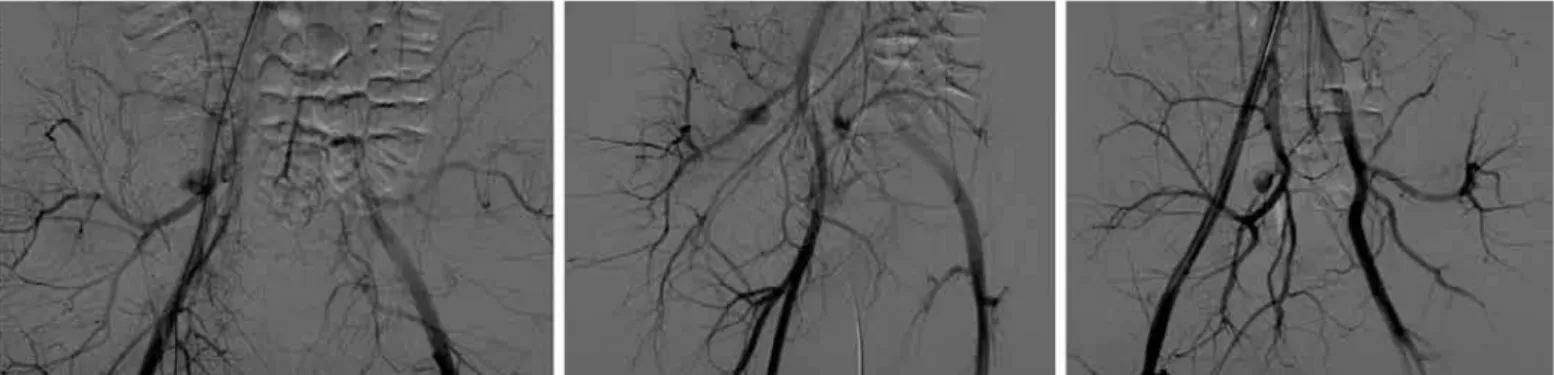
cause of the decrease in the patient’s hemoglobin levels (Figure 2). At this point, the patient was transferred to Hospital de São João, for vascular surgical care. Com-puted angiotomography was performed on an emergency basis, and this confirmed that a voluminous hematoma was present in the dorsal-lumbar-sacral region. An im-age of a sac measuring 37 mm with contrast inside (caliber of 21 mm), which suggested the presence of a pseudoaneurysm in a branch of the right internal iliac artery (Figure 3).
Embolization of the false aneurysm was scheduled three days later, but this attempt did not succeed be-cause the lesion was only poorly visible. One week later, the patient underwent arteriography, which did not show any evidence of the false aneurysms that had previously been encountered, thus showing spontane-ous resolution (Figure 4).
During the hospital stay, clinical stability was main-tained, without any evidence of worsening of the lum-bar hematoma or signs of peritoneal irritation, deep vein thrombosis or long-limb ischemia. There was hemody-namic stability and the hemoglobin level remained un-changed (Hb 12 g/dl).
DISCUSSION
In our sample, most of the pelvic fractures were stable and did not require specific treatment. However, a considerable proportion of such patients need speci-fic attention for treatment of pelvic fractures; if this is not undertaken, increased mortality and complications should be expected. Even in large trauma centers, more than 60% of pelvic fractures are stable and do not re-quire surgical stabilization. Among those that do need to be stabilized, most undergo external fixation and only 40% undergo internal fixation(3).
Pseudoaneurysms of branches of the iliac artery are rare and are related to trauma, pelvic fractures or Computed tomography scans over the chest, abdomen
90
Figure 3 –Images of the first angiography performed. Adjacent to the internal iliac artery, an image of a sac measuring 37 mm, with con-trast medium inside it (caliber of 21 mm), thus suggesting the presence of a pseudoaneurysm in a branch of the right internal iliac artery.
Figure 4 – Images from the last angiography performed, in which the occlusion of the previously diagnosed pseudoaneurysm can be seen.
Rev Bras Ortop. 2011;46(1):87-90
iatrogenic lesions(17). They are difficult to diagnose
because physical examination is almost impossible. They tend to be asymptomatic, unless rupture occurs, and this may occur several days or months after the initial trauma(14).
This case is of particular interest because the images obtained initially showed a small aneurysm of a branch of the internal iliac. The second angiography showed that the false aneurysm had decreased in size and the
last angiography showed that it had undergone throm-bosis, together with the small collateral branch, without extravasation of contrast medium on the angiography. In addition, while the patient was under observation, he remained stable, without any fall in hemoglobin and without changes to the D-dimers. The hematic infiltrate in the dorsal region went on gradually decreasing.
The natural history of pseudoaneurysms is unkno-wn. However, some pseudoaneurysms, like those that affect the femoral artery and the superior thyroid artery, may undergo spontaneous thrombosis. Such occurren-ces are related to the size of the aneurysm, the neck length of the pseudoaneurysm and the patient’s state of anticoagulation(12).
To our knowledge, this is the first report of spon-taneous thrombosis of a pseudoaneurysm in a branch of the iliac artery, in a patient with a pelvic fracture.
Thus, it is only in stable patients that clinical mo-nitoring to verify the evolution of the disease prior to treatment can be relied on.
REFERENCES
1. Bodden J. Treatment options in the hemodynamically unstable patient with a pelvic fracture. Orthop Nurs. 2009;28(3):109-14.
2. Parreira JG, Haddad L, Rasslan S. Lesões abdominais nos traumatizados com fraturas de bacia. Rev Col Bras Cir. 2002;29(3):153-60,
3. Chueire AG, Carvalho Filho G, Santos AF, Pockel KP. Fraturas do anel pélvico: estudo epidemiológico. Acta Ortop Bras. 2004;12(1):5-11.
4. Flint L, Babikian G, Anders M, Rodriguez J, Steinberg S. Definitive control of mortality from severe pelvic fracture. Ann Surg. 1990;211(6):703-6. 5. Riemer BL, Butterfield SL, Diamond DL, Young JC, Raves JJ, Cottington E, et
al. Acute mortality associated with injuries to the pelvic ring: the role of early patient mobilization and external fixation. J Trauma. 1993;35(5):671-5. 6. Wali MA. Internal iliac artery injury in a fractured pelvis. Ann Thorac Cardiovasc
Surg. 2003;9(5):337-9.
7. Mahendran B, Hynes N, Akhtar Y, Jawad A, Tawfik S, Courtney D, et al. En-dovascular management of traumatic iliac vessel disruption—Report of two cases. EJVES Extra. 2005;9(6): 131-4.
8. Saad NE, Saad WE, Davies MG, Waldman DL, Fultz PJ, Rubens DJ. Pseu-doaneurysms and the role of minimally invasive techniques in their manage-ment. Radiographics. 2005;25(Suppl 1):S173-89.
9. Astarci P, Alexandrescu V, Hammer F, Elkhoury G, Noirhomme P, Rubay J, et al. Late Presentation of Bleeding from a Traumatic Obturator Artery Aneurysm, Successfully Treated by Endovascular Means EJVES Extra. 2005;10(3):77-80. 10. Roblin P, Alexiou T, Sabharwal T, Reidy J, Ross DA. Successful stent-graft
placement for the treatment of a superior gluteal artery pseudoaneurysm in a patient following complex pelvic surgery. Br J Radiol. 2007;80(949):e7-10. 11. Melissano G, Venturini M, Baccellieri D, Calliari F, Del Maschio A, Chiesa R.
Distal embolization and proximal stent-graft deployment: a dual approach to endovascular treatment of ruptured superior gluteal artery aneurysm. Tex Heart Inst J. 2008;35(1):50-3.
12. Lee D, Legiehn GM, Munk PL. Pseudoaneurysm of the superior gluteal artery following polytrauma. Skeletal Radiol. 2007;36(9):875-8.
13. Barlas A, Aribal E, Yegen C. Pseudoaneurysm of the left gluteal artery after a pelvic fracture sustained during the Marmara earthquake: report of a case. Surg Today. 2001;31(8):751-3.
14. El Khoury M, Mesurolle B, Kao E, Mujoomdar A, Tremblay F. Spontaneous thrombosis of pseudoaneurysm of the breast related to core biopsy. AJR Am J Roentgenol. 2007;189(6):W309-11.
15. Silva RM, Cury RC, Donich R, Batista L, Sá F, Loureiro F, et al. Tratamento endovascular de um pseudo-aneurisma de um ramo de artéria ilíaca interna esquerda. Rev Angiol Cirurg Vasc. 2007;(3)-13-4.
16. Esra Özkavukcu, Erdem Çaylı, Cemil Yağcı, İlhan Erden. Ruptured il -iac aneurysm presenting as lumbosacral plexopathy. Diagn Interv Radiol. 2008;14(1):268.