Syncope in Patients with Right Ventricle Outflow Tract Premature
Beats and no Apparent Structural Cardiopathy
Ana Cristina Ludovice, Denise Tessariol Hachul, Francisco Carlos Darrieux, Silvana Cardoso Bastos, Eduardo Argentino
Sosa, Mauricio Ibrahim Scanavacca
Instituto do Coração do Hospital das Clínicas – FMUSP - São Paulo, SP, Brazil
objective: Study the prevalence of neurocardiogenic etiology in patients with unexplained syncope and ventricular premature beats, with right ventricle outflow tract morphology (RVOT) and no apparent structural cardiopathy.
Methods: Ninety patients (66 women, mean age 40.2±16.95 years of age) with monomorphic premature beats originated at RVOT were evaluated prospectively. Fifty-four (54) patients reported syncopes or near-syncopes associated to palpitations or not; twenty-seven (27) presented palpitations with no pre-syncope or syncope, and nine (9) were asymptomatic. All patients were submitted to echocardiogram, high resolution ECG and cardiac MRI to rule out structural cardiopathy, to exertion test to rule out adrenergic dependent ventricular tachycardia, and ECG prolonged outpatient monitoring (Holter and symptomatic events monitor) to correlate symptoms and ventricular arrhythmias. Investigation on the susceptibility to neurocardiogenic syncope was evaluated by Tilt Table Test (TTT). Groups were compared regarding gender, age, premature beats frequency and complexity, at exertion or not, TTT results and clinical course.
Results: In the syncope and pre-syncope groups, TTT was positive for 38% of cases, and in groups with palpitations and assymptomatics, it was positive for 11% (p = 0.0257). After recommendations and treatment of neurocardiogenic syncope, 85% of syncope and pre-syncope patients and positive TTT was asymptomatic along the 40-month follow-up. Two patients with syncope and negative TTT presented spontaneous, sustained ventricular tachycardia during clinic course.
Conclusion: The prevalence of neurocardiogenic syncope in patients with idiopathic RVOT premature beats is high. Patients with recurrent, unexplained syncope and idiopathic VE must be kept under investigation.
key words: Ventricular premature beats, neurocardiogenic syncope, ventricular arrhythmias, ventricular tachycardia.
Mailing Address: denise tessariol hachul •
Rua Joaquim Cândido de Azevedo Marques, 1205 05688-021 – São Paulo, SP, Brazil
Ventricular premature beats (VPB) are frequently seen in clinical practice. They occur in approximately 1% of individuals submitted to a routine ECG, and in 50% to 73% of those submitted to outpatient ECG monitoring1. Generally
speaking, they do not trigger serious clinical manifestations in individuals with normal heart at anatomic and functional evaluation2.
VPBs with right ventricle outflow tract morphology (RVOT) at ECG are frequently seen. Those patients may present recurring near-syncopes and syncopes, and most times it is not possible to establish a correlation between rhythm changes and the symptoms during outpatient ECG monitoring.
Recurrent syncope is an alarming symptom. Syncope etiology is many times erroneously associated to that arrhythmia in individuals irrespective of the clinical correlation between symptoms and electrocardiographic findings.
The neurocardiogenic syncope is the most common syncope etiology in individuals without apparent cardiopathy. It has not been reported a higher cardiovascular risk in that group of patients when compared to the normal population
3,4. Diagnosis is reached by suggestive history, by the
non-demonstration of arrhythmic causes and by positive TTT. The purpose of this study was to investigate the prevalence of neurocardiogenic syncope in patients with idiopathic RVOT premature beats.
Methods
Ninety patients were investigated prospectively: mean age 40.2 ± 16.95 (ranging between 7 and 79 years of age), being 66 of them females. Inclusion criteria included: the presence of idiopathic RVOT premature beats, characterized by left bundle branch block morphology and positive QRS complexes in D2, D3 and AVF at 12-lead ECG; isolated or repetitive (Figure 1), and normal heart. Exclusion criteria included: cardiopathy of any etiology (except mitral valve prolapse with no valvar insufficiency); spontaneous or induced sustained ventricular tachycardia; correlation between PB or nonsustained ventricular tachycardia (NSVT) with symptoms, positive signal-average ECG and polymorphic PB.
age: 37 ± 23 (6 to 79, median = 27 years of age);
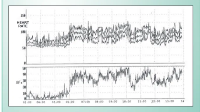
The comparison between groups in regard to clinical variations: age (0.3901), gender (0.1881) and number of ventricular PB under 24-hour Holter (0.2128) did not show significant difference. When the presence or absence of NSVT was investigated – at the 12-lead ECG, 24-hour Holter or ergometric test – no significant difference was found between groups (p = 0.3613). In the syncope group, two patients presented increase in ventricular PB on exertion; one of them presented paroxistic atrial fibrillation at peak of effort, which disappeared after exertion period was interrupted, with no clinical manifestation. Most patients presented decrease in number or absence of changes in ventricular PB behavior under physical stress. In the group with palpitations a higher number of PB under 24-hour Holter was observed as compared to the asymptomatics, and to the syncope and near-syncope group, although with no statistic difference (Figure 2).
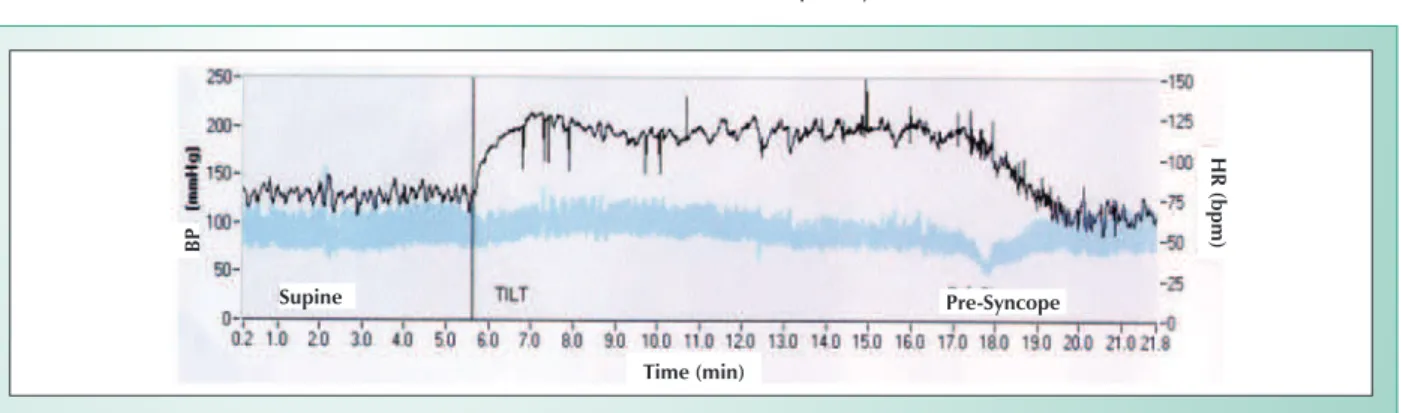
Tilt table test results - The groups reported differences regarding TTT positivity (Table I). While grouping patients with syncopes and near-syncopes and comparing them with groups with palpitations and asymptomatic, significant difference was observed regarding results for the TTT (Figure 3).
Thirty-eight percent of patients in the syncope and near-syncope group presented positive TTT for neurocardiogenic etiology whereas only 11% of patients with palpitations and asymptomatic presented the same result (p = 0.0257). Patients with positive TTT were younger than those with negative TTT (34.28 ± 16.01 versus 42.63 ± 15.63 years of age, respectively, with p = 0.0271).
Clinical course - Patients were followed for an average period of 37.7 ± 20.6 months (median = 40 months). Asymptomatic individuals were kept under no medication, and none reported clinical manifestation during follow-up.
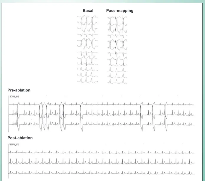
Patients with palpitations and correlation between symptoms and PB received beta-blockers or anti-arrhythmic drugs. Clinical control was reached in sixteen patients (59.3%). Eleven patients (40.7%) presented persistence of symptoms, and were submitted to radiofrequency ablation (Figure 4).
From the 21 patients with syncope and near-syncope and positive TTT, 8 (38%) presented remission of symptoms with general recommendations only. From the 13 (62%) patients who also received pharmacological treatment, only 3 presented recurrence of symptoms during the follow-up. Therefore,
Fig. 1 - Example of RVOT PB. Fig. 2 - 24-hour Holter showing PB frequency.
Heart rate
to rule out right ventricle arrythmogenic dysplasia through echocardiography (ECHO), cardiac MRI, radioisotopic ventriculography, and signal average ECG. To investigate the correlation between arrhythmia and symptoms, patients were submitted to 24-hour Holter monitoring, to loop recorder, and ergometric test.
All patients were submitted to Tilt Table Test (TTT) to evaluate their susceptibility to neurocardiogenic syncope. TTT was carried out in the morning, following prolonged baseline protocol, as previously described. Patients who had a negative prolonged baseline TTT were submitted to a pharmacological test with sublingual 1.25 mg of isosorbide dinitrate administration5. General diet and behavior recommendations
were given to all patients after TTT whenever there was a positive response. If symptoms persisted, pharmacological treatment was started after initial recommendations. Patients were evaluated every three months at the outpatient unit. Additional evaluation was carried out whenever syncope occurred. All patients agreed to participate in the research protocol, which was approved by the Institutional Scientific and Ethics Committee.
Statistical analysis - Sample size was designed to detect differences higher than 20% in number of patients with positive results for TTT among groups of symptomatic and asymptomatic patients, with significance level at 5% and test power at 80%. The groups of patients were compared as to quantitative variables by using variance analysis for data with normal distribution; Kruskal-Wallis test was used for data whenever the assumption of normality was not met. Pearson6
chi-square test and Fisher7 exact test were used for qualitative
variables in group comparisons. Multiple comparisons were carried out based on chi-square partitions 6-9.
Results
recruiting. Additionally, the presence of polymorphic PB on exertion was also an exclusion criterion. Therefore, this was a highly selected group. Negative results from the exams for the identification of those clinical syndromes are tranquillizing for patients with outflow PB and recurring syncope. However, the lack of a conclusive diagnosis for syncope etiology makes patients and families feel insecure, as the assisting doctor as well, who end up empirically prescribing anti-arrhythmic drugs.
The tilt table test - the method used to confirm vasovagal syncope diagnosis - is quite specific, although its positivity index does not exceed 60%, according to literature 11-13.
Some patients who have persisted in their unexplained syncope condition may have had the vasovagal etiology as well, although TTT low sensitivity has not allowed their identification. Under such condition, diagnosis can only be defined through data on the syncope during prolonged ECG monitoring by implantable or external events monitoring. An electrophysiologic study may be an additional method to elucidate some cases. In the present study, 2 cases of documented SVT during their course occurred in patients whose unexplained syncope persisted. That finding suggests that although the heart may be structurally normal, the investigation follow-up using electrophysiologic studies should be recommended for patients with frequent recurrences; whenever negative, prolonged monitoring of events must be considered 14-16.
Most patients in the present study showed to be kept asymptomatic during clinical follow-up. The action mechanism of beta blockers in symptom control is not clearly defined. Krittayaphong et al.17 have studied patients with RVOT PB
and have observed that atenolol helped to improve clinical symptoms and decrease arrhythmia frequency in the 24-hour Holter monitoring. But patients treated with placebo also showed improvement of symptoms, although no variations were reported for ventricular arrhythmia in the Holter monitoring. Such observation reiterates the idea that the symptoms reported by those patients are not fully related to ventricular arrhythmia, but rather, may be associated to autonomic tonus or psychogenic disorders. Hayashi et al18
have demonstrated that the predominance of sympathetic tonus plays a relevant role in triggering repetitive VE or SVT in patients with RVOT arrhythmia without structural cardiopathy, and that propranolol not only can reduce PB density but also its complexity.
85.7% of patients with positive TTT turned asymptomatic, and 14.3% presented improvement of symptoms during follow-up. None of those patients presented sustained ventricular tachycardia (SVT). However, 2 patients with syncope and negative TTT presented SVT in their clinical course. SVT was well tolerated and interrupted at the emergency unit. No death was reported.
Discussion
The present study demonstrates that patients with RVOT premature beats and normal heart who present unexplained, recurrent syncope must have vasovagal etiology investigated – and the prevalence is high (41%). Such information is relevant, since patients with recurring syncope and PB may present ventricular tachycardias – sustained and not sustained – as the cause of syncope, with distinctive prognostic and treatment10. In such cases, two clinical conditions potentially
associated with the risk of sudden death must be investigated and ruled out – arrhythmogenic ventricular displasia and catecholaminergic ventricular tachycardia – conditions under which the presence of syncope acts as a risk marker for the presence of sustained tachycardia and sudden death. In the present study, echocardiogram and cardiac MRI were carried out systematically, and used as exclusion criteria or patients with suspicion of arrythmogenic ventricular dysplasia. Radioisotopic ventriculography was also carried out for cases posing doubts; patients with enlarged RV or decreased ventricular function were excluded. Patients with catecholaminergic tachycardia typically present syncope on exertion and this was an exclusion criterion for patient
Fig. 3 - Positive Tilt Table Test in patient with RVOT PB. BP- blood pressure; HR- heart rate.
group ttt + (%) ttt – (%) total
Syncope 14 (41) 20 (59) 34 Pre-syncope 7 (35) 13 (65) 20 Palpitations 4 (15)) 23 (85) 27 Asymptomatic 0 9 (100) 9
Total 25 65 90
p(syncope versus pre-syncope) = 0.6246; p(palpitation versus asymptomatic) = 0.3902; p(syncope + pre-syncope versus palpitation + asymptomatic) = 0.0257.
table 1 - Results from ttt in the different groups of patients
In our materials, palpitations were the most recurring symptoms. Even the 2 patients with documented spontaneous, sustained VT during clinical course, VT was correlated to palpitations rather than with syncope or pre-syncope.
Conclusions
The prevalence of neurocardiogenic etiology in patients
with syncope and RVOT PB, with no structural cardiopathy is high and must always be investigated to identify patients with favorable and benign clinical course. For patients who are kept in their recurring, unexplained syncope condition invasive investigation (electrophysiologic studies) must be considered, as well as prolonged ECG outpatient unit monitoring.
1. Barret BN, Mandel WJ. The frequency and prognostic significance of electrocardiographic abnormalities in clinically normal individuals. Progr Cardiovasc Dis. 1981; 23: 299-319.
2. Seidl K, Hauer B, Zahn R, Senges J. High frequency catheter ablation as therapy of symptomatic ventricular extrasystole. Z Kardiol. 1997; 86: 211-20.
3. Sotoriades ES, Evans JC, Larson MG, et al. Incidence and prognosis of syncope. N Eng J Med. 2002; 12: 878-85.
4. Kapoor W. An Overview of the Evaluations and Management of Syncope. In: Syncope: Mechanisms and Menagement. Grubb BP, Olshansky B (Eds). Armonk, NY: Futura Publishing Co., 1998: PP 73-106. 38.
5. Scanavacca MI, Brito S, Maia I, el al. Diretrizes para avaliação e tratamento de pacientes com arritmias cardíacas. Arq Brasil Cardiol. 2002; 79(sup II): 1-50.
6. Rosner B. Fundamentals of Biostatistics. 2nd Ed. Massachusetts: PWS
References
Fig. 4 - Radiofrequency ablation of idiopathic RVOT PB.
Basal
Pre-ablation
Post-ablation
Publishers; 1986; 442-80.
7. Rosner B. Fundamentals of Biostatistics. 2nd Ed. Massachusetts: PWS
Publishers; 1986; 467-72.
8. Fleiss JL. Estatistical Method for Rate and Propotion. 2nd Ed. New York: John Wiley & Sons; 1981; 138-43.
9. Rosner B. Fundamentals of Biostatistics. 2nd Ed. Massachusetts: PWS Publishers; 1986: 47; 326-32.
10. Cox MM, Perlman BA, Mayor MR, et al. Acute and long term beta-adrenergic blockade for patients with neurocardiogenic syncope. J Am Coll Cardiol. 1995; 26: 1293.
11. Hachul DT, Sosa EA, Consolim F, et al. Valor diagnóstico do teste de inclinação na avaliação da síncope de origem indeterminada. Resultados preliminares. Arq Bras Cardiol. 1994; 62: 7-9.
12. Kapoor W, Smith M, Miller N. Upright tilt testing in evaluating syncope: a comprehensive literature review. Am J Med. 1994; 97: 78-88.
13. Sheldon R, Rose S, Flanagan P, et al. Risk factors for syncope recurrance after a positive tilt-table test in patients with syncope. Circulation. 1996; 93: 973-81.
14. Simantirakis EN, Chrysostomakis SI, Manios E, et al. Insertable loop recorder
in unmasking the causes of syncope. Pacing Clin Electiphysiol. 2000; 23 (10PtI): 1573-5.
15. Khran AD, Klein GJ, Norris C, et al. The etiology of syncope in pacients with negative tilt table and electrophysiological testing. Circulation. 1995; 92: 1819-24.
16. Grubb BP. Dysautonomic (Orthostatic) Syncope. In: Grubb BP, Olshansky B. (Eds). Syncope: Mechanisms and Management. Armonk, NY: Futura Publishing Co., 1998; PP 107-126.
17. Krittayaphong R, Bhuripanyo K, Punlee K, et al. Effect of atenolol on symptomatic ventricular arrhythmia without structural heart disease: a randomized placebo-controlled study. Am Heart Journal. 2002; 144: E10.