Radiofrequency ablation can reverse the structural
remodeling caused by frequent premature
ventricu-lar contractions originating from the right ventricuventricu-lar
outflow tract even in a ‘‘normal heart’’
Yuqiang Fang, Chunlan Wen, Li Yang, Xiaoqun Zhang, Wei Chu, Chunyu Zeng
The Third Military Medical University, Chongqing Institute of Cardiology, Daping Hospital, Department of Cardiology, Chongqing, China.
OBJECTIVE: The aim of this study was to evaluate whether frequent premature ventricular contractions originating from the right ventricular outflow tract remodel the cardiac structure and function in patients with a ‘‘seemingly normal heart’’ and whether radiofrequency ablation can reverse this remodeling.
METHODS:Sixty-eight patients with idiopathic frequent premature ventricular contractions originating from the right ventricular outflow tract and normal heart structure and function were enrolled in this study. The patients were divided into three groups according to the therapeutic method: radiofrequency ablation group (24 cases), anti-arrhythmia drug group (26 cases), and control group (18 cases without any treatment). Clinical Registration number: ChiCTR-ONRC-12002834
RESULTS:The basic patient characteristics were comparable between the three groups, except for the premature ventricular contraction rate, which was significantly lower in the control group. After six months of follow up, the premature ventricular contraction rate was significantly reduced in the radiofrequency ablation group, which was accompanied by a significant decrease in the following cardiac cavity inner diameters, as determined by echocardiography: right atrium (33.33¡3.78 vs. 30.05¡2.60 mm, p= 0.001), right ventricle (23.24¡2.40 vs. 21.05¡2.16 mm,p= 0.020), and left ventricle (44.76¡4.33 vs.41.71¡3.44 mm,p= 0.025). These results were similar in the anti-arrhythmia drug group, although this group exhibited a smaller extent of change (right atrium: 33.94¡3.25 vs. 31.27¡3.11 mm, p= 0.024; right ventricle: 22.97¡3.09 vs. 21.64¡2.33 mm, p= 0.049; left ventricle: 45.92¡6.38 vs. 43.84¡5.67 mm, p= 0.039), but not in the control group (p.0.05). There was a tendency toward improvement in the cardiac functions in both the radiofrequency ablation and anti-arrhythmia drug groups. However, these differences were not statistically significant (p.0.05).
CONCLUSIONS:These results indicate that radiofrequency ablation can potentially reverse the cardiac remodeling caused by frequent premature ventricular contractions even in structurally normal hearts and that frequent premature ventricular contractions should be abated even in structurally normal hearts.
KEYWORDS: Cardiac Remodeling; Radiofrequency Ablation; Idiopathic Frequency Premature Ventricular Contraction; Right Ventricular Outflow Tract.
Fang Y, Wen C, Yang L, Zhang X, Chu W, Zeng C. Radiofrequency ablation can reverse the structural remodeling caused by frequent premature ventricular contractions originating from the right ventricular outflow tract even in a ‘‘normal heart’’. Clinics. 2013;68(10):1312-1317.
Received for publication onApril 1, 2013;First review completed onApril 19, 2013;Accepted for publication onMay 12, 2013 E-mail: [email protected]
Tel.: 86 02368757801
& INTRODUCTION
Arrhythmia, especially tachycardia, is commonly encoun-tered in patients with cardiovascular disease and even in healthy subjects (1). Persistent tachycardia can induce
dilated cardiomyopathy, and the suppression of it can reverse the changes in cardiac structure and function in these patients (2,3). One of tachycardia, frequent premature ventricular contraction (FPVC), has been generally consid-ered as benign (4), although some recent studies suggest that long-term FPVC may cause progressive ventricular dysfunction and dilatation and may induce dilated cardio-myopathy (tachycardiocardio-myopathy) or even heart failure (4-8). The suppression of FPVCs with radiofrequency ablation (RFA) can reduce the inner diameter of the cardiac cavity and improve function significantly in these patients (5-9). However, the patients in these studies have abnormal cardiac function or cardiac cavities. Few studies have
Copyrightß2013CLINICS– This is an Open Access article distributed under the terms of the Creative Commons Attribution Non-Commercial License (http:// creativecommons.org/licenses/by-nc/3.0/) which permits unrestricted non-commercial use, distribution, and reproduction in any medium, provided the original work is properly cited.
No potential conflict of interest was reported.
investigated the effect of FPVCs on the ‘‘seemingly structurally normal heart.’’
In this study, we hypothesized that, in patients with a normal heart, idiopathic FPVCs originating from the right ventricular outflow tract (RVOT) may remodel the cardiac structure and function but that RFA or anti-arrhythmia drugs could potentially reverse this remodeling. To the best of our knowledge, we are the first to report treatment of patients with a normal heart with FPVCs originating from the RVOT.
& PATIENTS
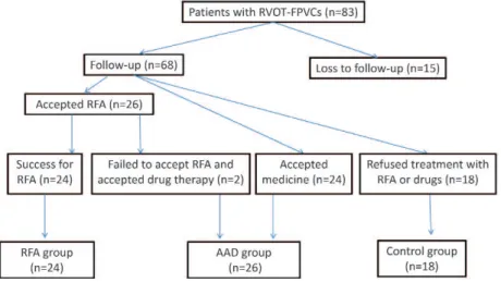
Eighty-three patients, hospitalized from September 2009 to June 2012, with RVOT-FPVCs were enrolled in this study. The monomorphic FPVC load of these patients was more than 10% of the total heart rate or more than 5% of the total heart rate with ten monomorphic sustained or unsustained ventricular tachycardia events per day (4,6). The study population included patients with or without symptoms. The patients with symptoms, including palpitation and debilitation, went to the hospital with the expectation of alleviating their symptoms. Some of the patients without symptoms wanted to eliminate the PVCs due to their fear of complications, whereas others did not intend to accept drug or RFA therapy. The patients were divided into different groups according to their treatment preference. Fifteen of the patients were excluded due to loss during follow-up; thus, a total of 68 patients (32 males and 36 females) with ages ranging from 21-66 years (mean age 46.2¡14.66 years) were enrolled in this study. These patients were divided into three groups according to the treatment method (Figure 1): the RFA group (n = 24) included patients who underwent successful ablation, the AAD group (n = 26) included patients who initially chose therapy with an anti-arrhythmia drug (n = 24) or who initially chose ablation but failed ablation and then chose drug therapy (n = 2), and the control group (n = 18) included those who refused ablation and drug therapy. In the AAD group, the patients received 0.2 g of Cordarone (Sanofi-Aventis, Hangzhou city, Zhejiang province, China) once per day or metoprolol succinate sustained-release tablets (AstraZeneca AB, Wuxi city, Jiangshu province, China) at a dose of 23.75–47.5 mg per day. If these doses were well tolerated and there were
no more than 1,000 PVCs per day (10), the doses were continued; otherwise, the doses were increased according to the tolerance of the patient or until there were no more than 1,000 PVCs per day (10,11).
All patients underwent extensive baseline evaluation to rule out any structural disease. The evaluation included a clinical history concerning the onset of symptoms, 12-lead electrocardiography (ECG), exercise testing, 24-h Holter monitoring, and echocardiography. Moreover, patients with certain diseases and conditions, including rheumatic heart disease, endocrine disease, hyperthyroidism, thyroid dys-function, arrhythmogenic drug intake, and electrolyte disturbances, were excluded from this study.
Antiarrhythmic drugs were discontinued for at least six half-lives before the study. The PVC burden was evaluated by PVC percentage, which was calculated as the number of PVCs divided by the total number of beats in 24 h. Informed consent forms regarding the RFA therapy and potential complications were signed by all patients. The study protocol conformed to the ethical guidelines of the 1975 Declaration of Helsinki as reflected in thea prioriapproval by the institutional human research committee. This study was registered with an internationally accredited site (ChiCTR-ONRC-12002834 - http://www.chictr.org/).
& DEFINITION OF RVOT-PVC AND EVALUATION OF CARDIAC STRUCTURE AND FUNCTION
Standard 12-lead surface ECG was performed before and during treatment. A right ventricular outflow tract-PVC was defined as follows: (1) PVC had a left bundle branch block (LBBB) morphology in V1,(2) the R/S transition zones (first
precordial lead with R/S ratio$1) in the PVCs were present in V2–V4, and (3) the R/S transition zones of the PVCs in the
precordial lead did not occur earlier than the R/S transition zones of the sinusal QRS on the same 12-lead surface ECG. Ventricular tachycardia was defined by standard electro-cardiographic criteria of at least five consecutive PVCs at a rate of more than 120 beats/min. Patients with atrial tachyarrhythmia, including atrial fibrillation, flutter, tachy-cardia and paroxysmal supraventricular tachytachy-cardia, were excluded from this study because of the possibility of tachycardia-induced LV dilation. The region of the LV outflow tract origin was excluded from this study.
Cardiac structure and function were evaluated by echo-cardiography at baseline and during follow-up according to the ASE/ESE method (ProSounda10, ALOKA, Japan and IE-33, Philips, Japan). Using two-dimensional and M-type Doppler echocardiography, we measured the end diastolic left ventricular internal diameter (normal range: 47¡4 mm), the left atrial internal diameter (normal range: 27–40 mm), the right basic ventricular internal diameter (normal range: 20–28 mm), the right atrial internal diameter (normal range: 29–44 mm), the inner diameter of the aorta (normal range: ,30 mm) and pulmonary artery (normal range: 12–26 mm), the inter-ventricular septal thickness (normal range: 6– 12 mm), and the left ventricular posterior thickness (normal range: 6–12 mm). The cardiac function parameters were also measured, including the left ventricular ejection fraction (LVEF, normal range: 55–80%), left ventricular fractional shortening (LVFS, normal range: 30–45%) and E/A ratio (normal range:.1).
& MAPPING AND CATHETER ABLATION PROCEDURE
All procedures were performed after the signing of the informed consent form. The patients were studied in the fasting state without sedation. Antiarrhythmic drugs were discontinued for at least six half-lives before the procedure. Under local anesthesia, a 7-F deflectable quadripolar ablation catheter (Biosense Webster, Diamond Bar, California, USA) with a 4-mm-tip electrode was percuta-neously introduced into the right ventricle. Based on the 12-surface-lead electrocardiogram with spontaneous RVOT-PVC, pace mapping was conducted using bipolar pacing between the distal pair of the electrodes with a stimulation pulse width of two ms. If the culprit PVCs were not found during the procedure, isoproterenol administration and/or programmed electrical stimulation with a digital stimulator (LEAD 7000B, Sichuan Jinjiang Electronic Science and technology Co., Ltd, China) was performed to induce the PVCs as previously described (5). An optimal pace map was defined as a match of all 12 surface leads when comparing the R/S ratio and subtle notching in the QRS complex during pacing. An identical match was necessary in at least 11 of 12 leads. The RFA was performed based on an optimal pace map for 60-90 s with a preset temperature of 50-60˚C and a power limit of 50 Watts. A successful ablation was defined as the non-recurrence and non-inducibility of culprit PVCs with or without isoproterenol administration at the rate of 0.2-0.6mg/min and/or programmed electrical stimulation for at least 30 min after ablation. All 12 surface electrocardiograms and the bipolar intra-cardiac electro-grams (filtered at 30-400 Hz) were recorded and stored using a 48-channel acquisition system (LEAD 7000B, Sichuan Jinjiang Electronic Science and technology Co.,
Ltd, China). During the procedure, intravenous heparin was administered as a 100 IU/kg bolus dose followed by additional boluses of 1,000 IU every hour. Procedural success was defined as the lack of recurrence of culprit PVCs within 72 h after the procedure under electrocardio-gram monitoring.
& FOLLOW-UP
All the patients were followed up every month for the next six months by 24-h Holter monitoring tests and the assessment of cardiac chamber size and function.
& STATISTICAL ANALYSIS
Continuous variables are presented as the mean ¡ SD and were compared using Student’s t-test or one-way ANOVA. Echocardiographic measurements before and after treatment were compared with the paired t-test. Ap-value ,0.05 was considered statistically significant.
& RESULTS
General characteristics of each group
The general characteristics of each group are listed in
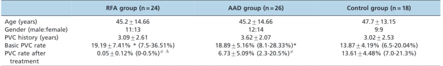
Table 1. In this study, the PVCs had been present for 0.5–10 years (mean 3.09¡2.61 years). The PVC rate was 8978– 36978/24 h, and 6.5–36.51% (mean 19.19¡7.41%) of the total QRS complexes were affected. A total of six patients had transient ventricular tachycardia (VT) (the morphology of the VT was the same as that of the PVC, and the number of VT events was greater 10 or the persistent time of VT was more than 10 s). There were no differences in the age, gender, or history of the patients with PVCs. However, the PVC rate was higher in the RFA (19.19¡7.41%) and AAD (18.89¡5.16%) groups compared with the control group (13.87¡4.19%). Of the 26 patients who received RFA therapy, 24 (92.31%) were successfully ablated. The two unsuccessfully ablated patients were transferred to the AAD group.
Effect of RFA on PVC burden and cardiac cavity diameters
At the six-month follow-up, the PVC rate was only 0.05¡0.12% in the RFA group, which was lower than that in the AAD (6.73¡5.09%) and control (13.61¡4.48%) groups. Among the patients with PVCs in the RFA group, 21 had less than 100 PVCs/day, two patients had 100-300 PVCs/ day, and only one patient had 561 PVCs/day. Compared with the control group, the number of PVCs was signifi-cantly lower in the AAD group (Table 1).
The diameters of the cardiac cavities were normal in all three groups at baseline, and there were no significant
Table 1 -General characteristics of each group.
RFA group (n = 24) AAD group (n = 26) Control group (n = 18)
Age (years) 45.2¡14.66 45.2¡14.66 47.7¡13.15
Gender (male:female) 11:13 12:14 9:9
PVC history (years) 3.09¡2.61 3.62¡2.07 3.02¡2.53
Basic PVC rate 19.19¡7.41% * (7.5-36.51%) 18.89¡5.16% (8.1-28.33%)* 13.87¡4.19% (6.5-20.04%) PVC rate after
treatment
0.05¡0.12% (0-0.5%)#D
6.73¡5.09% (2.3-20.5%)#
13.61¡4.48% (7.0-21.3%)
*
differences between the cavities, although the diameters of the right and left atrium and right and left ventricle were larger in the RFA and AAD groups compared with the control group. However, some cardiac cavity diameters decreased significantly and were smaller in the RFA group for 6 months follow-up (right atrium: 33.33¡3.78 mm vs.
30.05¡2.60 mm, p= 0.001; right ventricle: 23.24¡2.40 mm
vs.21.05¡2.16 mm,p= 0.020; left ventricle: 44.76¡4.33 mm
vs.41.71¡3.44 mm,p= 0.025). Similar results were obtained in the AAD group for 6 months follow-up (right atrium: 33.94¡3.25 mmvs.31.27¡3.11 mmp= 0.024; right ventricle: 22.97¡3.09 mm vs. 21.64¡2.33 mm, p= 0.049; and left ventricle: 45.92¡6.38 mm vs. 43.84¡5.67 mm, p= 0.039). There were no significant differences in the control group (p.0.05) (Table 2). After treatment, the diameters of the RA, RV, LA, and LV were significantly smaller in the RFA and AAD groups compared with the control group (Table 2,
p,0.05), and the diameters of the RA, LA, and LV were significantly smaller in the RFA group than in the AAD group (Table 2,p,0.05).
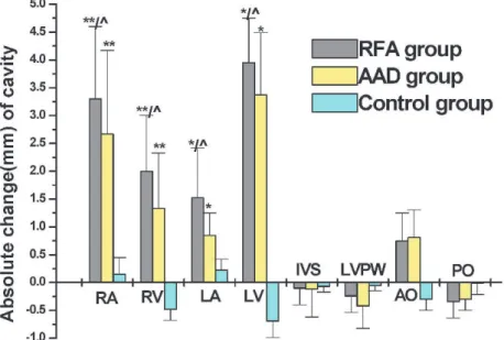
To examine the changes in the cardiac cavities, we plotted the absolute changes (Figure 2). The results indicated that the greatest amount of RFA change occurred in the right atrium (3.3¡0.4 mm), followed by the right ventricle (2.0¡0.2 mm) and left ventricle (3.0¡0.22 mm), and the least amount of change occurred in the left atrium (1.52¡0.15 mm). There was no obvious change in the inter-ventricular septal thickness, left ventricular posterior thickness, or inner diameter of the aortic root and pulmonary artery. The results were similar in the AAD group, but no significant change was observed in the control group.
Effect of RFA on cardiac function
The examination of cardiac function indicated no sig-nificant difference with regard to the LVFS, LVEF, and E/A ratio between the three groups before or after treatment. There was a tendency toward improved cardiac function in both the RFA group (LVFS: 34.33¡4.87 vs. 36.23¡2.7
p= 0.066; LVEF: 63.71¡4.94 vs. 66.24¡6.20, p= 0.093; E/A ratio: 1.34¡0.20 vs. 1.31¡0.14, p= 0.290) and AAD group (LVFS: 34.42¡5.07 vs. 35.96¡4.96 p= 0.098; LVEF: 64.04¡5.45 vs. 66.13¡6.80, p= 0.133; E/A ratio: 1.36¡0.31
vs.1.33¡0.35,p= 0.256). However, the differences were not significant (p.0.05;Table 3).
& DISCUSSION
FPVC is one of the most common types of arrhythmia in patients without structural heart diseases, and it is observed in 1-4% of the general population (12). Although FPVCs can cause palpitations, they are considered relatively benign because of the good prognosis after long-term follow-up (4,13). However, some recent reports undermine this popular belief. Chugh (9) reported that, in a case of dilated cardiomyopathy and FPVC, RFA therapy resulted in the disappearance of the FPVCs, the return of the dilated ventricle to a normal size, and a significant improvement in cardiac function. Kuroki (3) observed that FPVCs increased pulmonary venous flow regurgitation, pulmonary capillary wedge pressure, right atrial pressure, and left ventricular end-diastolic pressure. After ablation, these aberrant para-meters were normalized. Yarlagadda (5) observed that the LVEF increased to a normal level six months after successful RFA in 27 patients with monomorphic FPVCs. Bogun (6) found an inverse correlation between the LVEF and PVC rate and that among 22 patients who received successful RFA and a six-month follow-up, the LVEF increased from 0.34 to 0.59 in 18 patients but decreased from 0.34 to 0.25 in four patients. Facchini (14) observed a subclinical but significant increase in left ventricular dimensions in FPVC patients compared with the control group. Because of these findings, the European Heart Association (EHA) now includes tachycardiomyopa-thy (TCM) caused by FPVC in their 2008 guidelines (15) and considers it as an indication for an RFA procedure (11).
The inverse relationship between cardiac function and PVC rate was further confirmed by Beaufort-Krol (13), who performed a long-term follow-up of children with FPVCs and an anatomically normal heart. The results indicated that the PVCs originating from the left ventricle disappeared in most patients, whereas the PVCs originating from the right ventricle lasted to adulthood and had the possibility of further developing into TCM. However, it was previously unknown whether RFA therapy would have an effect on the structurally normal hearts with FPVCs. Thus, in this study, we enrolled patients with a structurally normal heart and FPVCs originating from the RVOT. After RFA or AMM treatment, cardiac cavity size was significantly decreased in diameter, accompanied by a reduction in FPVC rate, which may indicate the reversal of cardiac remodeling. Although no significant difference existed between cardiac function
Table 2 -Changes in cardiac cavity diameter in each group.
Diameter of Cardiac
Cavity RFA group (n = 24) AAD group (n = 26) Control group (n = 18) Pre-RF Post-RF p-value Pre-treat Post-treat p-value Basic Follow-up p-value RA (mm) 33.33¡3.78 30.05¡2.60*& 0.001 33.94
¡3.25 31.27¡3.11& 0.024 32.89
¡2.73 32.74¡2.49 0.251 RV (mm) 23.24¡2.40 21.05¡2.16& 0.020 22.97¡3.09 21.64¡2.33& 0.049 22.39¡2.89 22.87¡3.08 0.192
LA (mm) 33.45¡4.12 31.95¡3.06*& 0.092 32.82
¡5.04 32.17¡4.36& 0.192 32.83
¡3.76 32.61¡3.64 0.272 LV (mm) 44.76¡4.33 41.71¡3.44*& 0.025 45.92¡6.38 43.84¡5.67& 0.039 43.17¡3.56 43.86¡3.03 0.219
IVS (mm) 9.67¡1.66 9.71¡1.31 0.467 9.74¡1.46 9.77¡1.58 0.454 9.42¡1.18 9.49¡1.13 0.428 LVPW (mm) 8.22¡1.14 8.56¡1.28 0.201 8.19¡1.35 8.61¡1.62 0.106 8.37¡1.13 8.40¡1.14 0.472 AO (mm) 29.72¡2.67 28.95¡1.83 0.138 30.02¡3.37 29.18¡2.74 0.159 29.11¡1.99 29.50¡2.22 0.297 PA (mm) 20.62¡1.99 20.9¡1.76 0.312 20.65¡1.78 20.76¡1.85 0.251 20.89¡2.21 20.94¡2.07 0.470
*
p,0.05vs. AAD group;p,0.05vs. control group;
Thep-value in this table indicates the significance between pre-treatment and post-treatment.
before and after treatment with RFA, a tendency toward improvement was observed. This result may indicate that FPVCs play a remodeling role in cardiac cavities and function and that RFA may reverse the remodeling even in structurally normal hearts.
The fact that no significant difference was observed in cardiac function may be attributed to the small number of patients included in this study. Therefore, multicenter studies with a larger group of patients who receive a long-term follow-up investigation need to be conducted in the future. Moreover, the absence of randomization is another limitation of this study. Therefore, a randomized, double-blinded, and multicenter study with long-term fellow-up is expected to be performed in the future to confirm the results of this pilot study.
In conclusion, in patients with FPVCs originating from the RVOT and with a structurally normal heart, RFA reduces the FPVC rate and the size of the cardiac cavities and has a tendency to improve cardiac function. These results suggest that FPVCs should be reduced by RFA even in structurally normal hearts.
& ACKNOWLEDGMENTS
This study was supported in part by grants from the National Basic Research Program of China (973 Program, 2008CB517308, 2012CB517801), Natural Science Foundation Project of CQ (CSTC, 2009BA5044, 2009BB5332), National Natural Science Foundation of
China (30925018, 31130029, 81070259), and National Institutes of Health (R01HL092196).
& AUTHOR CONTRIBUTIONS
Fang Y contributed to the mapping and ablation of PVCs and writing the article. Wen C contributed to the treatment of PVCs with antiarrhythmic drugs. Yang L contributed to the cardiac evaluation by echocardiography. Zhang X contributed to the data collection. Chu W contributed to Holter evaluation of PVCs. Zeng C contributed to the design of the project.
& REFERENCES
1. Nagashima M, Matsushima M, Ogawa A, Ohsuga A, Kaneko T, Yazaki T, et al. Cardiac arrhythmias in healthy children revealed by 24-hour ambulatory ECG monitoring. Pediatr Cardiol. 1987;8(2):103-8, http://dx. doi.org/10.1007/BF02079464.
2. Omichi C, Tanaka T, Kakizawa Y, Yamada A, Ishii Y, Nagashima H, et al. Improvement of cardiac function and neurological remodeling in a patient with tachycardia induced cardiomyopathy after catheter abla-tion. J Cardiol. 2009;54(1):134-8.
3. Kuroki K, Tada H, Seo Y, Ishizu T, Igawa M, Yamasaki H, et al. Prediction and mechanism of frequent ventricular premature contrac-tions related to haemodynamic deterioration. European Journal of Heart Failure. 2012;14(10):1112-20, http://dx.doi.org/10.1093/eurjhf/hfs095. 4. Gaita F, Giustetto C, Di Donna P, Richiardi E, Libero L, Brusin MC, et al.
Long-term follow-up of right ventricular monomorphic extrasystoles. J Am Coll Cardiol. 2001;38(2):364-70, http://dx.doi.org/10.1016/S0735-1097(01)01403-6.
5. Yarlagadda RK, Iwai S, Stein KM, Markowitz SM, Shah BK, Cheung JW, et al. Reversal of cardiomyopathy in patients with repetitive mono-morphic ventricular ectopy originating from the right ventricular
Figure 2 -The absolute change in cardiac cavity diameters in each group. The cavity changes are indicated as absolute changes in the RFA group. Positive changes indicate a reduction in the diameters of the cardiac cavities. *p,0.01vs. control group,‘p,0.05vs. AAD
group. RA: right atrium; RV: right ventricle; LA: left atrium; LV: left ventricle; IVS: interventricular septum; LVPW: left ventricular posterior wall; AO: aortic opening; PO: pulmonary artery.
Table 3 -Variation of cardiac function in each group.
Cardiac function RFA group (n = 24) AAD group (n = 26) Control group (n = 18) Pre-RF Post-RF p-value Pre-treat Post-treat p-value Basic Follow-up p-value LVFS 34.33¡4.87 36.23¡2.70 0.066 34.42¡5.07 35.96¡4.96 0.098 34.61¡3.38 34.13¡3.45 0.408 LVEF 63.71¡4.94 66.24¡6.20 0.093 64.04¡5.45 66.13¡6.80 0.133 64.83¡4.30 64.17¡4.51 0.331 E/A ratio 1.34¡0.20 1.31¡0.14 0.290 1.36¡0.31 1.33¡0.35 0.256 1.36¡0.17 1.30¡0.13 0.164 Thep-value is a comparison between the pre-treatment and post-treatment or between the baseline values and the follow-up values of cardiac function.
outflow tract. Circulation. 2005;112(8):1092-7, http://dx.doi.org/10. 1161/CIRCULATIONAHA.105.546432.
6. Bogun F, Crawford T, Reich S, Koelling TM, Armstrong W, Good E, et al. Radiofrequency ablation of frequent, idiopathic premature ventricular complexes: comparison with a control group without intervention. Heart Rhythm. 2007;4(7):863-7, http://dx.doi.org/10. 1016/j.hrthm.2007.03.003.
7. Taieb JM, Maury P, Shah D, Duparc A, Galinier M, Delay M, et al. Reversal of dilated cardiomyopathy by the elimination of frequent left or right premature ventricular contractions. J Interv Card Electrophysiol. 2007;20(1-2):9-13, http://dx.doi.org/10.1007/s10840-007-9157-2. 8. Sternick EB, Correa F, Negri R, Scarpelli RB, Gerken LM. Reversible
cardiomyopathy provoked by focal ventricular arrhythmia orginating from the base of the posterior papillary muscle. J Interv Card Electrophysiol. 2009;25(1):67-72, http://dx.doi.org/10.1007/s10840-008-9341-z. 9. Chugh SS, Shen WK, Luria DM, Smith HC. First evidence of premature
ventricular complex induced cardiomyopathy: a potentially reversible cause of heart failure. J Cardiovasc Electrophysiol. 2000;11(3):328-9, http://dx.doi.org/10.1111/j.1540-8167.2000.tb01802.x.
10. Sekiguchi Y, Aonuma K, Yamauchi Y, Obayashi T, Niwa A, Hachiya H, et al. Chronic hemodynamic effects after radiofrequency catheter ablation of frequent monomorphic ventricular premature beats. J Cardiovasc Electrophysiol. 2005;16(10):1057-63, http://dx.doi.org/10. 1111/j.1540-8167.2005.40786.x.
11. Aliot EM, Stevenson WG, Almendral-Garrote JM, Bogun F, Calkins CH, Delacretaz E, et al. European Heart Rhythm Association (EHRA); Registered Branch of the European Society of Cardiology (ESC); Heart Rhythm Society (HRS); American College of Cardiology (ACC); American Heart Association (AHA). EHRA/HRS consensus on catheter ablation of ventricular arrhythmias. Heart Rhythm. 2009;6(6):886-933, http://dx.doi.org/10.1016/j.hrthm.2009.04.030.
12. Kostis JB, McCrone K, Moreyra AE, Gotzoyannis S, Aglitz NM, Natarajan N, et al. Premature ventricular complexes in the absence of identifiable heart disease. Circulation. 1981;63(6):1351-6, http://dx.doi. org/10.1161/01.CIR.63.6.1351.
13. Beaufort-Krol GC, Dijkstra SS, Bink-Boelkens MT. Natural history of ventricular premature contractions in children with a structurally normal heart: does origin matter? Europace. 2008;10(8):998-1003, http://dx.doi. org/10.1093/europace/eun121.
14. Facchini M, Malfatto G, Ciambellotti F, Chianca R, Bragato R, Branzi G, et al. Increased left ventricular dimensions in patients with frequent nonsustained ventricular arrhythmia and no evidence of underlying heart disease. J Cardiovasc Electrophysiol. 1999;10(11):1433-8, http://dx. doi.org/10.1111/j.1540-8167.1999.tb00202.x.