Taxonomy and distribution of the green algal genus
Halimeda
(Bryopsidales, Chlorophyta) in Brazil
1MARIA ELIZABETH BANDEIRA-PEDROSA
1, SONIA M.B. PEREIRA
1and EURICO C. OLIVEIRA
2,3(received: July 11, 2003; accepted: February 19, 2004)
ABSTRACT– (Taxonomy and distribution of the green algal genus Halimeda (Bryopsidales, Chlorophyta) in Brazil). Halimeda is a genus of calcified coenocytic green algae with a well known ecological importance in some tropical areas. Bleached calcified segments of Halimeda may accumulate in large deposits of economic potential as is the case in the northeastern coast of Brazil. In a survey of the genus in Brazil based on recent collections and examination of abundant material deposited on Brazilian herbaria we identified seven species: Halimedacuneata Hering, H.discoidea Decaisne, H. gracilis Harvey ex J. Agardh, H. incrassata (Ellis) Lamouroux, H. opuntia (Linnaeus) Lamouroux, H. simulans Howe and H. tuna (Ellis & Solander) Lamouroux. These species are described in detail, with emphasis on diagnostic characters. Our study has shown that the shape and size of the utricula in surface view, under scanning electron microscopy, can be utilized to discriminate some species. Fertile specimens of Halimeda cuneata and H. discoidea are reported for the first time in the region. Data on vertical and geographical distribution are presented for each species and the southern limit of the genus in the western Atlantic was extended.
Key words - Brazil, Bryopsidales, Halimeda, Halimeda cuneata
RESUMO – (Taxonomia e distribuição do gênero de algas verdes Halimeda (Bryopsidales, Chlorophyta) no Brasil). Halimeda é um gênero de algas verdes de talo cenocítico e calcificado que desempenha importante papel ecológico em regiões tropicais. Segmentos calcificados e branqueados de Halimeda podem-se acumular em grandes depósitos com potencial econômico, como ocorre na costa nordeste do Brasil. O levantamento do gênero no Brasil, baseado em coletas recentes e em abundante material depositado em herbários brasileiros, mostrou a presença de sete espécies: Halimedacuneata Hering, H.discoidea Decaisne, H. gracilis Harvey exJ. Agardh, H. incrassata (Ellis) Lamouroux, H. opuntia (Linnaeus) Lamouroux, H. simulans Howe e H. tuna (Ellis & Solander) Lamouroux, as quais são descritas em detalhe, com ênfase nos caracteres diagnósticos. Nossos estudos mostraram que a forma e tamanho dos utrículos em vista frontal, vistos em microscopia eletrônica de varredura, permite a distinção entre algumas espécies. Exemplares férteis de Halimeda cuneata e H. discoidea são descritos pela primeira vez para a costa brasileira. Para cada espécie é indicada a distribuição vertical e geográfica na costa brasileira, extendendo-se o limite sul de distribuição do gênero no Atlântico ocidental.
Palavras-chave - Brasil, Bryopsidales, Halimeda, Halimeda cuneata
Introduction
The genus Halimeda Lamouroux is characterized
by a coenocytic thallus constructed of a system of interwoven bifurcated siphonous filaments that expand into utricles at the thallus surface. Macroscopically, the thallus is characterized by a series of green articulated coin shaped segments made rigid by the impregnation of calcium carbonate as aragonite (Gilmartin 1960, Borowitska & Larkum 1977).
Species recognition has been based mainly on the gross morphology of the segments, branching pattern, position and shape of holdfasts. Anatomy provides additional distinctive characters such as the size and shape of the utricles and structure of the node joining two successive segments (Barton 1901, Taylor 1950, Hillis 1959, Hillis-Colinvaux 1980). Hillis-Colinvaux (1980) divided the genus into five sections based on the patterns of nodal structure: Rhipsalis, Opuntia, Halimeda, Micronesicae and Crypticae. Hillis et al.
(1998) based on a partial sequence of the 18S rDNA found a good correlation between several morphological characters and the molecular data, but recognized only three lineages: Rhipsalis, Opuntia and Halimeda. More
recently Kooistra et al. (2002) extended considerably
the sequencing study of Hillis et al. (1998) including SSU
and ITS 1 and 2 regions; although they recognized the relevance of nodullary siphons structure and
1. Universidade Federal de Pernambuco, Departamento de Biologia, Rua Dom Manoel de Medeiros, s/n, 52171-900 Recife, PE, Brazil.
2. Universidade de São Paulo, Instituto de Biociências, Caixa Postal 11461, 05422-970 São Paulo, SP, Brazil.
M.E. Bandeira-Pedrosa et al.: The genus Halimeda in Brazil
364
arrangement, they rejected the sectional treatment of Hillis-Colinvaux (1980).
The genus is broadly distributed in the tropics, being represented by 33 species (Hillis et al. 1998). The
following species have been reported from Brazil:
Halimeda discoidea Decaisne, H. gracilis Harvey ex
J. Agardh, H. incrassata (Ellis) Lamouroux, H. opuntia
(Linnaeus) Lamouroux, H. simulans Howe and H. tuna
(Ellis & Solander) Lamouroux (Horta & Oliveira 2002). However their circumscription is not always clear and varies with the author.
This study is a critical revision of the species that occur in Brazil based on the analysis of abundant material. Information on scanning electron microscopy (SEM) of thallus surface is presented for the first time and a map showing the geographical distribution of the species along the Brazilian coast is provided, extending the southern limit of the genus in the American Atlantic.
Material and methods
Recent material was collected from the intertidal and infralittoral zones during low tide by skin diving from 1997 to 2000 on several places of the northeastern coast of Brazil, and dredged during an extensive national program to survey the Brazilian platform (REVIZEE). We also studied material collections deposited in Brazilian herbaria (ALCB, IPA, JPB, PEUFR, RFA, SP, SPF and UFP) and the lectotype of Halimeda cuneata at BM. For the sake of brevity only a few selected specimens of the long list of material studied are included here.
For light microscopy, specimens were preserved in 4% Formalin and decalcified in 20% HCl. Dried herbarium specimens were immersed in 50% glycerol for 24 hours and then processed as described for liquid preserved specimens. Longitudinal and cross sections were made with steel blades. Nodal regions were dissected with needles, and the preparations were mounted in a 50% glycerol solution. Structure sizes are reported as the mean of ten measurements, with the extreme values indicated between brackets.
For scanning electron microscopy (SEM), small portions of mature segments were pre-fixed in the field in 2.5% glutaraldehyde in 0.1M sodium phosphate buffer, pH 7.0, and kept on ice. The material was then rinsed in the same buffer, post-fixed in 2% osmium tetroxide, dehydrated in an ethanol series (30% to 100%), dried in hexamethildezilasane (HMDS), sputter-coated with gold and observed in a MEV Phillips XL30.
Results
Although aware that the sections established by Hillis-Colinvaux (1980) are not supported by molecular
data according to Kooistra et al. (2002) we decided to
keep this traditional arrangement for the time being.
Section Rhipsalis J. Agardh ex De Toni
Characterized by having nodal medullary siphons fused all together for a short extension and with a sequence of pits. This section is represented in Brazil by Halimeda incrassata and H. simulans, both included
in “lineage 1” of Kooistra et al. (2002).
Halimeda incrassata (Ellis) Lamouroux, Nouv. Bull.
Sci. Soc. Philom. 3:186. 1812. ≡ Corallina incrassata
Ellis, Philosophical Transactions 57: pl.17, figs. 20-27. 1767.
Type locality: Jamaica Figures 1, 20, 24, 25.
Thallus erect, single or gregarious, strongly calcified at the base and less so at the upper segments; color light-green, becoming whitish on drying; up to 27 cm long; branching mainly di- to tetrachotomous; holdfast bulbous; the first two basal segments cylindrical and the third one usually flabellate, about 10 mm wide and 8 mm long; upper segments subcylindrical, cuneate or reniform, 9 mm wide by 6 mm long (figure 1). Cortex with up to five layers of utricles; external utricles hexagonal in outline, 72 (52-92) µm in diameter in surface view (figure 20) and 81 (59-103) µm long in cross-section holding together even after decalcification (figure 24); every secondary utricle 53 (44-74) µm long, gives rise to two primary ones; nodal medullary siphons interwoven and fused all together for not more than 100 µm, with conspicuous pores (figure 25).
Selected material: BRASIL: CEARÁ: Guajiru, Trairi, 21-XII-1991, N.P. Dantas (PEUFR17866). RIO GRANDE DO NORTE: 05º04’S, 35º25’W, 16 m, 26-II-1980, G.C. Teixeira (PEUFR5082); 05º30’S, 35º04’W, 29 m,
2-II-1997, REVIZEE (PEUFR35121). PERNAMBUCO: Tamandaré, 10-III-1999, M.E. Bandeira-Pedrosa & M.D. Santos (PEUFR35117); 07º47’S, 34º29’ W, 52 m,
17-X-1995, REVIZEE, (PEUFR35124). PARAÍBA: 07º18’00”S, 34º33’00”W, 30 m, 14-II-1980, F.R. Lima
(PEUFR6464); Monumento, Cabedelo, 28-VII-68,
S.M.B. Pereira (SPF27553). ALAGOAS: 09º20’S, 35º05’W, 45 m, 9-IX-1965, s.col. (SPF25967).
from the subtidal are more robust and with the typical reniform segments, whereas the intertidal ones have subcylindrical segments.
Halimeda simulans Howe, Bull. Torrey Bot. Club
34:503. 1907.
Type locality: Puerto Rico Figures 2, 22, 26, 27.
Thallus erect, single or gregarious, lightly calcified at the base and less so at the upper segments; color dark-green when alive, becoming light-green on drying; up to 14 cm long; branching mainly di-trichotomous; holdfast bulbous; the first two basal segments short, subcylindrical, subcuneate; upper segments discoid to reniform, with imbricated aspect and up to 12 mm wide and 9 mm long (figure 2). Cortex with up to four layers of utricles; external utricles keep together even after decalcification (figure 26), hexagonal in outline, and similar to H. incrassata, in surface view, but smaller
with 38 (18-48) µm in diameter (figure 22) and 34 (22-48) µm long in cross section. Every secondary utricle supports 2-4 primary ones; secondary utricles 62 (55-92) µm long. Medullary siphons interwoven, with short nodal fusions, forming a single group with inconspicuous pores (figure 27).
Selected material: BRASIL: PERNAMBUCO: Fernando de Noronha Archipelago: 03º27’S, 35º02’W, 58 m, 4-VI-1998, REVIZEE (PEUFR35027); 04o46’S,
35º24’W, 39 m,16-X-1967, Almirante Saldanha
(SPF25972); Itamaracá, 8-VIII-1968, P. Montouchet
(SPF29381); Recife, Boa Viagem 28-VIII-1963, D.B. Andrade (SPF29718); 07º26’S, 34º30’W, 42 m,
12-V-1998, REVIZEE (PEUFR35026). BAHIA: Barra
do Gil, Itaparica, 9-VIII-1998 M.E. Bandeira et al.
(PEUFR35126); Itaparica, Mar Grande, 7-VII-2000,
M.C. Accioly (PEUFR35025); 13º38’S, 38º45’W, 50 m,
19-X-1997, REVIZEE (PEUFR36497).
This species was found in the state of Pernambuco, Fernando de Noronha Archipelago and the state of Bahia (figure 42), being collected from the intertidal up to a depth of 63 m. In Bahia this species occurs in dense intertidal populations among Penicillus capitatus
Lamouroux, Caulerpa spp., Udotea spp. and Halimeda opuntia. Intertidal specimens are more vigorous and
calcified than the subtidal ones. Williams & Blomquist (1947) were the first to report the occurrence of
H. simulans in Brazil (coast of Pernambuco and
Fernando de Noronha Archipelago), which was considered as uncertain by Oliveira Filho (1977), in the absence of material for confirmation. Later on, Ugadim
& Pereira (1978) found it among material dredged from Pernambuco, which is now confirmed.
Section Opuntia J. Agardh ex De Toni
This section is characterized by having nodal medullary siphons fused in pairs for a short distance. It is locally represented by only one species, Halimeda opuntia, which is included in “lineage 5” of Kooistra et al. (2002).
Halimeda opuntia (L.) Lamouroux, Nouv. Bull. Sci.
Soc. Philom. 3:186. 1812. ≡ Corallina opuntia L., Syst.
Nat. p.805. 1758. Type locality: Jamaica Figures 3-5, 21, 28-30.
Thallus erect, forming compact or loosely prostrate tufts, strongly calcified; color light-green to whitish up to 34 cm long; branching polystichous, attached to the substrate by multiple holdfasts; segments variable in shape, usually trilobate, flattened to cylindrical, sometimes twisted, auriculated to crenulated ca. 18 mm in diameter and 9 mm long; remaining segments discoid to cuneate, with up to 12 mm wide and 9 mm long (figures 3-5). Cortex with 3-5 layers of utricles dichotomously branched (figure 28); external utricles rounded to polygonal, lightly coalescent after decalcification, 29 (15-44) µm in diameter, in surface view (figure 21), and 30 (26-41) µm long in cross section. Every secondary utricle supports 2 primary ones. Nodal medullary siphons interwoven, fused in pairs, seldom in groups of 3-4, with short nodal fusions (figures 29, 30). Selected material: BRASIL: CEARÁ: Guajiru, Trairi, 21-XII-1991, N.P. Dantas (PEUFR17867). RIO GRANDE DO NORTE: 05º09’05”S, 35º08’05”W, 28 m, 5-II-1980, G.C. Teixeira (PEUFR5016); 05º30’S, 35º04’W, 29 m,
2-II-1997, REVIZEE (PEUFR35078); 04º47’S, 35º20’W,
44 m, 13-X-1995, REVIZEE (PEUFR35079).
PERNAMBUCO: Jaboatão dos Guararapes, Candeias, 21-I-1962, I. Pontual (PEUFR1215); Tamandaré,
Campas, 3-XII-1990, J.A.P. Angeiras & A.S. Lopes
(PEUFR20564); Itamaracá, Jaguaribe, 18-IX-1997, M.E. Bandeira-Pedrosa & M.F.B. Oliveira (PEUFR35042);
Ipojuca, Serrambí, 16-X-1997, M.E. Bandeira-Pedrosa & A.L.M. Cocentino (PEUFR35045); Porto de Galinhas,
15-XII-1997, M.E. Bandeira-Pedrosa & A.L.M. Cocentino (PEUFR35051); Tamandaré, 10-III-1999, M.E. Bandeira-Pedrosa (PEUFR35061); Paulista,
Nossa Senhora do Ó, 24-XI-1957, A. Lima (IPA11370);
M.E. Bandeira-Pedrosa et al.: The genus Halimeda in Brazil
366
PARAÍBA: Cabo Branco, 27-V-1998, M.E. Bandeira-Pedrosa & A.I. Kanagawa (PEUFR35063). ALAGOAS: Francês, 26-IV-1998, M.E. Bandeira-Pedrosa & E.A.C. Guedes (PEUFR35064); Barra de São Miguel,
26-IV-1998, M.E. Bandeira-Pedrosa & E.A.C. Guedes
(PEUFR 35065); Paripueira, 28-IV-1998, M.E. Bandeira-Pedrosa & E.A.C. Guedes (PEUFR35127); Pajuçara,
29-I-1965, E.C. Oliveira (SP96476). BAHIA: Itaparica,
M.E. Bandeira-Pedrosa et al. (PEUFR35073); Salvador,
Forte, 6-VIII-1998, M. Accioly (PEUFR35077); Itaparica,
Coroa, 28-XI-1981, Y. Ugadim (SPF25975); Salvador,
Pituba, I-1977, E.C. Oliveira (SPF51423).
Adittional material studied (H.M.S. Challenger Exp.): ADMIRALTY ISLES, H.N. Moseley 1876 (BM);
BAHIA: Barra Grande-Brazil, H.N. Moseley 1876
(BM); PERNAMBUCO: Fernando de Noronha Archipelago,
H.N. Moseley 1876 (BM).
This is the most common and widespread species of Halimeda worldwide, being considered pantropical.
In Brazil the it is more common in the intertidal, but was also dredged at 44 m, from Ceará State to Rio de Janeiro (figure 42). Its morphology is variable: on reef flats tufts are more compact and imbricated with twisted segments, whereas on reef vertical walls tufts are more loose, branching tend to become distichous and segments are auriculate assuming the cordate form described by Barton (1901).
This species may have a key ecological role due to its high biomass and percentage of substrate cover. In some places it forms dense beds inhabited by a large diversity of invertebrates; in others their dead segments accumulate in large amounts and are sporadically utilized as a source of carbonates.
Section Halimeda
This section is characterized by short nodal medullary siphons extensively fused in groups of 2-3. The following species are present in Brazil: Halimeda cuneata, H. discoidea, H. tuna and H. gracilis. The
first three are included in the “lineage 3” and the last one in “lineage 4” of Kooistra et al. (2002).
Halimeda cuneata Hering in Krauss, Flora p.209. 1846.
Type: SOUTH AFRICA: NATAL: Dulham, Krauss s.n.,
1839 (Lectotype BM!, designated by Hillis-Colinvaux 1980)
Figures 11-17, 31-34.
Thallus erect, single, lightly calcified; color dark-green, becoming white or yellowhish on drying; up to
25 cm long; branching sparse to dense, usually dichotomous but also trichotomous to irregular in places and with up to 8 segments on a series without furcation (figures 11-14); holdfast basal and small; the first two basal segments are cylindrical or subcylindrical; upper segments are flat, cuneate to trapezoid, sometimes discoid with a smooth and shiny surface measuring up to 25 mm wide by 20 mm long; most of the segments are joined by a sort of short stipe, 1 mm long and 6 mm in diameter (figure 15), composed by densely interwoven, torulose and thick-walled filaments (figure 16). Cortex with three layers of utricles, seldom four; external utricles goblet shaped, keeping firmily together after decalcification; fused utricles were seen occasionally; external utricles are polygonal in surface view (figure 17) with 52 (22-66) µm in diameter; in cross section they measure 104 (74-144) µm long; secondary utricles suport 2-4 primary ones (figure 31); nodal medullary siphons interwoven and fused in groups of 2-3 for a variable extent (figures 32, 33); above the node the medullary filaments have thick walls and are pigmented, and may have a small tuft of short utricles above the point of filament fusions (figure 32). Gametophores are produced from the primary utricles and have 4-7 pedunculated gametangia, 370 µm long and 140 µm in diameter (figure 34).
Selected material: BRASIL: RIO GRANDEDO NORTE: 05º 09’05”S, 35º11’00”W, 20 m, 5-II-1980, G.C. Teixeira (PEUFR5017). PARAÍBA: 06º33’S, 34º47’05”W, 26 m, 4-VI-1981, F.R. de Lima
(PEUFR6510). BAHIA: Itaparica, Mar Grande, 6-X-1964, A.B. Joly & Y. Ugadim (SPF630); Salvador,
Amaralina, 4-X-1964, A.B. Joly & Y. Ugadim
(SPF1829); Ilhéus Grande, 15-I-1965, E.C. Oliveira
(SPF54706); Itapoã, 20-IV-1987, G. Mitchell
(RFA4991); Forte, 6-VIII-1998, M.E. Bandeira-Pedrosa & M.C. Accioly (PEUFR35028); Itaparica,
Mar Grande, 9-VIII-1998, M.E. Bandeira-Pedrosa & M.C. Accioly (PEUFR35032); Salvador, Itapoã,
29-VI-2000, M.E. Bandeira-Pedrosa & M.C. Accioly
(PEUFR35035). ESPÍRITO SANTO: Ponta da Fruta, 19-VIII-1978, E.C. Oliveira (SPF 26021); Piuma,
Acaiaca, 18-VIII-1978, E.J. Paula (SPF26022); Ilha
do Francês, 30-V-1986, prof. 15 m, E.C. Oliveira
(SPF28801); Itaipava, 17-III-1988, E.C. Oliveira
(SPF52193); Manguinhos, 20-I-1973, E.C. Oliveira & L. Behar (SPF53490); Ponta da Fruta, 4-VII-1985,
fértil, E.J .Paula (SPF53739); Santa Cruz, 23-VI-1986, G. Mitchell (RFA4605); Vitória, Camburí,
22-VI-1994), C. Nassar (RFA9256).
M.E. Bandeira-Pedrosa et al.: The genus Halimeda in Brazil
368
M.E. Bandeira-Pedrosa et al.: The genus Halimeda in Brazil
370
M.E. Bandeira-Pedrosa et al.: The genus Halimeda in Brazil
372
NATAL: Richards Bay, W.G. Rump, 1929 (BM); Port
Elizabeth, G.F. Papenfuss, 1955 (BM).
This species, so far known only from the Indo-Pacific, was recently found in Brazil (E. Bandeira-Pedrosa et al., unpublished data). In Brazil this species
is common on reef flats from Rio Grande do Norte through Espírito Santo states (figure 42) from the intertidal to 26 m depth. More developed specimens were found at the exposed edges of the reef formation. Segment morphology is rather variable on the reef plateau, and may make its recognition in the field difficult, especially from Halimeda discoidea. This species has
also being locally confused with H. tuna. Segments with
typical morphology are found in more protected areas and in pools. Fertile specimens were found only in the state of Espírito Santo, in July.
Halimeda discoidea Decaisne, Ann. Bot. 18:96. 1842.
Type locality: “Kamtschatka” [true provenance unknown].
Figures 6, 7, 18, 35-37.
Thallus erect, single, lightly to moderately calcified; color light-green to green yellowish becoming whitish on drying; up to 11 cm long; branching sparse to dense, di-trichotomous; holdfast single and small; segments discoid to reniform, with a smooth surface, about 46 mm wide and 25 mm long (figures 6, 7). Cortex with 2-3 layers of utricles 65 (74-92) µm long in cross-section (figure 35) and 57 (37-77) µm in diameter in surface view, often laterally fused in pairs (figure 18), holding together strongly even after decalcification; secondary utricles subglobose, 121 (92-210) µm wide; nodal medullary siphons interwoven and fused in groups of 2-3 for a short distance (figure 36). Gametophores branched, produced from secondary utricles on the surface or margin of the fertile segments, bearing 5-8 globose to pear-shaped gametangia about 103 µm wide and 159 µm long (figure 37).
Selected material: BRASIL: CEARÁ: Trairi, Guajiru, 21-XII-1991, N.P. Dantas (PEUFR17865); 04º56’S,
35º19’ W, 32 m, 14-V-1998, REVIZEE (PEUFR35083);
01º43’S, 37º07’W, 54 m, 6-VI-1998, REVIZEE
(PEUFR35086); 03º50’S, 37º37’W, 166 m, 12-X-1995,
REVIZEE (PEUFR35092). RIO GRANDE DO NORTE: 05º17’00”S, 35º08’00”W, 34 m, 22-I-1980, F.R. Lima
(PEUFR4979). PARAÍBA: 07º15’05”S, 34º36’00”W, 28 m, 2-IV-1981, G.C. Teixeira (PEUFR5700);
07º18’S, 34º31’W, 35 m, 22-I-1981, A.I. Kanagawa
(JPB10247). PERNAMBUCO: 07º26’S, 34º30’W, 42 m, 12-V-1998, REVIZEE (PEUFR35081); Fernando de
Noronha, Baía de Sueste, 7-III-1993, S.M.B. Pereira
(PEUFR21630); Fernando de Noronha, Baía de Sueste, 2-XI-1985. E.C. Oliveira & V. Eston
(SPF51496). SERGIPE: V-1999, PETROBRAS/UFSE
(PEUFR35097); VIII-1999, PETROBRAS/UFSE
(PEUFR35098). BAHIA: Sta Cruz de Cabrália, Coroa Alta, 3-I-1979, E.C. Oliveira (SPF51158); Salvador,
Itapoã, 29-VI-2000, M.E. Bandeira-Pedrosa & M.C. Accioly (PEUFR 35080). ESPÍRITO SANTO: 17º04’S, 36º53’W, 250-60 m, 14-XI-1997, REVIZEE
(PEUFR35093); 17º47’S, 35º52’W, 63 m, 13-XI-1997
REVIZEE (PEUFR35095).
Additional material studied: BRASIL: BAHIA: Barra Grande, H.M.S. Challenger Exp., H.N. Moseley, 1876
(BM).
General morphology present some variation on populations of different depth; intertidal specimens tend to be more calcified and densely branched, with smaller and thicker segments. This species was found from Ceará state through Espírito Santo State (figure 42) from the intertidal to 166 m depth. It was found in the State of Sergipe for the first time. Coloration is darker on specimens from the intertidal than from the subtidal specimens. This species often occurs together with
H. opuntia. Fertile material was found only once, in
November, 1997, in deep water.
Halimeda gracilis Harvey ex J. Agardh, Acta
Universitets Lund 23:82. 1887. Type locality: Sri Lanka. Figures 8, 19, 38-40.
Thallus prostrate, moderate to strongly calcified; color whitish, becoming grayish on drying; up to 24 cm long; branching di-trichotomous; attachment by multiple holdfasts; segments subcylindrical, cuneate to reniform, with a smooth and shiny surface, margin undulate, about 18 mm wide and 11 mm long (figure 8); segments friable before decalcification, with utricles lightly coalescent after decalcification; cortex with two, rarely three layers of utricles, rounded in outline, 52 (44-67) µm in diameter in surface view (figure 19), and 72 (67-92) µm long in cross section. Every secondary utricle supports up to 8 primary ones; secondary utricles clavate (figure 38), 74 (56-92) µm wide and 170 (166-333) µm long. Medullary siphons interwoven, with extensive paired nodal fusions, eventually with three filaments fused together (figures 39, 40).
Selected material: BRASIL: CEARÁ: 01º43’S, 37º07’W, 54 m, 6-VI-1998, REVIZEE (PEUFR35112);
M.E. Bandeira-Pedrosa et al.: The genus Halimeda in Brazil
374
35103). RIO GRANDEDO NORTE: 04º51’S, 36º22’W, 30m, 27-III-1980, G.C. Teixeira (PEUFR4500); 05º17’S,
35º08’W, 34 m, 25-XI-1979, F.R. Lima (PEUFR4975).
PARAÍBA: João Pessoa, Tambaú, 26-IX-1980, (JPB10221) A.I. Kanagawa; 07º34’00”S, 34º42’00”W,
20 m, 21-I-1981, A. Fernandes (PEUFR6513).
PERNAMBUCO: Ipojuca, Serrambí, 12-VII-1987, M.C. Accioly (PEUFR2784), 07º28’S, 34º32’W, 36 m,
31-I-1997, REVIZEE (PEUFR35108). SERGIPE: V-1999
PETROBRAS/UFSE (PEUFR35115); VIII-1999, PETROBRAS/UFSE (PEUFR35116). BAHIA: 13º38’S, 38º45’W, 50 m, 19-X-1997, REVIZEE (PEUFR36471);
15º34’S, 38º51’W, 50 m, 25-X-1997, REVIZEE
(PEUFR36498); 17º04’S, 36º53’W, 250 m, 14-XI-1997,
REVIZEE (PEUFR36478). ESPÍRITO SANTO: 19º28’S, 38º22’W, 94 m, 27-II-1996, REVIZEE (PEUFR36474).
RIODE JANEIRO: 20º35’S, 40º05’W, 50 m, 24-II-1982,
Orion (SPF29386); 20º38’S, 40º01’W, 57 m, 27-II-1996, REVIZEE (PEUFR36472).
Additional material studied: SRI LANKA: W.H. Harvey 72 (Isotype and photo at BM)
This species was found from Ceará through Rio de Janeiro states (figure 42), from the intertidal to 250 m depth. Its occurrence at 250 m is not enough evidence that it was actually growing at that depth. It is here reported for the states of Sergipe and Bahia for the first time. Specimens from deep water have larger segments that resemble Halimeda tuna, whereas
intertidal ones are closer to H. opuntia. This probably
explain the difficulties many authors had in identifying this species, as can be seen in many herbaria.
Halimeda tuna (Ellis & Solander) Lamouroux, Nouv.
Bull. Sci. Soc. Philom 3:186. 1812. ≡ Corallina tuna
Ellis & Solander, Natur. Hist. Zooph. p.111, pl.20, fig.e. 1786.
Type locality: Mediterranean Sea Figures 9, 10, 23, 41.
Thallus erect, single, moderate to lightly calcified; color light to dark-green, up to 10 cm long; branching sparse to dense, di-trichotomous; holdfast single and small; basal segments strongly calcified, subcuneate to subcylindrical, remaining segments discoid to subcuneate, with a rough surface, up to 20 mm wide and 12 mm long (figures 9, 10). Cortex with usually three, but up to four layers of utricles (figure 41); external utricles keep lightly coalescent after decalcification; polygonal in outline in surface view, with cell walls thicker than the other species (figure 23); 44 (29-55) µm in diameter, in surface view, and 108 (59-196) µm long
in cross section; every secondary utricle supports 6 or more primary ones; secondary utricles 48 (44-52) µm wide and 111 (85-177) µm long. Medullary siphons interwoven, with extensive or short nodal fusions in groups of 2-3 as in H. cuneata and H. discoidea
(figures 32, 36).
Selected material: BRASIL: MARANHÃO: Parcel Manoel Luís, at 10 m, 29-V-2000, A.C.L.L. Castro & M.M. Ferreira-Correia (PEUFR38008); 01º36’S,
38º10’W, 51 m, 18-IX-1995, REVIZEE (PEUFR35039).
CEARÁ: Guajiru, Trairi, 21-XII-1991, N.P .Dantas
(PEUFR17868). RIO GRANDEDO NORTE: 04º 50’00”S, 35º41’00”W, 30 m, 2-III-1980, S.M.B. Pereira & G.C. Teixeira (PEUFR4451). PARAÍBA: Ponta do Seixas, 18-XII-1980, F.A.F. Carvalho (JPB10220).
PERNAMBUCO: Ipojuca, Serrambi, 15-X-1986, M.C. Accioly & S.M.B. Pereira (PEUFR12785); Campas,
Tamandaré, 16-III-1991, J.A.P. Angeiras
(PEUFR20568); Itamaracá, Jaguaribe, 15-X-1997, M.E. Bandeira-Pedrosa & M.F. Oliveira-Carvalho
(PEUFR30523); Tamandaré, 5-V-2000, M.E. Bandeira-Pedrosa (PEUFR35037). ALAGOAS: Ponta Verde, 4-II-1965, E.C. Oliveira (SPF635); Garça Torta,
27-IV-1998, M.E. Bandeira-Pedrosa & E.A.C. Guedes
(PEUFR30528). BAHIA: Coroa Vermelha, 19-IV-1988,
C. Nassar (RFA4363). RIODE JANEIRO: 18º01’S, 35º53’W, 60 m, 13-XI-1997, REVIZEE (PEUFR36481).
Adittional material studied: FRANCE: Cape Ferrat, Mediterranean, VI-2000, E.C. Oliveira.
(PEUFR38016).
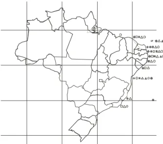
Figure 42. Geographic distribution of Halimeda species on the Brazillian coast: = Halimeda cuneata, = H. discoidea,
= H. gracilis, = H. incrassata, = H. opuntia,
Discussion
One of the problems in identifying species of
Halimeda is that gross morphology varies among
populations of the same species growing in different habitats, making this criterium unreliable as was already remarked by Barton (1901) and by subsequent authors (e.g. Hillis 1959, Stark et al. 1969, Hillis-Colinvaux
1980). This was fully suported by our observations based on a large sample of specimens from a broad range of depths. This plasticity is certainly the explanation for the confusions we found in local literature (e.g. Pereira
1974, Dantas 1994) and in herbaria. Identification at species level is possible only by a combination of several criteria including habitat, gross morphology and anatomy of mature segments and nodes. Particularly useful are the patterns of nodal medullary siphons fusion, in addition to the size, shape and distribution of the utricles as remarked long ago (Barton 1901). Nodal structure seems to vary less and, therefore, is more useful at the section level. Gametangia arrangement may also help (Kamura 1966, Hillis-Colinvaux 1980) as we have seen in
H.discoidea and H. cuneata, but they are quite rare
and undescribed for some taxa (Hillis-Colinvaux 1980). The utilization of the shape and size of the utricula in surface view under SEM proved to be rewarding
Key for the identification of Halimeda species reported from Brazil
1. With a bulbous holdfast; nodal medullary siphons fused in a single group (section Rhipsalis)
2. Basal segments usually cylindrical, but also subcuneate or flabellate; primary utricles measuring
over 50 µm in diameter ...H. incrassata
2. Basal segments subcuneate or reniform, imbricate; primary utricles measuring less than 50 µm
in diameter ...H. simulans
1. With one or several non-bulbous holdfasts; nodal medullary siphons fused in groups of 2-3. 3. Attachment by several holdfasts; nodal medullary siphons fused in pairs for a short extension
(not more than 100 µm) (section Opuntia) ... H. opuntia
3. Attachment by single or multiple holdfasts; nodal medullary siphons fused in groups of 2-3 for more than 100 µm (section Halimeda)
4. Thallus flaccid and prostrate, attached by multiple holfasts; secondary utricles clavate ...H. gracilis
4. Thallus erect with a single conspicuous holdfast; secondary utricles not clavate
5. Segments discoid; secondary utricles inflated, sub-globular ... H. discoidea
5. Segments sub-cylindrical, cuneate to reniform; secondary utricles not inflated
6. Basal segments cylindrical, stipe-like; nodal region pedunculate or cushion-like ...H. cuneata
6. Basal segments subcuneate; nodal region sessile ... H. tuna
This species was found from the state of Maranhão through the State Rio of Janeiro (figure 42) from the intertidal to 60 m depth. Intertidal populations
are more calcified and with a smaller holdfast, with thicker, smaller and rougher segments, usually dark-green to brownish.
allowing species discrimination in some cases if associated with other characters (figures 17-23).
A good example of morphological variability is given by Halimeda opuntia, which is well known for its large
variation in branching pattern and segment morphology. This species can form extensive mats with entangled branches as well as loose tufts with distichous branching, in which case it can be easily confused with H. gracilis.
Its segments may be flat, subcylindrical, contorted, auriculate, trilobate and crenulate depending on where it is growing. Because of this variability Barton (1901) proposed to divide this taxon into four forms: f. cordata,
f. elongata, f. opuntia and f. triloba. Taylor (1960)
accepted f. cordata and f. triloba and added f. minor
Vickers; however, this division is not supported by our observations for we observed a morphological continuous in large samples and even some specimens with mixed characters.
Halimeda cuneata, H. discoidea, H. gracilis,and H. tuna, all from section Halimeda Hillis-Colinvaux
(1980), are very similar and put together under the
H. tuna complex (Hillis 1959). Halimeda cuneata
M.E. Bandeira-Pedrosa et al.: The genus Halimeda in Brazil
376
difficulties to species identification may be related to hybridization. However, this is based only on morphology and have no experimental support. This hypothesis has already been suggested by Behar (1972) commenting on the difficulties in distinguishing some specimens of
Halimeda cuneata from H. discoidea. Within this
closely related group, H. discoidea can be distinguished
microscopically by the presence of inflated or subglobose secondary utricles. However, in H. discoidea the typical
form is attributed to intertidal specimens, whereas the deep-water specimens have been described as
H. discoidea var. platyloba (Taylor 1960). The same
applies for H. tuna, where the deep-water specimens
have been described as H. tuna var. platydisca or f. platydisca (Taylor 1960).
Identification of specimens from deep water is much more complicated because of morphological plasticity (cf. Stark et al. 1969). For example, deep water
plants of H. gracilis exhibited larger, reniform to
subcuneate segments, easily mistaken for the deep-water form of H. tuna. This deep-water form of H. gracilis
was described as H. gracilis f. lata by Taylor (1950).
The recent introduction of gene sequencing as a tool in the classification of Halimeda brings a new set
of information (Hillis et al. 1998, Kooistra et al. 2002).
This in great part supports the classification based on morphological characters, but not always. For instance in the sectional division of Hillis-Colinvaux (1980)
H. gracilis appear within the section Halimeda (lineage
3 of , Kooistra et al. 2002) but in this last work it appears
in linneage 4. Molecular data of Kooistra et al. (2002)
showed that H. tuna from the Atlantic and from the
Mediterranean were not nearest neighbor. That paper also casts doubts on the supposed pantropical distribution of several species and suggests allopatric speciation through vicariance in the tropical Atlantic and Indo-Pacific.
On what concerns the distribution of Halimeda in
Brazil the seven reported species are concentrated on the northeast coast (figure 42). It is worth noting that all species are present on the coast of Bahia state (figure 42). This fits well with the distribution of reefs in Brazil, the largest concentration being in the state of Bahia as well. The southern limit of the genus is in the Cabo Frio region, Rio de Janeiro State (Zeller 1876, Taylor 1930, 1931, Oliveira Filho 1977), just a little north of a well known area of upwelling.
However, it is on the coast of Pernambuco that the largest biomass of Halimeda spp. seems to be
concentrated, especially due to the thick mats of
H. opuntia. In this region, dead segments of Halimeda
form extensive deposits that have been sporadically utilized commercially (Kempf 1970, 1980, Pereira 1974, Oliveira Filho 1981).
As to vertical distribution the genus is well known for having species that go deep in the sublittoral (e.g. Littler & Littler 2000, Stark et al. 1969). In Brazil, Halimeda gracilis and H. discoidea were found at
depths of over 160 m, whereas H. tuna, H. incrassata, H. simulans and H. opuntia were found up to 60 m.
We have a sample of H. discoidea dredged from 260 m;
however, there is no guaranty that this species was actually growing at that depth.
In conclusion although we have cleared up some confusions on the identification of the Halimeda species
in Brazilian herbaria and extended the distribution of several taxa along the coast, we realised that due to morphological convergence in response to habitat and herbivore pressure, the next step would be to check our morphological hypothesis with molecular biology techniques.
Acknowledgements – Dr. Zenilda Bouzon for help with SEM observations and Dr. Ana Malinska for the utilization of the Scanning Electron Microscope facilities at the Universidade Federal de Santa Catarina. Professor Michael J. Wynne corrected the English text and made valuable suggestions to improve the manuscript.
References
BEHAR, L. 1972. Clorofíceas do litoral Sul do Estado do Espírito Santo. I. Siphonocladales e Siphonales. Dissertação de mestrado, Universidade de São Paulo, São Paulo.
BARTON, E.S. 1901. The genus Halimeda. Siboga-Expeditie Monographe, Leiden 60:1-32.
BOROWITZKA, M.A. & LARKUM, A.D.W. 1977. Calcification in the green alga Halimeda.V. An ultrastructure study of the thallus development. Journal of Phycology 13:6-16.
DANTAS, N.P. 1994. Estudos taxonômicos dos representantes da ordem Caulerpales (Chlorophyta) da Praia de Guagirú (Estado do Ceará - Brasil). Dissertação de mestrado, Universidade Federal de Pernambuco, Recife.
GILMARTIN, M. 1960. The ecological distribution of the deep water algae of Eniwetok. Ecology 41:210-221. HILLIS, L.W. 1959. A revision of genus Halimeda (order
Siphonales). Publications of the Institute of Marine Sciences 6:321-403.
HILLIS, L.W., ENGMAN, J.A. & KOISTRA, W.H.C.F. 1998. Morphological and molecular phylogenies of Halimeda (Chlorophyta, Bryopsidales) identify three evolutionary lineages. Journal of Phycology 34:669-681.
HORTA, P. & OLIVEIRA, E.C. 2002. Algas marinhas bênticas do Brasil http://www.ib.usp.br/algamare-br (acesso em 10/10/2003).
KAMURA, S. 1966. On the sexual reproduction of two species of Halimeda (Chlorophyta). Bulletin of Arts and Sciences of the University of Ryukyus, Mathematical and Natural Sciences 9:302-313.
KEMPF, M. 1970. Nota preliminar sobre os fundos costeiros da região de Itamaracá (norte do Estado de Pernambuco-Brasil). Trabalhos Oceanográficos da Universidade Federal de Pernambuco 9-11:95-110.
KEMPF, M. 1980. Perspectiva de exploração econômica dos fundos de algas calcárias da plataforma continental do nordeste do Brasil. Trabalhos Oceanográficos da Universidade Federal de Pernambuco 15:139-164. KOOISTRA, W.H.C.F., COPPEJANS, E.G.G. & PAYRI, C.
2002. Molecular systematics, historical ecology, and phylogeography of Halimeda (Bryopsidales). Molecular Phylogenetics and Evolution 24:121-13.
LITTLER, D.S. & LITTLER, M.M. 2000. Caribbean reef plants. OffShore Graphics Inc., Washington.
OLIVEIRA FILHO, E.C. 1977. Algas marinhas bentônicas do Brasil.Tese de livre docência, Universidade de São Paulo, São Paulo.
OLIVEIRA FILHO, E.C. 1981. Marine phycology and exploitation of seaweeds in South America. In Proceedings of the International Seaweed Symposium (T. Levring, ed.). W. de Gruyter, Berlin v.10, p.97-112.
PEREIRA, S.M.B. 1974. Cloroficeas da Ilha de Itamaracá e arredores (Estado de Pernambuco-Brasil). Dissertação de mestrado, Universidade de São Paulo, São Paulo. STARK, L.M., ALMODOVAR, L. & KRAUS, R.W. 1969.
Factors affecting the rate of calcification in Halimeda opuntia (L.) Lamouroux and Halimeda discoidea Decaisne. Journal of Phycology 4:305-312.
TAYLOR, W.R. 1930. Algae collected on the “Hassler”, “Albatross” and Schmitt Expeditions: I. Marine algae from Brazil. American Journal of Botany 16:621-630. TAYLOR, W.R. 1931. A synopsis of the marine algae of Brazil.
Revue Algologique 5:279-313.
TAYLOR, W.R. 1950. Plantas of Bikini and other northern Marshall Islands. University of Michigan Press, Ann Arbor.
TAYLOR, W.R. 1960. Marine algae of the eastern tropical and subtropical coasts of the Americas. University of Michigan Press, Ann Arbor.
UGADIM, Y. & PEREIRA, S.M.B. 1978. Deep-water marine algae from Brazil collected by the Recife Commission. I. Chlorophyta. Ciência e Cultura 30:839-842.
WILLIAMS, L.G. & BLOMQUIST, H.L. 1947. A collection of marine algae from Brazil. Bulletin of the Torrey Botanical Club 74:383-397.