Clinicoradiological Session
Case 2 / 2010 - Five-Month-Old Male Infant with Subpulmonary
Interventricular Communication and Aortic Coarctation
Edmar Atik, Fabiana M. Passos, Alexandre S. Cauduro
Hospital Sírio Libanês, São Paulo, SP - Brazil
Mailing Address: Edmar Atik •
Rua Dona Adma Jafet, 74 conj.73 - Bela Vista - 01308-050 - São Paulo, SP - Brasil
E-mail: [email protected], [email protected]
Manuscript received July 30, 2009; revised manuscript received November 12, 2009; accepted November 12, 2009.
Key words
Congenital cardiopathies; ventricular septal defect, aortic coarctation, pulmonary artery /abnormalities.
trunk and the pulmonary arteries demonstrates the presence of pulmonary flow directly from the left ventricle to the right ventricular outflow tract, as in the subpulmonary location of the ventricular septal defect.
Differential diagnosis
All other acyanotic congenital cardiopathies with left-to-right shunt must be recalled, such as interatrial communication and patent ductus arteriosus, as long as they present a marked hemodynamic effect, which occurs, however, at more advanced ages. This alteration of marked dilatation of the pulmonary trunk is also seen in cyanotic cardiopathies with pulmonary hyperflow, such as total anomalous pulmonary venous connection.
Diagnostic confirmation
The clinical elements were decisive for the diagnosis of the ventricular septal defect and aortic coarctation. The echocardiogram confirmed the presence of a ventricular defect with a direct connection with the pulmonary valve in a subpulmonary location, measuring 10 mm in diameter, in addition to the aortic coarctation in the isthmus region, measuring 3 mm in diameter, in relation to 7 mm of the ascending aorta and 5 mm of the aortic arch. The right ventricle was not dilated, considering the preferential flow from the left ventricle to the pulmonary artery tree. The left ventricle was enlarged and the left ventricular function was preserved (LVEF: 68%, Ao: 14, LA: 18, LV: 27) (Fig.2).
Management
At surgery, the resection of the aortic coarctation in termino-terminal anastomosis with the aortic arch resulted in an adequate diameter of the region. The 10-mm subpulmonary or committed ventricular septal defect was closed through an incision in the pulmonary trunk, using a bovine pericardial patch. The heart failure condition was resolved.
Comments
The marked dilatation of the pulmonary trunk is seldom detected in infants, even in those with cardiopathies with marked pulmonary hyperflow. This anatomical confirmation, in this case, was due to the direct flow from the left ventricle to the pulmonary artery tree, through the ventricular septal defect. Moreover, this defect, when doubly committed to the great vessels, pathogenically explains the concomitant existence of aortic coarctation, considering the increased
Clinical data
The patient remained asymptomatic up to three months before, when he started to present fatigue even at rest, which grew progressively worse.
Physical examination: the patient was tachydyspneic, had normal skin color, ample pulses in both upper extremities and decreased in the lower extremities. Weight was 5,130g. The blood pressures in the right upper limb and right lower limb were 150/80 and 100/80 mmHg, respectively. HR: 130 bpm. The aorta was not palpated at the suprasternal notch. The patient presented slight precordial impulses at the left sternal border and the ictus cordis was diffuse and palpated at the 5th intercostal space on the hemiclavicular line. Heart sounds were hyperphonetic and there was a holosystolic murmur of ++ intensity, on the sternal border, irradiating to the lower sternal border and the mitral area. The liver was not palpated.
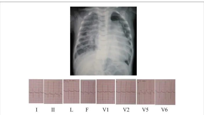
The electrocardiogram (Fig.1) showed sinus rhythm and biventricular overload, with RS complexes in all precordial leads and normal ventricular repolarization. Moreover, there were signs of left atrial overload. ÂQRS was at 90o, ÂP at +30o and ÂT at +50o.
Radiographic image
The image shows enlargement of the cardiac silhouette at the expense of the ventricular arch and greatly increased pulmonary vascular network in the hila and pulmonary periphery. The markedly increased mid-arch dilatation was also noteworthy (Fig. 1).
Diagnostic impression
This image is compatible with the diagnosis of a cardiopathy that accompanies a left-to-right shunt, such as ventricular septal defect. The increased dilatation of the pulmonary
Clinicoradiological Session
Arq Bras Cardiol 2010; 94(3) : e26-e27
Figure 1 -Chest X-ray showing the enlarged cardiac silhouette and pulmonary vascular network. The marked dilatation of the mid-arch is noteworthy, due to the pulmonary trunk enlargement. The electrocardiogram demonstrates signs of biventricular overload.
Figure 2 -The echocardiogram shows the aortic coarctation in the isthmus region, after the emergence of the left subclavian artery, measuring 3-mm in diameter in suprasternal view, in A; the subpulmonary ventricular septal defect, measuring 10 mm in diameter in cross-sectional view, at the left sternal border, in B; and dilatation of the left cavities in apical four-chamber view, in C. .
shunting from the left ventricle through the ventricular septal defect to the pulmonary trunk, still during fetal life. This dynamics helps the ductus arteriosus to be located after
the aortic coarctation. The surgical correction prevents an unfavorable evolution related to the LV enlargement and dysfunction.