Image
Mailing Address: Marcelo Luiz Campos Vieira •
Rua Cardoso de Melo, 463 / 21- Vila Olímpia - 04548-002 – São Paulo, SP – Brazil
E-mail: [email protected]
Manuscript received March 15, 2011; revised manuscript received March 15, 2011; accepted April 14, 2011.
Introduction
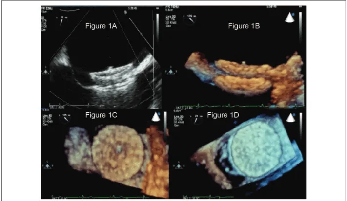
Female patient, 37 years old, with pervious foramen ovale and a history of transient ischemic attack, underwent successful implantation of Amplatzer septal occluder
for atrial septal defect closure. Transesophageal three-dimensional echocardiography (3D) allowed viewing the prosthesis from multiple levels of analysis (Figure 1).
Keywords
Foramen ovale; prosteses and implants; echocardiography, three-dimensional.
Amplatzer Septal Occluder for Closing Foramen Ovale: View Through
3D Echocardiography
Marcelo Luiz Campos Vieira e Carlos A. Pedra
Hospital Israelita Albert Einstein, São Paulo, SP – BraZil
Figure 1 - 2D transesophageal echocardiogram (Figure 1A) and 3D transesophageal echocardiogram (Figure 1B — side view, Figure 1C — view from the left atrium; Figure 1D — view from the right atrium) for a view of the Amplatzer septal occluder for closure of foramen ovale.