Case Report
This study reports the case of an anomalous implantation of VVI pacemaker electrode in the left ventricle (LV) diagnosed during routine evaluation, two years after implantation. The patient is a 65-year-old woman with Chagas disease. Electrocardiogram (ECG) revealed a pattern of right branch block. Profile chest X-ray showed electrode with posterior curvature path. In transthoracic echocardiography, the diagnosis revealed that the catheter penetrated the right atrium, crossed the atrial septum, descended through the left atrium and mitral valve orifice and deployed on the LV wall. The following related aspects are addressed: potential deployment routes, clinical, radiological, electrocardiographic and echocardiographic pictures, complications and treatment options.
Pacemaker Electrode Misplaced in the Left Ventricle
André Luiz Cerqueira de Almeida, Viviane Machicado Cavalcante, Márcio Diego Castro Teixeira, Gustavo Rocha
Costa de Freitas
Universidade Estadual de Feira de Santana, Hospital EMEC - Feira de Santana, BA - Brazil
Key words
Pacemaker, artificial; electrodes, implanted; left ventricle; atrioventricular block.
Mailing address: André Luiz Cerqueira de Almeida •
Rua Alto do Paraguai, 280 - 44042-310 - Feira de Santana, BA - Brazil E-mail: [email protected]
Manuscript received November 02, 2008; revised manuscript received May 24, 2009, accepted on December 23, 2009.
focus grade II/VI. Electrocardiogram (ECG) performed during this investigation showed pacemaker spikes and ventricular depolarization of right bundle branch block (RBBB) (Fig. 1). Profile chest X-ray showed pacemaker electrode with posterior curvature path which, together with the RBBB finding on ECG, suggested electrode misplaced in the left ventricle (LV) (Fig. 2). The patient was referred to transthoracic echocardiogram, which showed that the electrode reached the right atrium (RA), crossed the oval fossa in the interatrial septum (probably through a patent foramen ovale), crossed the left atrium, went down the mitral valve orifice and deployed in the LV lateral wall (Fig. 3 and 4).There was no thrombotic material attached to the electrode. Doppler revealed discreet mitral regurgitation. Global and regional left ventricular contractile functions were preserved. As the patient had been with the pacemaker for two years and was asymptomatic and with normal ventricular function, it was decided to maintain the electrode in the position found and anticoagulation with Warfarin (INR between 2.5 and 3.5). After five years of monitoring, the patient remains clinically stable, with no signs or symptoms of neurological or systemic embolic event.
Discussion
Misplacement of pacemaker electrode in the LV is a recognized but underdiagnosed and underreported complication of PM implantation3.
The electrode can be implanted incorrectly due to improper puncture of the subclavian artery, causing it to penetrate the LV retrogradly across the aortic valve4. In other situations, the catheter is placed in the LV after reaching the RA, going through the interatrial septum (either piercing the septal membrane or going through a patent foramen ovale, or through a pre-existing atrial septal defect) and going down the mitral valve orifice2,3,5. It may also penetrate the interventricular septum1,6 or, more rarely, the atrioventricular septum and be deployed in the LV7. Besides these situations, the electrode can be implanted abnormally inside the coronary sinus8. The main route reported has been through the interatrial septum, which is comparable to our case, probably because of incomplete closure of the foramen ovale, a fact present in 15% to 25% of the adult population1.
Patients with PM catheter at normal position in the right ventricle (RV) often present with paradoxical splitting of second heart sound, which similar to what occurs in individuals with left bundle branch block (LBBB). Presence of fixed splitting of second heart sound during inspiration and expiration indicates that the electrode is misplaced in the LV5.
Introduction
Electrode pacemakers misplaced in the left ventricle is an uncommon complication associated with pacemaker implants1. Although it is an underdiagnosed event, some cases have been reported in the literature emphasizing the possibility of complications, especially thromboembolic events1-3. This article reports the case of electrode pacemaker misplaced in the left ventricle in a female patient with Chagas disease.
Case report
Figure 1 -12-lead electrocardiogram with RBBB pattern.
The 12-lead ECG and chest X-ray profile provide important information that may lead to clinical suspicion of PM misplacement in the LV. Situations in which a catheter is correctly inserted in the RV apex are expected to present ECG with LBBB pattern. Presence of RBBB leads to suspicions that the electrode is misplaced in the LV. Occasionally, however, a RBBB pattern can be observed while the electrode is at normal position in the
Case Report
Almeida et al
Pacemaker electrode misplaced in LV
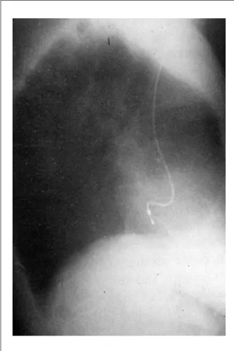
Figure 2 -Proile chest X-ray shows the posterior path of the pacemaker
electrode.
Figure 3 -Apical four-chamber echocardiogram showing the pacemaker electrode path: it enters the right atrium, crosses the interatrial septum, goes through the left atrium and mitral valve oriice, and deploys in the left ventricular wall.
Another quick and simple test to determine the electrode position is the chest radiograph. The posteroanterior projection is not very helpful in these situations. A profile assessment is recommended. If the electrode is at normal position in the RV, it will appear at an anterior position in this projection. If it was deployed in the LV or in the coronary sinus, the electrode path will be held backward1-3.
Therefore, while physical examination, ECG and chest X-ray are useful in suspecting misplacement of pacemaker electrode in the LV, they are not able to deliver an accurate diagnosis2. Transthoracic echocardiography is the test of choice to confirm the exact position of the electrode and trace its deployment route2,5. The transesophageal echocardiogram should be done if the chest echocardiogram is not clarifying2.
Misplacement of PM electrode in the LV may result in thrombus formation, which increases the risk of morbidity1-3. Cerebral thromboembolic complications are the most feared, and neurological findings may range from mental confusion to stroke with severe neurological sequelae2,3. Vascular complications are also described as secondary on access via the subclavian artery. The most common ones are bleeding with hematoma formation, loss of brachial and radial pulse, and arterial thrombosis3. However, many patients remain asymptomatic for long periods and are occasionally diagnosed in routine assessments1-3.
Figure 4 -Echocardiogram subcostal view showing the pacemaker electrode path: it enters the right atrium, crosses the interatrial septum, goes through the left atrium and mitral valve oriice, and is deployed in the left ventricular wall.
of thrombus formation in the catheter path1. Those who remain asymptomatic may opt for anticoagulant therapy or electrode removal1,3. In case of cerebral embolic event, the possibility of catheter extraction should be considered. If this is not possible, chronic anticoagulation with Warfarin should be started, keeping the INR between 2.5 and 3.51-3. Arterial implant should be referred for catheter removal3,4. Antiplatelet therapy leads a very high risk of thromboembolic event, making its use inadvisable1.
Conclusions
Misplacement of PM electrode in the left ventricle is an uncommon condition, but it is easily presumed through ECG because of the universal RBBB presentation. We recommend routine performance of 12-lead ECG immediately after pacemaker implantation. If ECG presents RBBB pattern, the electrode path should be checked through profile chest X-ray and echocardiography. If electrode misplacement in
left cavities is early confirmed, it should be repositioned immediately, especially because of the ease of doing so without the need to perform sternotomy. If the diagnosis is delivered late, the electrode should be removed or chronic anticoagulation with Warfarin should be adopted maintaining the INR between 2.5 and 3.5.
Potential Conflict of Interest
No potential conflict of interest relevant to this article was reported.
Sources of Funding
There were no external funding sources for this study.
Study Association
This study is not associated with any post-graduation program.
References
1. Van Gelder BM, Bracke FA, Oto A, Yildirir A, Haas PC, Seger JJ, et al. Diagnosis and management of inadvertently placed pacing and ICD leads in the left ventricle: a multicenter experience and review of the literature. Pacing Clin Electrophysiol. 2000; 23: 877-83.
2. Arnar DO, Kerber RE. Cerebral embolism resulting from a transvenous pacemaker catheter inadvertently placed in the left ventricle: a report of two cases confirmed by echocardiography. Echocardiography. 2001; 18: 681-4. 3. Sharifi M, Sorkin R, Sharifi V, Lakier JB. Inadvertent malposition of a
transvenous-inserted pacing lead in the left ventricular chamber. Am J Cardiol. 1995; 76 (1): 92-5.
4. Reising S, Safford R, Castello R, Bosworth V, Freeman W, Kusumoto F. A stroke of bad luck: left ventricular pacemaker malposition. J Am Soc Echocardiogr. 2007; 20 (11):1316.e1-3.
5. Ananthasubramaniam K, Alam M, Karthikeyan V. Abnormal implantation of permanent pacemaker lead in the left ventricle via a patent foramen ovale: clinical and echocardiographic recognition of a rare complication . J Am Soc Echocardiogr. 2001; 14 (3): 231-3.
Case Report
Almeida et al
Pacemaker electrode misplaced in LV
7. Ergun K, Cagli K, Sahin O, Deveci B, Golbasi Z, Sasmaz H. Atrioventricular membrane perforation: a very rare complication of transvenous pacemaker implantation. J Am Soc Echocardiogr. 2005; 18 (1): 71-4.
8. Yamada T, Plumb VJ, McElderry HT, Epstein AE, Kay GN. Left ventricular lead implantation in an unusual anatomy of the proximal coronary sinus. J Interv Card Electrophysiol. 2007; 18 (2): 191-3.
9. Yang Y-N, Yin W-H, Young MS. Safe right bundle branch block pattern during permanent right ventricular pacing. J Electrocardiol. 2003; 36 (1): 67-71.