CASE REPORTS
Echocardiographic assessment of a cardiac
lymphoma: beyond two-dimensional imaging
Anto
´nio Gaspar
1*, Nuno Salome
´
1, Se
´rgio Nabais
1, Aida Branda
˜o
1, Alda Simo
˜es
1, Catarina Portela
2,
Alberto Salgado
1, Anto
´nio Pereira
1, and Adelino Correia
11Department of Cardiology, Hospital de S. Marcos, Apartade 2242, 4701-965, Braga, Portugal; and2Department of Oncology,
Hospital de S. Marcos, Braga, Portugal
Received 11 March 2009; accepted after revision 6 June 2009; online publish-ahead-of-print 1 July 2009
Lymphoma is usually recognized as the third most frequent metastatic malignancy involving the heart. In recent years, the incidence of cardiac lymphoma has increased, mainly because of HIV-infected patients. We present a case of secondary cardiac lymphoma in an HIV patient presenting with heart failure. Transthoracic echocardiography showed increased left ventricular (LV) wall thickness and an extensive mass in the right cavities with involvement of the tricuspid annulus (Figure 1). Doppler tissue imaging (DTI) showed reduced systolic and diastolic velocities at mitral and tricuspid annulus, compatible with systolic and diastolic myocardial dysfunction, likely owing to infiltration. After 2 weeks of chemotherapy, repeated exam showed significant reduction of the tumour mass and of the LV wall thickness, as well as normalized systolic and diastolic velocities at mitral and tricuspid annulus, as assessed by DTI. Use of transthoracic echocardiography, mostly two-dimensional imaging, has been described for several years for the diagnosis of cardiac involvement as well as for the assess-ment of tumour regression in response to chemotherapy. The present case report highlights the poten-tial utility of other echocardiographic modalities, particularly DTI, for the assessment of cardiac lymphoma but also for monitoring the tumour response to adequate therapy.
KEYWORDS
Cardiac lymphoma; Infiltration; Transthoracic
echocardiography; Doppler tissue imaging
Introduction
When considering malignant tumours affecting the heart, secondary malignancies are far more frequent than primary tumours. Lymphoma is usually recognized as the third most frequent metastatic malignancy involving the heart, when considering absolute numbers (following lung and breast carcinomas), being the second most frequent, when considering relative incidence of metastasis (following melanoma).1Cardiac involvement by malignant lymphoma is more common than usually assumed, with autopsy series documenting cardiac involvement in nearly 20–25% of non-Hodgkin lymphomas (NHL).2–4 Moreover, the incidence of cardiac lymphomas has increased in recent years, mainly because of HIV infection.5,6
Assessment of a cardiac lymphoma is usually based on two-dimensional imaging evaluation. However, other echo-cardiographic modalities, such as Doppler tissue imaging (DTI), can offer additional information about potentially infiltrative malignancies such as lymphoma.
We present a case of secondary cardiac lymphoma in an HIV-infected patient.
Case report
A 68-year-old man was admitted with fatigue, shortness of breath, and peripheral oedema with 1 week of evolution. He also presented a 3-month history of postprandial fullness and 40-kg weight loss in approximately 7 weeks. He was known to be HIV-positive since 2002. In 2006 he was started on antiretroviral therapy because of his low CD4-positive lymphocyte count (112 CD4-positive lympho-cytes per microlitre) and a high viral load (176 copies/mL).
He was also a diabetic (type 2) and had ulcerative collitis. On physical examination, he presented muscle wasting, with bilateral malleolar oedema and absence of breath sounds in the lower half of the right hemithorax. His electro-cardiogram showed sinus tachycardia with right axis deviation, Sokolow-Lyon criteria for left ventricular (LV) hypertrophy and T wave inversion in inferior leads. The chest X-ray revealed pleural effusion on the right side. Cyto-pathological examination of the fluid revealed 84% of
*Corresponding author. Tel:þ351 915301131; fax:þ351 253209091.
E-mail address: [email protected]
Published on behalf of the European Society of Cardiology. All rights reserved.&The Author 2009. For permissions please email: [email protected].
nucleated cells to be atypical lymphoid cells, and immuno-phenotypic characteristics suggested large B-cell NHL.
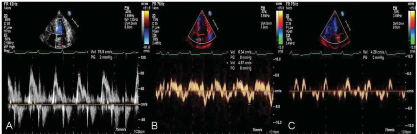
Transthoracic echocardiography showed increased LV wall thickness and an extensive mass in the right cavities with involvement of the tricuspid annulus (Figure 1A and B; see Supplementary data online,Videos 1 and 2). LV systolic func-tion was assessed using the ejecfunc-tion fracfunc-tion (EF) calculafunc-tion by the modified Simpson’s rule approach (EF¼50%) and peak mitral annular systolic velocity (S0) determination
(S0
¼4,87 cm/s) in the lateral area (Figure 2B). Integrating both results, the patient had preserved EF but impaired LV longitudinal systolic function as assessed by DTI. DTI also demonstrated severe right ventricular (RV) function impair-ment with a peak tricuspid annular systolic velocity (St) of 4.28 cm/s (Figure 2C). Inferior vena cava (IVC) appeared dilated, with a respiratory variability of,50%; minor tricus-pid regurgitation allowed the estimation of pulmonary systo-lic artery pressure (PSAP) in 50–55 mmHg. DTI revealed an
E/E0 ratio of 12, which was in accordance with moderate
diastolic dysfunction as assessed by pulse-wave Doppler of the transmitral flow [pseudo-normal pattern (Figure 2A)], pulmonary vein flow (systolic wave velocity inferior to
diastolic wave velocity), and mitral annulus DTI (reduced annular velocities).
Transoesophageal echocardiography confirmed the pres-ence of an extensive mass involving the right cavities and the pericardium, with minimal pericardial effusion (Figure 3A). A little mass was visualized in the right atrial appendage (Figure 3B). Attending to its localization, differ-ent echogenicity and mobility, it was considered consistdiffer-ent with a thrombus. This finding, in addition to the clinical picture, right-axis deviation on ECG, and transthoracic infor-mation (namely RV dysfunction and pulmonary hyperten-sion), raised the hypothesis of concomitant pulmonary embolism. In addition, pulmonary embolism could have been, at least partially, responsible for the reduction in DTI velocities of the tricuspid annulus. A computed tomogra-phy (CT) scan was promptly performed and discarded the presence of pulmonary embolism. Cerebral, thoracic, and abdominal CT scan confirmed that no other organ was found to be involved by the lymphoma.
The patient was started on a chemotherapeutic regimen with cyclophosphamide, vincristine, adriamycin, and predni-solone associated to rituximab, a monoclonal antibody
Figure 1 First transthoracic echocardiogram; two-dimensional imaging of parasternal view (A) and apical four-chamber view (B) showing increased left ventricular wall thickness and an extensive mass in the right cavities with involvement of the tricuspid annulus.
Figure 2 First transthoracic echocardiogram; pulse-wave Doppler of the transmitral flow (A) showing a pseudo-normal pattern; DTI at the mitral annulus with reduced peak mitral annular systolic and diastolic velocities (S0andE0) in the lateral area (B), as well as reduced peak tricuspid annular systolic velocity (St) (C).
against the protein CD 20. Oral anticoagulation was also initiated, given the possible thrombus in the right atrial appendage. Additional medical treatment consisted of diuretics (furosemide and spironolactone), an angiotensin-converting enzyme inhibitor, and oral anti-diabetic agents.
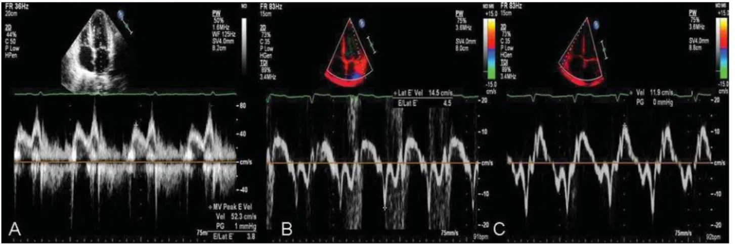
His clinical condition improved significantly 2 weeks later. The new echocardiogram showed significant reduction of the tumour mass and of the LV wall thickness (Figure 4A and B; see Supplementary data online,Video 3). LVEF was normal (EF¼58%) and DTI revealed normalized mitral annulus vel-ocity with S0
¼8 cm/s (in the lateral area of the mitral annulus) (Figure 5B). A significant improvement of right ven-tricular systolic function was also evidenced by DTI with St¼ 11.9 cm/s (Figure 5C). In addition to the important reduction in LV wall thickness, the echocardiographic exam revealed improvement in diastolic function, with transmitral flow showing relaxation anomaly and an E/E0 ratio of 4
(Figure 5A and B). A decrease in PSAP (40 mmHg) was observed as well as a non-dilated IVC with normalized respir-atory variability. These findings favoured the hypothesis of an extensive infiltration of the left and right ventricles by lymphoma cells with consequent impairment of LV and right ventricular contractility and relaxation. Based on the
clinical and echocardiographic evolution of the patient, the same chemotherapeutic regimen was maintained.
The patient’s treatment was completed after 4 months of chemotherapy and he underwent another transthoracic echocardiogram. No mass was visualized and the remaining exam was unchanged when compared with the previous one. On his last visit (8 months later), he was doing well and there was no evidence of recurrence.
Discussion
As referred previously, the incidence of cardiac lymphomas has considerably increased, mainly in HIV-infected patients.5,6 It is the second most frequent malignancy in HIV-infected patients, after Kaposi’s sarcoma.7,8 The inci-dence of NHL among HIV-positive patients has been reported to be 2.9%, a nearly 60 times higher incidence when com-pared with general population.9,10
The clinical presentation of cardiac lymphoma can be varied: heart failure, pericardial effusion, arrhythmias, or on the opposite, no cardiac manifestations.5The possibility of cardiac involvement in patients with lymphoma should always be considered, particularly in the presence of HIV
Figure 4 Transthoracic echocardiogram after 15 days of chemotherapy; two-dimensional imaging of parasternal view (A) and apical four-chambers view (B) showing significant reduction of the tumour mass and also of the left ventricular wall thickness.
Figure 3 Transoesophageal echocardiography showing an extensive mass in the right cavities (A) and most likely a thrombus in the right atrial appendage (B).
infection.9Cardiac lymphoma most commonly presents as a nodular or polypoid mass with variable myocardial infiltration.8
When assessing a cardiac lymphoma, two-dimensional imaging allows the accurate evaluation and quantification of masses and ventricular wall thickening. In addition, Doppler study, particularly DTI, appears to be useful in asses-sing myocardial impairment owing to infiltration. Klein
et al.11,12 demonstrated the correlation between Doppler-filling patterns and the degree of amyloid infiltration as measured by LV wall thickness, when evaluating cardiac amy-loidosis. The existence of such correlation could be hypoth-esized for the infiltrative component of cardiac lymphoma. However, it should be noted that the ability of Doppler study, namely E/E0, to predict LV-filling pressures in cardiac
lymphoma remains to be established. DTI was also very useful to quantify RV systolic dysfunction in our patient since other methods, such as tricuspid annular plane systolic excursion (TAPSE), were unavailable because of marked ana-tomical deformation of the tricuspid annulus.13
Besides allowing a better visualization of the cardiac mass and its relationship with cardiac structures, transoesopha-geal echocardiography allowed the distinction of a probable thrombus, from the neoplasm, in the right atrial appendage. Prognosis of HIV-associated cardiac NHL is generally poor. Although clinical improvement and significant tumour regression has been described with combination chemother-apy, early post-chemotherapy death may occur in conse-quence of massive pulmonary emboli, refractory heart failure, and cardiac arrhythmias.7,14
In conclusion, this case highlights not only the utility of echocardiography imaging, but also the potential utility of its various modalities and techniques, such as two-dimensional imaging, DTI, and transoesophageal echocardio-graphy among others, to assess cardiac involvement by lymphoma. Echocardiography imaging is also helpful to monitor the response to the chemotherapeutic regimen insti-tuted, and to define possible adjuvant therapies, such as anticoagulation in the presence of concomitant thrombus.
Supplementary data
Supplementary data are available at European Journal of Echocardiographyonline.
Conflict of interest:none declared.
References
1. McAllister HA Jr, Hall RJ, Cooley DA. Tumors of the heart and pericardium.Curr Probl Cardiol1999;24:57–116.
2. Robert WC, Glancy DL, De Vita WT. Heart in malignant lymphoma: a study of 196 autopsy cases.Am J Cardiol1968;22:85–107.
3. McDonnell PJ, Mann RB, Buckley BH. Involvement of the heart by malignant lymphoma. A clinicopathological study. Cancer 1982;49: 944–51.
4. McAllister HA, Fenoglio JJ. Tumors of the cardiovascular system. In: Hartman WH, Cowan WR eds.Atlas of Tumor Pathology. Washington, DC: Armed Forces Institute of Pathology; 1978. Second series: fascicle 15. 5. Holladay AO, Siegel RJ, Schwartz DA. Cardiac malignant lymphoma in
acquired immune deficiency syndrome.Cancer1992;70:2203–7. 6. Maric I, Washington S, Schwartz A, Anandan V, Karcher D. Human
herpesvirus-8-positive body cavity-based lymphoma involving the atria of the heart. A case report.Cardiovasc Pathol2002;11:244–7. 7. Kaplan L, Afridi N, Holmvang G, Zukerberg L. Case 31-2003: a 44-year-old
man with HIV infection and a right atrial mass.N Engl J Med 2003;
349:14.
8. Rerkpattanapipat P, Wongpraparut N, Jacobs L, Kotler M. Cardiac mani-festations of acquired immunodeficiency syndrome. Arch Intern Med
2000;160:602–8.
9. Khan N, Ahmed S, Wagner P, Rumley R, Movahed A. Cardiac involvement in non-Hodgkin’s lymphoma: with and without HIV infection. Int J
Cardiovasc Imag2004;20:477–81.
10. Beral V, Peterman T, Berkelman R, Jaffe H. AIDS-associated non-Hodgkin lymphoma.Lancet1991;338:884–5.
11. Klein AL, Oh JK, Miller FA, Seward JB, Tajik AJ. Two-dimensional and Doppler echocardiographic assessment of infiltrative cardiomyopathy.
J Am Soc Echo1988;1:48–59.
12. Klein AL, Hatle LK, Burstow DJ, Seward JB, Kyle RA, Bailey KRet al.
Doppler characterization of left ventricular diastolic function in cardiac amyloidosis.J Am Coll Cardiol1989;13:1017–26.
13. Lindqvist P, Calcutteea A, Henein M. Echocardiography in the assessment of right heart function.Eur J Echocardiogr2008;9:225–34.
14. Rolla G, Bertero MT, Pastena G, Tartaglia N, Corradi F, Casabona Ret al.
Primary lymphoma of the heart. A case report and review of the litera-ture.Leuk Res2002;26:117–20.
Figure 5 Transthoracic echocardiography after 15 days of chemotherapy; pulse-wave Doppler of the transmitral flow (A) showing relaxation anomaly; DTI at the mitral annulus with normalized peak mitral annular systolic and diastolic velocities (S0andE0) in the lateral area (B), as well as normalized peak tricuspid annular systolic velocity (St) (C).