Review Article
The Utility of Transcranial Doppler in the Acute Ischemic Stroke
Viviane Flumignan Zétola and Marcos Christiano Lange
Internal Medicine Department, Neurology Service - Cerebrovascular Diseases, Paraná State Federal University (UFPR) - Curitiba, PR, Brazil
Mailing Address: Viviane flumignan zétola •
Rua General Carneiro, 181 – sala 1236 – 80060-900 – Curitiba, PR
E-mail: [email protected]
Manuscript received March 5, 2006; revised manuscript received April 13, 2006; accepted May 9, 2006. The first clinical trials on the utility of Transcranial Doppler
(TCD) in acute ischemic stroke (AIS) were conducted still in the pre-thrombolysis stage. Middle cerebral artery
(MCA) mean flow velocity (MV) below 20 cm/sec or 50% below contralateral MCA within 24 hours translates worse
prognostics factors1. Intravenous thrombolysis in the management of AIS brought forth an even higher utility of
TCD in the acute phase – to add to emergency evaluation – thus allowing the identification of intracranial occlusion
and acting as a unique tool for continuous monitoring of vessel recanalization2. More recent study designs suggest
that changes found in TCD should be added to inclusion criteria for the use of intravenous thrombolytic, since that therapeutic regimen is potentially indicated and beneficial in cases of encephalic ischemia with intracranial artery branch occlusion3-5.
TCD has been absorbed by major university services
in our country, as well as by hospitals that assist patients with cerebrovascular diseases6. In addition to being non-invasive, TDC has other major advantages: it is low-cost,
dynamic, fast for specific cases, and portability. It may be carried out and repeated many times, by the bed side, and may also stand for therapeutic potential7-9. Major difficulties posed by the method are: it belongs to the group of operator-dependent complementary exams, and the lack of studies on interpretation criteria that can be uniformly accepted7. In regard to acute AIS – concurrently to imaging exams such as computed axial tomography (CAT
Scan) and nuclear magnetic resonance (NMR) – TCD has
been innovating concepts and treatments, contributing for better assistance to patients who are thrombolysis candidates and non-candidates. Research on micro emboli
– a unique tool – and high sensitivity for the detection and
follow-up of intracranial stenoses have turned TCD into a routine exam for cerebrovascular disease patients3,10. The present review will focus on the assistance for acute cerebrovascular diseases (CVD). It is important to point out
that major studies are related to the analysis of MCA, whose
topography corresponds to most ischemic cerebral events. Whenever possible, non-invasive extracranial investigation should be carried out with ultrasound of cervical vessels
– carotid, vertebral and subclavian arteries – to help
interpret and therefore increase accuracy of TCD11.
Thrombolysis
Although patients with acute AIS can still count on few services for treatment in our country, the tendency for
therapy improvement is imperative and promising. ‘ Post-hoc’ studies have made it clear that although most patients submitted to thrombolysis have shown better prognostics for improvement when compared to placebo, a significant group still does not report benefits, thus showing selective therapeutic effect based on certain characteristics of acute AIS12,13. When reviewing pioneer coronary studies, one notices that the core factor related to thrombolysis is circulation close to thrombosed vessel, which is graded through flow on
arteriography - TIMI – ‘thrombolysis in myocardial ischemia’
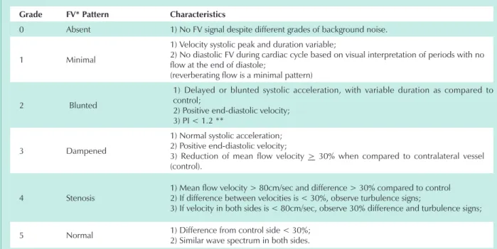
14.Using that classification as a model, in 2001 Demchuk and
collaborators described a systematic analysis of MCA in AIS
through TCD – called ‘thrombolysis in brain ischemia‘ - Table 13. The purpose is to determine residual flow velocity as well as its correlation with AIS severity, functional independence
after index ischemic event included. Major limitation is the lack of flow in the vessel - ‘grade 0’, to be distinguished from
the difficulty in ultrasound transmission through squamous
portions of the temporal bone, found in approximately 11%
of patients, as well as from the inexistence of a golden pattern for method comparison4,6. Counting on a well-trained team and making use of a standard protocol, TCD may be carried
out in less than 15 minutes, therefore not interfering in the
therapeutic window for thrombolysis15.
A number of experimental studies have already demonstrated that ultrasound makes fibrinolytic agents activities easier due to improvement in medication transportation, change in fibrin structure, and higher tPA-fibrin binding8. The ‘CLOTBUST’ study has demonstrated a possible potentialization of thrombolysis when concurrent to TCD in cases where MCA occlusion evolution is recorded
as under 3 hours. The group under constant monitoring of
IV thrombolytic infusion reported higher recanalization and improvement of functional independence when compared to patients not constantly monitored through TCD5.On the other hand, a recent study - ‘TRUMBI’ – where authors reviewed the correlation between ultrasound low intensity (300Khz)
and potential thrombolysis had early discontinuation due to higher incidence of symptomatic intraparenchymatose
Key words
Ultrasonography, transcranial ultrasonography Doppler, cerebrovascular accident.
Review Article
zétola & lange
The UTIlITy Of TRAnSCRAnIAl dOPPleR In The ACUTe ISCheMIC STROKe
Arq Bras Cardiol 2006; 87(6): 725-728
haemorrhage in the study group16. Further studies are required for method validation. The IV microbubbles solution has demonstrated to even increase the effect obtained from the association of TCD and the thrombolytic17.
From all the information provided by the method, TCD also allows recognizing early recanalization of MCA occluded after thrombolytic infusion irrespective of clinical recovery, a condition known as stunned brain15,18. The pattern of recanalization – if spontaneous – must also be distinguished
from intracranial stenosis, which may be presented with similar findings at first exam. Sequential exams are crucial for definition, since in recanalization flow velocities return to values close to normal within variable time frame.
Monitoring of microembolism
TCD identifies microembolic signs (MES) in intracranial circulation. Such study is possible, since ultrasound distinguishes signal characteristics through embolyc materials
– solid or gaseous – from erythocyte flow velocity. Those differences are present in the wave spectrum – intensity and frequency – and in sonority emission19.
Embolism may result from cardiac sources, from major arteries – aorta, carotids and vertebral arteries -, intracranial
arteries, and paradoxically reach cerebral circulation
through a venous-arterial shunt – patent oval foramen or
arteriovenous fistula20. The cardioembolic source stands for 20 to 30% of all AISs, with most patients presumptively identified following TOAST criteria, through the combination
of clinical symptomatology associated to mid and high risk factors from complementary cardiovascular exams findings
- ECG, transthoracic or transaesophagic ECG – and imaging
exams – cranial CAT Scan and NMR21. However, none of those methods allows direct identification of the active embolic phenomenon as TCD does. Methodology includes
simultaneous monitoring of both MCAs for at least 30 minutes,
with fixed transducers in order to reduce movement artifacts.
With two possible embolic sources – cardiogenic and carotid plaque – the identification of MES contributes with higher
diagnosis accuracy and support for therapy decision making. MES detection, in addition, acts as a predictor for new cerebral ischemic event recurrence20,22-24.
Research lines have been established with monitoring of surgical and endovascular procedures to correlate the presence of MES and post-surgery neurologic changes25-29. Such procedure may in the future identify crucial sites for surgical manipulation in carotid stenoses.
TCD technical advance will contribute for deeper knowledge of embolic phenomenon pathosphysiology, and will most likely be of great help in showing the most appropriate therapy. At this point in time we could say that studies on micro embolism are useful for patients with non-defined AIS, and which is of probable cardioembolic etiology, as well as for carriers of atheroscleroses of large vessels30,31.
Intracranial stenoses
Intracranial disease corresponds to approximately 8% of
AISs, depending on gender and race32,33. Diagnosis is frequently reached through arteriography, the gold standard, whose risks and invasive procedure do not allow large scale use, therefore, with figures probably underestimated. Advances in
diagnosis techniques – as angioresonance, angiotomography and TCD – tend to confirm the relevance of that etiology as
grade fV* Pattern Characteristics
0 Absent 1) No FV signal despite different grades of background noise.
1 Minimal
1) Velocity systolic peak and duration variable;
2) No diastolic FV during cardiac cycle based on visual interpretation of periods with no flow at the end of diastole;
(reverberating flow is a minimal pattern)
2 Blunted
1) Delayed or blunted systolic acceleration, with variable duration as compared to
control;
2) Positive end-diastolic velocity;
3) PI < 1.2 **
3 Dampened
1) Normal systolic acceleration;
2) Positive end-diastolic velocity;
3) Reduction of mean flow velocity > 30% when compared to contralateral vessel
(control).
4 Stenosis
1) Mean flow velocity > 80cm/sec and difference > 30% compared to control 2) If difference between velocities is < 30%, observe turbulence signs;
3) If velocity in both sides is < 80cm/sec, observe 30% difference and turbulence signs;
5 Normal 1) Difference from control side < 30%; 2) Similar wave spectrum in both sides.
*FV: Flow Velocity ** PI: Pulsatility Index
Table 1 - TIbI assessed through dTC in patients at AIS acute stage
Review Article
zétola & lange The UTIlITy Of TRAnSCRAnIAl dOPPleR In The ACUTe ISCheMIC STROKe
Arq Bras Cardiol 2006; 87(6): 725-728
References
1. Halsey Jr JH, Tan MJ. Evaluation of acute stroke. In: Newell DW, Aaslid R (eds). Transcranial Doppler. New York: Raven Press; 1992. p. 145-51.
2. Baruzzi AC, Knobel E, Cirenza C, Kihara EN, Souza VC, Massaro A, et al. Use of tissue plasminogen activator factor for acute ischemic stroke. Arq Bras Cardiol. 1997; 68: 347-51.
3. Arenillas JF, Molina CA, Montaner J, Abilleira S, González-Sánchez MA, Álvarez-Sabín J. Progression and clinical recurrence of symptomatic
middle cerebral artery stenosis: a long-term follow-up transcranial Doppler
ultrasound study. Stroke. 2001; 32: 2898-904.
4. Demchuk AM, Burgin WS, Christou I, Felberg RA, Barber PA, Hill MD, et al. Thrombolysis in brain ischemia (TIBI) transcranial Doppler flow grades
predict clinical severity, early recovery, and mortality in patients treated with
intravenous tissue plasminogen activator. Stroke. 2001; 32: 89-93.
5. Alexandrov AV, Molina CA, Grotta J, Garami Z, Ford SR, Alvarez-Sabin J, et
al. Ultrasound-enhanced systemic thrombolysis for acute ischemic stroke.
N Engl J Med. 2004; 351: 2170-8.
6. Zétola VF, Lange MC, Muzzio JA, Marchioro I, Nóvak EM, Werneck LC. Doppler transcraniano na prática neurológica. Arq Neuropsiquiatr. 2006; 64: 100-3.
7. Demchuk AM, Alexandrov AV. Acute ischemic stroke. In: Alexandrov A (ed). Cerebrovascular ultrasound in stroke prevention and treatment. New York: Blackwell Publishing/Futura; 2004. p. 170-80.
8. Francis CW. Ultrasound-enhanced thrombolysis. Echocardiography. 2001; 18: 239-46.
9. Egido JA, Sánchez C. Neurosonology in cerebral ischemia: future application of transcranial Doppler in acute stroke. Cerebrovasc Dis. 2001; 11(suppl 1): 15-9.
10. Segura T, Serena J, Castellanos M, Teruel J, Vilar C, Dávalos A. Embolism in acute middle cerebral artery stenosis. Neurology. 2001; 56: 497-501.
11. Chernyshev OY, Garami Z, Calleja S, Song J, Campbell MS, Noser CA, et al. Yield and accuracy of urgent combined carotid/transcranial ultrasound testing in acute cerebral ischemia. Stroke. 2005; 36: 32-7.
12. del Zoppo GJ, Poeck K, Pessin MS, Wolpert SM, Furlan AJ, Ferbert A, et al.
Recombinant tissue plasminogen activator in acute thrombotic and embolic
stroke. Ann Neurol. 1992; 32: 78-86.
13. Caplan LR, Mohr JP, Kistler JP, Koroshetz W. Should thrombolytic therapy be the first-line treatment for acute ischemic stroke? N Engl J Med. 1997; 337: 1309-13.
14. Hackworthy RA, Sorensen SG, Fitzpatrick PG, Barry WH, Menlove RL,
Rothbard RL, et al. Dependence of assessment of coronary artery reperfusion during acute myocardial infarction on angiographic criteria and interobserver
variability. Am J Cardiol. 1988; 62: 538-42.
15. Alexandrov A. Ultrasound identification and lysis of clots. Stroke. 2004; 35(Suppl 1): 2722-5.
16. Daffertshofer M, Gass A, Ringleb P, Sitzer M, Sliwka U, Els T, et al.
Transcranial low-frequency ultrasound-mediated thrombolysis in brain ischemia: increased risk of hemorrhage with combined ultrasound and
tissue plasminogen activator: results of a phase II clinical trial. Stroke. 2005; 36: 1441-6.
17. Molina CA, Ribo M, Rubiera M, et al. Microbubble administration
accelerates clot lysis during continuous 2-MHz ultrasound monitoring in stroke patients treated with intravenous tissue plasminogen activator.
Stroke. 2006; 37: 425-9.
18. Alexandrov AV, Hall CE, Labiche LA, Wojner AW, Grotta JC. Ischemic
stunning of the brain. Early recanalization without immediate clinical
improvement in acute ischemic stroke. Stroke. 2004; 35: 449-52. 19. Segura T, Serena J. Detección de microembolias mediante DTC. In: Molina
CA, Serena J, Alvarez Sabín J (eds). Manual de Doppler transcraneal. Madrid: Aula Medica Ediciones; 2000. p. 51-60.
20. Markus H. Microembolic signal detection in cerebrovascular disease. In: Babikian VL, Wechsler LR, Toole JF (eds). Transcanial Doppler ultrasonography. 2nd edition. Woburn: Butterworth-Heinemann; 1999. p. 167-74. 21. Adams HP Jr, Bendixen BH, Kappelle LJ, Biller J, Love BB, Gordon DL, et
al. Classification of subtype of acute ischemic stroke. Definitions for use
in a multicenter clinical trial. TOAST. Trial of Org 10172 in Acute Stroke Treatment. Stroke. 1993; 24: 35-41.
22. Batista P, Oliveira V, Ferro JM. The detection of microembolic signals in
patients at risk of recurrent cardioembolic stroke: possible therapeutic
relevance. Cerebrovasc Dis. 1999; 9: 314-9.
23. Markus HS, MacKinnon A. Asymptomatic embolization detected by Doppler
ultrasound predicts stroke risk in symptomatic carotid artery stenosis. Stroke.
2005; 36: 971-5.
24. Valton L, Larrue V, le Traon AP, Massabuau P, Geraud G. Microembolic signals
and risk of early recurrence in patients with stroke or transient ischemic
attack. Stroke. 1998; 29: 2125-8.
25. Spencer MP, Thomas GI, Nicholls SC, Sauvage LR. Detection of middle
cerebral artery emboli during carotid endarterectomy using transcranial
Doppler ultrasonography. Stroke. 1990; 21: 415-23. an independent risk for CVA, although literature still needs
validated criteria34,35.
TCD stands as an ancillary diagnosis tool with increased sensitivity and specificity when strict criteria are complied with36. Sensitivity lies between 70 and 90%, and specificity between 90 and 95% for arteries in the anterior circulation,
and a little lower for arteries in posterior circulation, where
sensitivity is between 50 and 80%, and specificity is between 80 and 96%37. It is important to point out that in practice, mean velocity analysis to identify intracranial analysis is not enough. It must be combined with other parameters such as asymmetry, segmental elevations, spectral analysis, and
knowledge on extracranial circulation. Major confusion
factor in interpreting velocities is collateral or vasodilation circulation as compensatory mechanisms in cases associated to extracranial stenoses38. A finding compatible with intracranial stenosis in AIS acute phase must be distinguished
from ‘vanishing stenosis’. The term defines the process of
recanalization of an occluded vessel that behaves as stenosis hemodynamically speaking. Evolution exams are required for diagnosis differentiation10,39.
When compared to other emergent, non-invasive methods, TCD contributes with the advantage of also monitoring micro emboli at stenosis site, frequently been observed in our practice, and which may influence therapy decision making40.
Therefore, the use of TCD in AIS acute phase allows
complementary diagnosis – still underestimated in practice – as support to imaging exams in etiology and pathophysiology,
therefore guiding neurologists in their therapy conduct, and most likely estimating prognosis for those patients.
Potential Conflict of Interest
No potential conflict of interest relevant to this article was reported.
Review Article
zétola & lange
The UTIlITy Of TRAnSCRAnIAl dOPPleR In The ACUTe ISCheMIC STROKe
Arq Bras Cardiol 2006; 87(6): 725-728
26. Siebler M, Sitzer M, Rose G, Bendfeldt D, Steinmetz H. Silent cerebral
embolism caused by neurologically symptomatic high-grade carotid stenosis.
Event rates before and after carotid endarterectomy. Brain. 1993; 116 (Pt 5): 1005-15.
27. Markus HS, Thomson N, Brown MM, Thomson ND. Asymptomatic cerebral
embolic signals in symptomatic and asymptomatic carotid artery disease.
Brain. 1995; 118: 1005-11.
28. Siebler M, Nachtmann A, Sitzer M, Rose G, Kleinschmidt A, Rademacher J, et al. Cerebral microembolism and the risk of ischaemia in asymptomatic high-grade internal carotid artery ischaemia. Stroke. 1995; 26: 2184-6. 29. Malheiros SM, Massaro AR, Gabbai AA, Pessa CJ, Gerola LR, Branco JN, et
al. Is the number of microembolic signals related to neurologic outcome in
coronary bypass surgery? Arq Neuropsiquiatr. 2001; 59: 1-5.
30. Alexandrov AV, Neumyer MM. Diagnostic criteria for cerebrovascular
ultrasound. In: Alexandrov A, ed. Cerebrovascular ultrasound in stroke
prevention and treatment. New York: Blackwell Publishing/Futura; 2004. p. 107-8.
31. Ringelstein EB, Droste DW, Babikian VL, Evans DH Grosset DG, Kaps M, et
al. Consensus on microembolus detection by TCD. International Consensus
Group on Microembolus Detection. Stroke. 1998; 29: 725-9.
32. Sacco RL, Kargman DE, Gu Q, Zamanillo MC. Rate-ethnicity and
determinants of intracranial atherosclerotic cerebral infarction. Stroke.
1995; 26: 14-20.
33. Wityk RJ, Lehman D, Klag M, Coresh J, Ahn H, Litt B. Race and sex differences in the distribution of cerebral atherosclerosis. Stroke. 1996; 27: 1974-80.
34. Wong KS, Li H, Chan YL, Ahuja A, Lam WWM, Wong A, et al. Use of
transcranial Doppler ultrasound to predict outcome in patients with
intracranial large-artery occlusive disease. Stroke. 2000; 31: 2641-7.
35. Chimowitz MI, Kokkinos J, Strong J, Brown MB, Levine SR, Silliman S, et al.
The Warfarin-Aspirin symptomatic in,tracranial disease study. Neurology.
1995; 45: 1488-93.
36. Felberg RA, Christou I, Demchuk AM, Malkoff M, Alexandrov AV. Screening
for intracranial stenosis with transcranial Doppler: the accuracy of mean flow
velocity tresholds. J Neuroimaging. 2002; 12: 1-6.
37. Sloan MA, Alexandrov AV, Tegeler CH, Spencer MP, Caplan LR, Feldman
E, et al. Assessment transcranial Doppler ultrasonography report of the Therapeutics and Technology Assessment Subcommittee of the American
Academy of Neurology. Neurology. 2004; 62: 1468-81.
38. Rorick MB, Nichols FT, Adams RJ. Transcranial Doppler correlation with angiography in detection of intracranial stenosis. Stroke. 1994; 25: 1931-4.
39. Diehl RR, Samii C, Diehl A. Dynamics and embolic activity of
symptomatic intra-cranial cerebral artery stenoses. Acta Neurol Scand.
2002; 106: 173-81.
40. Gao S, Wong KS, Hansberg T, Lam WW, Droste DW, Ringelstein EB.
Microembolic signal predicts recurrent cerebral ischemic events in acute
stroke patients with middle cerebral artery stenosis. Stroke. 2004; 35: 2832-6.