Arq Neuropsiquiatr 2012;70(8):637-644 643
Clinically nonfunctioning pituitary
adenoma growth after radiosurgery
Crescimento de adenoma pituitário clinicamente não funcionante após radiocirurgia
Rafael Loch Batista, Andrea Cecilia Toscanini, Andrea Glezer, Mário Gilberto Siqueira, Salomon Benabou, Erick Talamoni Fonoff, Wagner Malagó Tavares, Manoel Jacobsen Teixeira, Malebranche Berardo Carneiro da Cunha Neto
Departamento de Neurocirurgia Funcional da Divisão de Neurocirurgia Funcional do Hospital das Clínicas da Universidade de São Paulo (USP), São Paulo SP, Brazil.
Correspondence: Rafael Loch Batista; Hospital das Clínicas da Universidade de São Paulo; Alameda Joaquim Eugênio de Lima 1.058/102; 01403-002 São Paulo SP - Brasil; E-mail: [email protected]
Conflict of interest: There is no conflict of interest to declare.
Received 29 February 2012; Received in final form 26 March 2012; Accepted 02 April 2012
Fig 1. Before radiosurgery. Cranial nuclear magnetic resonance scan from March, 2004, revealing probable tumor residues, measuring 2.5 x 2.0 x 1.5 cm, with greater involvement of the sphenoid sinus.
Radiosurgery (RS) is a minimally invasive technique suitable for lesions of the central nervous system (CNS), with <3 cm in diameter or volume <30 mL. Benign tumors of the CNS are candidates to perform RS1. It has emerged as a therapeutic option for clinically nonfunctioning pituitary adenomas (CNFPA), associated with efective control of tu-mor growth and few complications1. About 30% of pituitary adenomas are classiied as CNFPA, and patients with this tumor usually have clinical symptoms, such as headaches, visual loss, hypopituitarism and, less commonly, pituitary apoplexy2. hese tumors often develop slowly, but diagnosis tends to be late. Surgery is the primary treatment option, preferably transsphenoidal, complemented or not by radio-therapy (RT) or RS1. In cases of tumor residues, the thera-peutic approach remains controversial.
LETTERS
Arq Neuropsiquiatr 2012;70(8):637-644
644
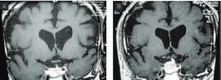
Fig 2. After radiosurgery. (A) Image from June, 2004 (three months after radiosurgery); (B) image from September, 2004 (six months after radiosurgery), both showing growth of the sellar lesion.
A
B
than 48 months, RS was able to control tumor growth in 93 to 96% of the cases1-3.
Complications of RS are optic neuropathy, lesions in adja-cent vascular structures, parenchymal brain injury, hypopitu-itarism, and neoplasms1-4. Stenosis or occlusion of the inter-nal carotid artery has already been reported5.
CASE
We reported a case of a patient with CNFPA who under-went RS and showed tumor growth after it.
A 59-year-old male patient, single, born in São Paulo, Brazil, reported that a cranial computed tomography (CCT) performed in 1992 by headache had identiied sellar and su-prasellar tumors with 2.0 x 2.0 cm. In 2001, the CCT was re-peated and revealed a slight increase (2.2 x 2.0 cm) of them. he patient denied headaches or visual loss.
Hormonal evaluation did not identify any changes. Nuclear magnetic resonance (NMR) scan showed sellar tu-mor with supra- and infrasellar extension, measuring 2.2 x 2.5 x 2.8 cm, isointense on T1 and hyperintense on T2, with compression of the optic chiasm. Transsphenoidal pituitary surgery was performed in 2001.
he anatomicopathologic diagnosed pituitary adenoma with discrete anaplasia and sites reagent for thyroid-stimu-lating hormone (TSH) in the cytoplasm.
Cranial NMR was repeated in 2001, 2002, and 2003, and scans revealed intrasellar content, with no signs indicating tumor residues. In 2004, NMR demonstrated residues with 2.5 x 2.0 x 1.5 cm.
RS was underwent in March, 2004 (Fig 1). NMR sellar was underwent in June and September, 2004 (Fig 2), with lesion growth in six months. In January, 2005, NMR revealed a sig-niicant reduction in tumor volume to about 1.5 cm.
A possible explanation to growth and subsequently re-gression after RS is the occurrence of apoplexy. Reports of pituitary apoplexy cases after RT have promoted a relation-ship between apoplexy and radiation1. Radiation is known to increase vascularization of pituitary adenomas, therefore leading to apoplexy.
RS is an alternative for the treatment of CNFPA, however, there are no reports of pituitary apoplexy or tumor growth after RS. A possible reabsorption of the apoplectic content leading to subsequent regression is likely. Tumor growth af-ter RS, probably due to pituitary apoplexy, may be a possible complication of this therapeutic modality.
1. Laws ER, Sheehan JP, Sheehan JM, Jagnathan J, Jane JA, Oskouian R. Stereotatic radiosurgery for pituitary adenomas: a review of the literature. J Neur-Oncol 2004;69:257-272.
2. Sanno N, Teramoto A, Osamura RY, et al. Pathology of pituitary tumours. Neurosurg Clin N Am 2003;14:25-39.
3. Laws ER, Vance ML. Radiosurgery for pituitary tumours and craniopharyngiomas. Neurosurg Clin North Am 1999;10:327-336.
4. Akabane A, Yamada S, Jokura H. Gamma knife radiosurgery for pituitary adenomas. Endocrine 2005;28:87-92.
5. Muramatsu J, Yoshida M, Shioura H, et al. Clinical results of LINAC-based stereotactic radiosurgery for pituitary adenoma. Nippon Igaku Hoshasen Gakkai Zasshi 2003;63:225-230.