Pulmonary alveolar proteinosis: four cases*
JOÃO CARLOS THOMSON1, MARINA KISHIMA2, MARIANA ULBRICHT GOMES3, MARIANO DE ALMEIDA MENEZES3, JOSÉ PERANDRÉ NETO3, PAULA TAPIA GOMES PEREIRA3
* *Study conducted at the Universidade Estadual de Londrina (UEL, State University of Londrina) School of Medicine -Londrina, Paraná, Brazil.
1. Postdoctoral fellowship in Thoracic Surgery from the Royal Brompton Hospital - London, England; Coordinator of the Postgraduate Commission for Master's and Doctoral Degrees of the Health Science Center of the Universidade Estadual de Londrina (UEL, State University of Londrina) - Londrina, Paraná, Brazil
2. Master's degree in Internal Medicine, Specialist in Pathology and Assistant Professor of Pathologic Anatomy at the Universidade Estadual de Londrina (UEL, State University of Londrina) School of Medicine - Londrina, Paraná, Brazil 3. Medical Researcher of the Universidade Estadual de Londrina (UEL, State University of Londrina) - Londrina, Paraná, Brazil Correspondence to: João Carlos Thomson. Rua Júlio César Ribeiro, 204 - CEP 86039-200, Londrina, PR, Brasil. E-mail: [email protected]
Submitted: 2 February 2005. Accepted, after review: 23 August 2005.
ABSTRACT
Objective: The aim of this study was to present the evolution of four patients presenting pulmonary alveolar proteinosis and treated at the State University of Londrina School of Medicine. We focus on the importance of whole-lung lavage as the treatment of choice. Methods: A retrospective study of four patients, three females and one male, 22 to 34 years old, presenting similar histories of progressive dyspnea and dry cough. The final diagnosis was established through open-lung biopsy. Three of the patients underwent whole-open-lung lavage in the Department of Surgery. The procedures were performed under general anesthesia and using a double-lumen endotracheal tube. Results: One patient presented spontaneous regression of the pulmonary alveolar proteinosis without the need for whole-lung lavage. In the other three cases, the number of lavages varied: in one patient, a single unilateral lavage resulted in complete remission of the bilateral process; in another patient, three lavages yielded no significant improvement; in the remaining patient, four lavages provided intervening periods of transient improvement. Conclusion: In the cases evaluated, whole-lung lavage proved an efficient treatment for pulmonary alveolar proteinosis. Although some patients presented a certain resistance to the procedure, it might lead to complete remission of the disease in others.
INTRODUCTION
Pulmonary alveolar proteinosis (PAP) is a disease characterized by the build-up of lipoprotein material inside the alveoli, significantly interfering with pulmonary gas exchange. It is a rare disorder, with an estimated prevalence of 0.37 cases for every 100,000 people.(1)
First described in 1958(2), the etiology of PAP remains obscure. It can be primary (90% of the cases), secondary to other conditions or caused by the inhalation of chemical agents.(1-3)
In recent years, a number of physiopathological hypotheses have been proposed and can be summarized as follows: pulmonary macrophage function defect; abnormal structure of the surfactant protein; cytokine production disorder; anomalous expression of the granulocyte-macrophage colony-stimulating factor or its receptors in the pulmonary alveoli macrophages and type II pneumocytes. All of these would lead to a catabolic defect and intra-alveolar build-up of surfactant proteins.(1,4-7)The clinical presentation varies, although the usual symptoms are dyspnea and cough. Fever, thoracic pain and hemoptisis are less common manifestations that can also occur, mainly in the presence of secondary pulmonary infection.(1,3,8) Although the physical examination is often normal, it is possible to observe the following: inspiratory breath sounds, digital clubbing and cyanosis (peripheral or central, depending on the seriousness of the disease).(3) Some nonspecific tests, such as arterial gasometry, lactic dehydrogenase, tumor markers, surfactant proteins (A, B and D) and imaging (chest X-ray and computed tomography of the chest), facilitate the diagnosis.(3) The diagnosis can be confirmed through bronchoalveolar lavage and transbronchial biopsy, given suggestive clinical and radiological conditions. However, the gold standard is open lung biopsy. (3,6-7,9)
Whole lung lavages still seem to be the most effective and safe alternative for the treatment of PAP.(3,8,10-15) Nevertheless, studies also indicate that segmental or lobular bronchoscopic lavages are promising therapies,(3-4,13) as is replacement therapy with granulocyte-macrophage colony-stimulating factor. Other treatments reported in the literature have produced varying results. These include treatment with corticosteroids, potassium iodide, streptokinase and trypsin.(3,6-7)
The aim of this article is to demonstrate our
experience in the management of PAP, relating four cases treated at our facility and their respective evolutions, using whole lung lavage as the basis for the treatment.
METHODS
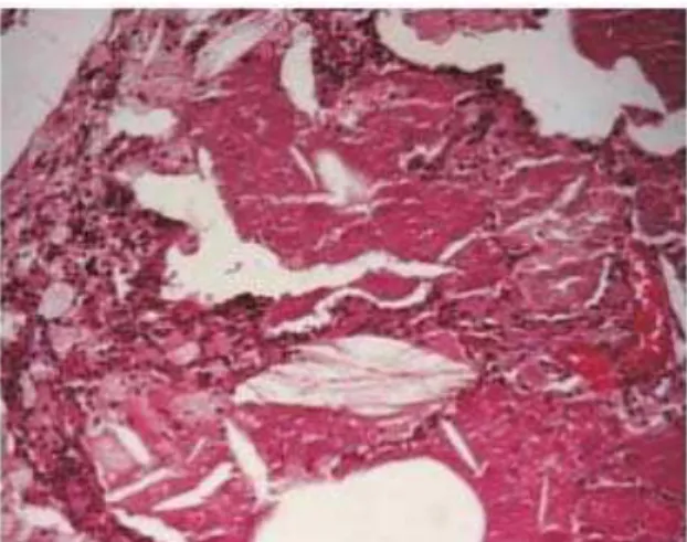
This was a retrospective study in which the medical records of four patients with PAP, diagnosed through open lung biopsy, were analyzed (Figure 1).
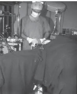
Three of the four patients (three females) were submitted to whole lung lavages, all carried out by the same surgeon, in operating rooms, according to the technique described by Ramirez-Rivera et al. in 1965, (14) under general anesthesia and double-lumen endotracheal tube intubation. In this technique, both lungs are initially well isolated, and the capacity of the lung to be infused is measured. Lavages are repeatedly made with heated saline solution, to which heparin (1.5 ml for each bottle of solution) and N-acetylcysteine are added. Using a gravity feed, the solution is slowly poured into the lung for a lavage. When the measured volume is reached, infusion is ceased and another passage is opened, which, due to gravity, combined with siphonage, drains the corresponding lung (Figure 2). An infusion of six to ten liters of solution is recommended for each lung, in case there are no events. The following controls are conducted: arterial gasometry, electrocardiogram and liquid inflow and outflow.
In all of the procedures carried out with our patients, we observed that the outflow of liquid was
at first opaque, becoming progressively more diluted over the course of the lavage (Figure 3).
The relevant tests and the thoracic control radiological images are presented along with each reported case.
CASE REPORT
Case 1
A 26-year-old, white female patient, who was a clerk and a nonsmoker, reported that, during
pregnancy, she began to present progressive dyspnea, dry cough, with little expectoration, and cyanosis (of the lips and of the extremities). After delivery, her condition worsened, and she experienced dyspnea at rest, increased cyanosis and weight loss (fourteen kilos since the onset of the symptoms). After a number of unsuccessful treatments (for tuberculosis and paracoccidioidomycosis, as well as generalized treatment with corticosteroids), the patient sought treatment at our facility, where the test results were inconclusive, and it was proposed that a pulmonary biopsy be performed. However, the patient refused the proposed procedures and was discharged at her request. One week later, the patient returned to the emergency room with acute respiratory failure, and was admitted to the intensive care unit. Upon an improvement in her condition, the patient agreed to be submitted to an open lung biopsy.
The anatomopathological examination of the lingular biopsy sample revealed amorphous material and acidophilus in the interior of the alveoli, as well as focal septal fibrosis, providing evidence of PAP.
With the diagnosis confirmed, two months of pharmacological outpatient treatment was carried out at another facility. However, there was no clinical improvement; the patient presented persistent cough, pulmonary suppuration and fever. Bronchopulmonary lavage (BPL) was then proposed and was conducted initially in the right lung, since, judging from the chest X-ray, it was the most affected (Figure 4A). Pre-BPL arterial gasometry demonstrated arterial oxygen tension (PaO2) of 39mmHg, arterial carbon dioxide tension (PaCO2) of 28.7 mmHg and arterial oxygen saturation (SaO2) at 69%.
Approximately 30 minutes after the beginning of the procedures, ventricular extrasystoles and tachycardia were observed. Due to worsening of the metabolic and cardiac profiles, the BPL was suspended after the infusion of 4850 mL of the solution and the return of 4950 mL.
The patient responded well to the treatment and was discharged from the hospital Outpatient follow-up was initiated, and clinical and radiographic improvement was observed without medication. Thirty day after the BPL, the patient was asymptomatic, acyanotic and able to perform household chores normally. Arterial gasometry carried out four months after the procedure Figure 2 - Image of the whole lung lavage procedure
revealed a PaO2 of 85 mmHg, PaCO2 of 24 mmHg and SaO2 at 95%.
A chest X-ray (Figure 4B) indicated favorable evolution, with a virtually normal image, even in the left lung, which had not undergone lavage
Case 2
A 34-year-old, white, female patient, who was a homemaker and a nonsmoker, presented for treatment with a six-month history of dry cough, unrecorded fever, respiratory-dependent chest pain and weight loss of five kilos in six months. At the time, she presented five tests showing negative results for acid-fast bacilli and had already undergone treatment for pneumonia due to related symptoms. Due to other diagnostic hypotheses, an open lung biopsy was indicated, the anatomopathological examination of which confirmed PAP. It is noteworthy that this test revealed the following: negative culture for fungus; negative (open-air) inspection for fungus; negative culture for Mycobacterium tuberculosis; and negative testing for acid-fast bacilli.
With the diagnosis confirmed, BPL was proposed and was performed in the right lung with a 5000 mL infusion of solution and an outflow of 4460 mL. Pre-operative arterial gasometry showed a PaO2 of 48 mmHg, PaCO2 of 35.4 mmHg and SaO2 at 84.1%. Post-operative arterial gasometry showed a PaO2 of 54 mmHg, PaCO2 of 33.9 mmHg and SaO2 at 89.1%. The patient showed great improvement, and, at that time, a new BPL was scheduled, this time in the left lung. However, given the significant improvement in her condition, and according to reports in the literature related to contralateral lung improvement after unilateral BPL,(3-10) the left lung lavage was postponed. It was decided that monitored outpatient treatment would be carried out and that, if necessary, another BPL would be scheduled.
After one year and three months, the patient returned, reporting dyspnea at the slightest effort and dry cough. The initial arterial gasometry presented a PaO2 of 34.4 mmHg, PaCO2 of 33.1 mmHg and SaO2 at 71%.
Due to the recurrence of the conditions, another BPL was necessary. The BPL was performed in the right lung with a 5500 mL infusion and 5410 mL of drainage. Some days after the procedure, since the patient still presented dry cough and dyspnea, a BPL of the left lung was performed, with a 5500
mL infusion of the solution and a return of 5450 mL. Arterial gasometry at discharge indicated that the PaO2 was 59.2 mmHg, the PaCO2 was 36.2 mmHg, and the SaO2 was at 91.5%.
After an uneventful two years, the dry cough and dyspnea returned. A BPL of the right lung was performed (5490 mL infusion and outflow of 5.310 mL). Seven days later, BPLs were performed in the left lung (4000 mL infusion and outflow of 3800 mL) and right lung (5100 mL infusion and outflow of 4580 mL).
Since then, the patient has received outpatient follow-up treatment and has shown significant improvement in her clinical profile, although a chest X-ray showed that the lesion remained.
Case 3
A 27-year-old, white, female patient, who was a homemaker, presented for treatment after being referred from a basic health clinic in order to investigate extensive bilateral pneumopathy. She complained of a dry cough for one year and d y s p n e a f o r o n e m o n t h , a c c o m p a n i e d b y respiratory-dependent chest pain in the left hemithorax and weight loss of thirteen kilos in two months. She reported no fever, expectoration or hemoptysis. General tests were carried out and were inconclusive. Rheumatologic tests and serology for the human immunodeficiency virus were negative. It was then decided that, in order to investigate the diffuse pulmonary interstitial process, a pulmonary biopsy should be conducted, the anatomopathological examination of which confirmed PAP.
Since PAP was confirmed, the routine procedure was to perform a BPL. A right-lung BPL was first
A B
performed, and then, after some days, a left-lung BPL was performed. During the left BPL, the patient presented frequent extrasystoles and a drop in oximetry. Therefore, the procedure was interrupted prematurely. Even after treatment had been initiated, the patient did not improve, presenting persistent dry cough, dyspnea and weakness. It was suggested that other causes of the pulmonary condition should be investigated, since the evolution was not that expected for the diagnosed disease.
Therefore, we opted for conducting another clinical investigation, including bronchoscopy with transbronchial biopsy, so as to determine whether there was a fungal infection. In the material collected in the bronchoscopy, bronchial brush with testing for fungus, testing for acid-fast bacilli and culture were all negative. Anatomopathological examination confirmed PAP.
As there was no improvement in the radiological or clinical profile, the patient presenting recurrent bronchial spasms, corticosteroid treatment (15 mg per day of prednisone) was introduced, continued supplementary oxygen was prescribed, and the patient was discharged from the hospital.
Case 4
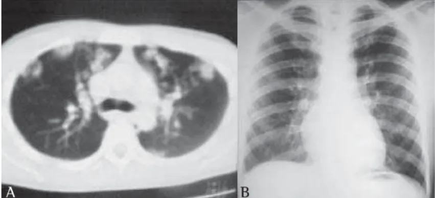
A 22-year-old, white, male patient was referred to our facility and presented cough without expectoration, daily fever and weight loss. A chest X-ray showed nodular bilateral lesions, confirmed by a computed tomography scan of the chest (Figure 5A). Routine tests were inconclusive, and an open lung biopsy was proposed. The result of the anatomopathological examination was consistent with PAP. Since the respiratory
symptoms were of low intensity, we opted for conservative treatment. Over the years, the condition of this patient has remained unchanged. He performs regular daily activities, and his chest X-rays present few alterations (Figure 5B).
DISCUSSION
A rare disease, the prevalence of PAP is estimated to be 0.37 cases for every 100,000 people.(1) It is seen predominantly in males (3:1), and 80% of all cases occur among individuals in their thirties or forties.(3) Among the patients treated for PAP at our facility, female patients were predominant, and the age varied from 22 to 34 years. The most common symptoms were cough, dyspnea and weight loss.(1,3,8) All diagnoses were c o n f i r m e d t h r o u g h a n a t o m o p a t h o l o g i c a l examination of material collected from open lung biopsies.
The treatment for PAP remains controversial. Despite the fact that countless other therapies are being used (segmental lavage, replacement of the granulocyte-macrophage colony-stimulating factor, corticosteroid therapy, potassium iodide, streptokinase and trypsin),(3,6,13) whole lung lavage, since its initial description,(14) has remained the treatment of choice, although it has been modified and improved upon by various authors.(3,8,10-15) Despite the factors considered negative by many authors, such as the need for general anesthesia and an experienced team, in addition to the possibility of hypoxemia and hemodynamic instability during the procedure, we chose BPL because we consider it to be effective, safe and easily executed at our facility.
Of the four patients diagnosed with PAP, we performed BPL on three of them, since, due to dyspnea, there was a significant limitation of daily activities.
Any case of PAP can be placed into one of the following categories: stable, though with persistent symptoms; progressive deterioration; and spontaneous improvement.(1,6) According to some authors(3), more than 60% of the patients presented a favorable response after two lavages of each lung. In the literature, it is also reported that few patients require more than six cycles of BPL, less than 15% of patients require lavage every six months to maintain pulmonary function, and less than 10% Figure 5 - A) Computed tomography of the chest at the time of diagnosis;
B) Chest X-ray years later, without treatment
do not respond to BPL.(3,6,8) We were able to observe such diverse evolutions in our patients, who presented, variously, the following: spontaneous remission, single lavage with clinical and radiological improvement of both lungs; repetitive lavages with remission of the symptoms in the intervals; and progressive deterioration, despite two lavages.
In relation to the procedure itself, the greatest r i s k s a r e r e p r e s e n t e d b y h y p o x e m i a a n d hemodynamic instabilities,(3) which occurred on two occasions with two different patients, in which case, we opted for premature interruption of the procedure. Other possible complications are hydropneumothorax, pleural collection and surgical emphysema, as well as endotracheal granuloma and stenosis (due to multiple procedures),(4) none of which were observed in any of our patients.
Since PAP is still a disease that is difficult to treat, whole lung lavage remains the treatment of choice.(3,8,10-15) New alternatives are needed, since BPL is not exempt from adverse effects and must be performed by an experienced team. Segmental lavages and replacement therapy with granulocyte-macrophage colony-stimulating factor seem promising,(3,5-7,13) although more studies are needed.
REFERENCES
1. Trapnell BC, Whitsett JA, Nakata K. Pulmonary alveolar proteinosis. N Engl J Med. 2003;349(26):2527-39. 2. Rosen SH, Castleman B, Liebow AA. Pulmonary alveolar
proteinosis. N Engl J Med. 1958;258(23):1123-42. 3. Shah PL, Hansell D, Lawson PR, Reid KB, Morgan C.
Pulmonary alveolar proteinosis: clinical aspects and
current concepts on pathogenesis. Thorax. 2000; 55(1):67-77.
4. Cheng SL, Chang HT, Lau HP, Lee LN, Yang PC. Pulmonary alveolar proteinosis: treatment by bronchofiberscopic lobar lavage. Chest. 2002;122(4):1480-85. Comment in: Chest. 2002;122(4):1123-4.
5. de Vega MG, Sánchez-Palencia A, Ramírez A, Cervera S, Aneiros J. GM-CSF therapy in pulmonary alveolar proteinosis. Thorax. 2002;57(9):837.
6. Barraclough RM, Gillies AJ. Pulmonary alveolar proteinosis: a complete response to GM-CSF therapy. Thorax. 2001;56(8):664-5.
7. Schoch OD, Schanz U, Koller M, Nakata K, Seymour JF, Russi EW, et al. BAL findings in a patient with pulmonary alveolar proteinosis successfully treated with GM-CSF. Thorax. 2002;57(3):277-80.
8. Luisetti M, Pochetti P, Rodi G, Corsico A, Beccaria M, Cerveri I. Patients with pulmonary alveolar proteinosis enrolled in the San Matteo Hospital program for whole lung lavage: baseline characteristics and follow-up. Chest. 2001;120(4 Suppl):270S.
9. Arcasoy SM, Lanken PN. Images in clinical medicine. Pulmonary alveolar proteinosis. N Engl J Med. 2002;347(26): 2133.
10. Kavuru MS, Popovich M. Therapeutic whole lung lavage: a stop-gap therapy for alveolar proteinosis. Chest. 2002;122(4):1123-24. Comment on: Chest. 2002;122(4): 1480-5.
11 . Selecky PA, Wasserman K, Benfield JR, Lippmann M. The clinical and physiological effects of whole-lung lavage in pulmonary alveolar proteinosis: a ten-year experience. Ann Thorac Surg. 1977;24(5):451-61. 1 2 . Bingisser R, Kaplan V, Zollinger A, Russi EW.
Whole-lung lavage in alveolar proteinosis by a modified lavage technique. Chest. 1998;113(6):1718-9.
1 3 . Wang BM, Stern EJ, Schmidt RA, Pierson DJ. Diagnosing pulmonary alveolar proteinosis. A review and an update. Chest. 1997;111(2):460-6.
14. Ramirez-Rivera J, Kieffer RF Jr, Ball WC Jr. Bronchopulmonary lavage in man. Ann Intern Med. 1965;63(5):819-28. 15. Morgan C. The benefits of whole lung lavage in pulmonary