ARTICLE
Epilepsy with onset at over 50 years of age: clinical
and electroencephalographic characteristics
Epilepsia com início após os 50 anos de idade: características clínicas e eletrencefalográficas
Glória Maria Almeida Souza Tedrus1, Lineu Corrêa Fonseca1, Elizardo Nogueira Junior2, Daniele Pazetto3
he elevated incidence of epilepsy with onsetat over 50 years of age, in relation to the total number of new cases of
epilepsy, has been ignored in the literature in recent decades1.
Studies have shown that epilepsy afects approximately 1 to 2% of the elderly population, and the incidence increases progres-sively with the advance in age. hese cases of epilepsy can occur due to an acute cerebral seizure or have no apparent precipitator2,3.
On the other hand, there is consensus in the literature that epileptic seizures (ES) are more diicult to diagnose in the elder-ly for various reasons such as the diiculty in obtaining an accu-rate clinical history, a frequently atypical ictal presentation, dii-culty in making a diferential diagnosis between an epileptic and non-epileptic event3-6 and due the occurrence of comorbidities5,6.
Although epilepsy is considered to be one of the com-monest neurological afections in the elderly, and despite the
need to take public health measures due to the progressive increase in the elderly population, particularly in developing countries, no national (Brazilian) publications and only a few international ones on epilepsy in this age range were found.
hus, the objective of the present study was to evaluate the clinical and electroencephalographic aspects of ES and epilepsies in patients with late-onset ES at over 50 years of age, also considering diferent age ranges.
METHODS
Patients
Fifty-ive patients, consecutively attended at the neu-rological clinic of the Celso Pierro Hospital and Maternity
1Department of Neurology, Faculdade de Medicina, Pontifícia Universidade Católica de Campinas (PUC-Campinas), Campinas SP, Brazil; 2Department of Neurology, Hospital e Maternidade Celso Pierro, PUC-Campinas, Campinas SP, Brazil;
3Scholarship student Reitoria, PUC-Campinas, Campinas SP, Brazil.
Correspondence: Lineu Corrêa Fonseca; Rua Sebastião de Souza 205 / sala 122; 13013-173 Campinas SP – Brasil; E-mail: [email protected]
Conflicts of interest: There is no conflict of interest to declare.
Received 04 April 2012; Received in final form 25 June 2012; Accepted 02 July 2012.
ABSTRACT
Epilepsy in older individuals has an elevated incidence. The objective of the present work was to evaluate the clinical, EEG and brain imaging aspects in patients showing late-onset epilepsy. Fifty-five patients with late-onset epilepsy (older than 50 years) were evaluated. They were composed of two groups according to the onset age of the epilepsy seizure (ES): 51–60 (G51-60) and over 60 (G60+) years. Focal ES predomi-nated although they were less frequent in G60+. The occurrence of status epilepticus was high and more frequent in G60+ whereas seizures in series predominated in G51-60. Symptomatic epilepsy was more frequent and the vascular etiology predominated. Epileptiform activity was associated with a greater number of ES, and background activity abnormalities were more frequent in G60+. In conclusion, epilepsy with onset at over 50 was predominantly focal and symptomatic, with a high occurrence of status epilepticus and of seizures in series.
Key words: epilepsy, aged, epileptic seizures, electroencephalography.
RESUMO
Epilepsia no idoso tem elevada incidência e peculiaridades pouco estudadas. O objetivo do presente trabalho foi avaliar aspectos clínicos e eletrencefalográficos de pacientes que apresentaram a primeira crise epiléptica (CE) tardiamente. Foram avaliados 55 pacientes com epilepsia tardia (com início após os 50 anos), divididos em dois grupos segundo a idade de início das CE: de 51–60 anos (G51-60) e após os 60 anos (G60+). Predominaram as CE focais, que foram menos frequentes em G60+. A ocorrência de status epilepticus foi elevada e mais frequente em G60+, enquanto as CE em série predominaram em G51-60. A epilepsia sintomática foi a mais frequente e de etiologia vascular. Atividade epileptiforme esteve associada ao maior número de crises epilépticas. Anormalidades da atividade de base ao EEG foram mais frequentes em G60+. Em conclusão, a epilepsia iniciada após os 50 anos é predominantemente focal e sintomática, com alta ocorrência de status epilepticus e CE em série.
of Pontifícia Universidade Católica de Canpinas (PUC-Campinas) between February 2010 and March 2011,had ep-ilepsy with onset at over 50 years of age, were included in this prospective study. he patients were classiied into two groups according to the ES onset age: from 51 to 60 (G51-60) and over 60 (G60+). he study was approved by the Ethics Committee for research in human beings of PUC-Campinas and the patients signed a consent form.
Procedures
he following procedures were carried out:
A clinical-neurological assessment evaluate socio-de-mographic and clinical aspects: age, gender, marital sta-tus, onset age, type and frequency of ES, evolution time of the epilepsy, use of anti-epileptic drugs (AED), anteced-ents of clinical and neurological diseases, clinical-neuro-logical examination.
Digital electroencephalogram was obtained at rest, during hyperpnea and with intermittent photo-stimula-tion, preferably on the day following an ES. The presence of alterations in the background activity, of non-specific abnormalities and of epileptiform activity (EA), charac-terized according to its location, extent and type, that is, spikes, complexes of slow-wave spikes or sharp spikes, was studied.
Brain imaging data were obtained for all the patients, us-ing MRI for 35 cases and the CT-scan for 20 cases.
Data analysis
he ES and epilepsies were classiied according to the criteria of the International Classiication of Epilepsies and
Epileptic Syndromes7.
To characterize the presentation of the ES, the following
deinitions proposed by Massengo et al.6 were used in this
study: status epilepticus (SE) — a more prolonged ES or a
se-quence of ES without complete recovery of consciousness between them — and ES in series — various ES in one day.
Controlled epilepsy was considered as that in which there had been no ES during the previous 12 months. About thera-py with AED, the patients were classiied as in a monothera-peutic regime or a polytheramonothera-peutic one (two or more AED).
Fisher’s exact or χ2 tests were used to compare the
pro-portions between the groups for the categorical variables, and the level of signiicance was p<0.05.
RESULTS
Fifty-ive patients (27 male cases) with a mean age of
67±8.0 were studied, of which 30 showed the ES onset
be-tween 51 and 60 years of age, and 25over 60.
hirty-two (58%) patients were married, 7 (12.7%) single and 16 (29%) either divorced or widowed.
Epileptic seizures
he mean onset age for the ES was 61 (51 to 83) and Table 1 shows the clinical data of the patients according to their onset age range (G51-60 and G60+ groups).
Focal ES occurred in 37 (67.3%) cases as follows: com-plex in 21 (38.2%) cases, motor/sensitive in 12, autonomic in 3, and psychic in 1 case. Generalized tonic-clonic (TCG) ES only occurred in 18 (32.7%) cases.
The proportion of patients with focal seizures, espe-cially complex focal seizures, was lower in G60+ than in G51-60 (χ2, p<0.05).
he irst ES was characterized by status epilepticus in 13
patients (23.6%) and ES in series in 5 cases (9.1%). During the period studied, 8 (14.5%) of the patients sufered anoth-er status epilepticus or ES in series episode.
As shown in Table 1, when comparing the groups
clas-siied according to onset age, a greater occurrence of status
epilepticus was found in the G60+ group, and of ES in series in the G51-60 group (Fisher’s exact test, p<0.05).
he postictal period was prolonged in 7 (12.7%) patients.
Electroencephalogram
Focal ES was registered in two patients.
On the interictal EEG, the background activity was nor-mal in 23 (41.8%) cases, abnornor-mal due to the presence of slow continuous activity in 19 (34.5%) cases and intermittent in 13 (23.6%) cases (Table 1).
he recording of continuous abnormality in the back-ground activity was more common in patients with an onset age of over 60 than in the G51-60 group (χ2, p<0.05).
EA, characterized by spikes, sharp waves or complexes of spikes and sharp wave — slow waves was recorded in 38 (69.1%) cases.
Epileptic syndromes
According to the ILAE criteria, focal symptomatic epilepsy was characterized in 37 (67,3%) cases, probably symptomatic in 17 (30,9%), and generalized idiopathic in 1 case (Table 1).
No signiicant diference was observed between G51-60 and G60+ groups with respect to the proportions of the dif-ferent epileptic syndromes.
Generalized idiopathic epilepsy
Symptomatic epilepsy
Symptomatic epilepsy was diagnosed in 37 (67.2%) of the cases, since the alterations in the imaging exams suggested a cause/efect relationship compatible with the localization of an ictal manifestation and other clinical aspects. Stroke was the most frequent etiology, corresponding to 28 (50.1%) of the cases (Table 1).
Vascular alterations were observed in the imaging exams and a history of an ischemic stroke in 21 (38.1%) of the cas-es. In three cases, ES occurred together with the installing of ischemic strokes, and in two SE with the subsequent recur-rence of the ES during the period of the study.
An association was found between the occurrence of strokes and the register of abnormality in the background ac-tivity by slow continuous or intermittent acac-tivity (χ2, p<0.05).
he presence of vascular diseases characterized by old ischemic areas was observed in the neuroimaging exams in 7 (12.7%) cases, without any history of prior clinical manifesta-tion, but suggesting a causal relationship with the ES.
he etiological diagnosis was sequel of traumatic brain in-jury in four of the cases, neurocysticercosis of the vesicular type in two cases, and arteriovenous malformation, neuroibroma-tosis and degenerative vascular disease in one case each.
One of the patients with symptomatic epilepsy showed two types of ES, suggesting diferent epileptic syndromes of which one was post-stroke symptomatic epilepsy and the other possibly temporal lobe epilepsy. Initially this patient had two episodes of GTC-type ES in sequence to the instal-lation of an ischemic stroke, with a hemiparesis, aphasia and right homonymous hemianopsia. he CT-scan showed
a left parietal-occipital ischemic area, and in the follow-ing 24 months the patient had one more GTC. However, 16 months after the stroke, the patient started having short episodes of “feeling bad” with “dizziness that was diicult to describe”, during which he would make non-comprehensi-ble sounds, following which his responsiveness was under-mined. hese episodes occurred with an irregular frequen-cy, sometimes several in the same day. On carrying out an investigation using the MRI, an area of left occipital gliosis, areas of subcortical microangiopathy, a volumetric reduc-tion in T1 and an increase in the T2 signal of the left hippo-campus, were observed.
Other brain imaging indings were observed in six other patients with symptomatic epilepsy, not correlated with the localization of the EA or ES, that is, intracranial calciications suggestive of neurocysticercosis in ive patients, and in one case, basal ganglia calciications calciications, compatible with Fahr’s disease.
Probably symptomatic focal epilepsies
A diagnosis of focal, probably symptomatic epilepsy was made in 17 (30.9%) of the cases. Focal ES occurred in 14 of
these patients and status epilepticus in 2 cases.
In nine cases, the following alterations in the brain imag-ing exam could not be correlated with the ES: difuse cortical atrophy in ive cases, and focal atrophy or intracranial calcii-cations in two cases each. In eight patients, the brain imaging exams were normal.
Alterations in background activity were observed on the EEG in four cases, and an ES was recorded in one case.
Table 1. Clinical data of 55 patients with epilepsy, according to the epileptic seizures onset age.
Clinical data
Epileptic seizures onset age range
p-value* Whole group
n=55 (100%)
51 to 60 n=30 (54.5%)
Over 60 n=25 (45.5%)
Focal epileptic seizure 37 (67.3) 24 (80.0) 13 (52.0) 0.028**
Complex 21 (38.2) 16 (53.3) 5 (20.0)
0.025**
Others 16 (29.1) 8 (26.7) 8 (32.0)
Generalized tonic-clonic seizure 18 (32.7) 6 (20.0) 12 (48.0)
Epileptic seizures in series 5 (9.1) 4 (13.3) 1 (4.0)
0.047**
Status epilepticus 13 (23.6) 3 (10.0) 10 (40.0)
Prolonged post-ictal period 7 (12.7) 4 (13.3) 3 (12.0) 0.883
EEG background activity
Normal 23 (41.8) 12 (40.0) 11 (44.0)
0.027
Intermittent slow activity 13 (23.6) 11 (36.7) 2 (8.0)
Continuous slow activity 19 (34.5) 7 (23.3) 12 (48.0)
EEG epileptiform activity 38 (69.1) 22 (73.3) 16 (64.0) 0.456
Epileptic syndromes
Generalized idiopathic 1 (1.8) 1 (3.33) 0 (0.0)
0.175
Probably symptomatic 17 (30.9) 12 (40.0) 5 (20.0)
Symptomatic 37 (67.2) 18 (60.0) 19 (76.0)
Etiology of vascular diseases 28 (50.1) 14 (46.7) 14 (56.0) 0.425
Number of seizures below 10 34 (61.8) 16 (53.3) 18 (72.0) 0.323
Monotherapy with antiepileptic drug 46 (83.6) 22 (73.3) 23 (92.0) 0.033**
Epilepsy controlled 43 (78.1) 25 (83.3) 18 (72.0) 0.310
Status epilepticus or seizures in series and the epileptic syndrome
As can be seen in Table 2, status epilepticus or seizures
in series occurred with greater frequency in patients with symptomatic focal epilepsy (56.8%) than in those with
prob-ably symptomatic focal epilepsy (11.8%) (χ2, p=0.023).
Number, treatment and control of the ES
Table 1 shows the data referring to the treatment and control of the ES.
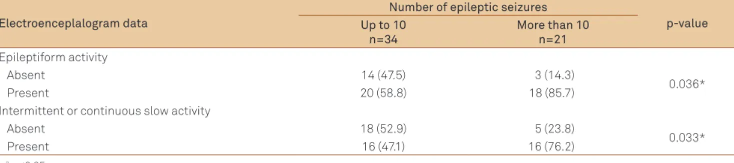
here were less than 10 ES in 34 (61.8%) cases and the epilepsy was under control in 43 (78.1%) cases. An associa-tion between a number of ES greater than 10, the inding of EA and continuous or intermittent abnormality of the back-ground activity, was found (Table 3).
Monotherapy with AED was used in 46 (83.6%) patients, and in a greater proportion in the G60+ group than in the G51-60 group (Fisher’s exact test, p<0.05).
DISCUSSION
Epileptic seizures
In the present study with patients showing an ES onset age over 50, focal seizures were shown to predominate (67% of the patients), in agreement with other studies, which
ini-tially found focal seizures in about 70% of the cases1,8.
It was shown that focal seizures, including the complex ones, occurred in a smaller proportion in patients with an ES onset age over 60 than in those with an onset age between 51 and 60. here were possibly diferent seizure genesis mecha-nisms occurring in the older group, but a greater diiculty in obtaining adequate descriptions of the seizures could also have been a factor in this age range.
Complex seizures have been referred to as being more common in the elderly and appear as luctuations in
responsiveness, associated with automatisms. Such episodes can be diicult to characterize, leading to confusion with de-mentias, memory lapses and even with episodes of mental
confusion9. hus, the diagnosis of ES may be retarded or may
even not be made, as occurs in about 30% of the cases3,6,9-11.
Some authors have suggested that complex ES in the el-derly present a ictalsemiology distinct from that found in the young adult, since in the elderly the localization most fre-quently found involves the frontal and parietal lobes and not the temporal lobe4,12.
As in the literature, an elevated frequency of status
ep-ilepticus and of ES in series was found in the present se-ries. More than 30% of the ES in the elderly appeared as
status epilepticus or seizures in series, the occurrence
be-ing twice that found in other age ranges2,13-15.
Status epilepticus de novo frequently occurs in the elderly with no prior history of ES and epilepsy, and represents a consid-erable risk for the subsequent development of epilepsy, as also a worse cognitive prognosis and increase in mortality of up to 40%16.
In this series, status epilepticus was found to a greater
ex-tent in those patients with an onset of epilepsy at over 60, whereas seizures in series occurred predominantly in those with an onset between 51 and 60 years of age. It is possible that this inding is connected to diferences in the charac-teristics of inhibition of the epileptogenic mechanisms con-nected to age. No similar report was found in the literature.
his research showed that SE and seizures in series oc-curred more frequently in symptomatic epilepsy than in probably symptomatic epilepsy.
In the elderly, prolonged acute confusional state with a pleomorphic clinical presentation and no associated motor manifestation can be ictal manifestations of non-convulsive SE, and wrongly diagnosed as vascular and psychiatric
disor-ders or even as symptomatic of dementia3,12,17,18.
Another relevant clinical aspect of the present series was the report of a prolonged post-ictal period in 13% of the
Table 2. Epileptic syndromes and status epilepticus or epileptic seizures in series.
Status epilepticus or seizures in series Epileptic syndrome* Total
Symptomatic Probably symptomatic
Present 16 (43.2) 2 (11.8) 18
Absent 21 (56.8) 15 (88.2) 36
Total 37 17 54
*χ2, p=0.023
Table 3. Number of epileptic seizures and electroencephalogram data.
Electroenceplalogram data
Number of epileptic seizures
p-value Up to 10
n=34
More than 10 n=21
Epileptiform activity
Absent 14 (47.5) 3 (14.3)
0.036*
Present 20 (58.8) 18 (85.7)
Intermittent or continuous slow activity
Absent 18 (52.9) 5 (23.8)
0.033*
Present 16 (47.1) 16 (76.2)
cases, characterized by mental confusion, focal motor dei-cit, aphasia or psychiatric disorder. Reports can be found in the literature of a post-ictal phase lasting for up to 24 hours in approximately 14% of patients, the majority of which char-acterized by confusional states9,19 or as Todd’s paralysis10,11,19.
A prolonged post-ictal period makes the diagnosis of ES diicult and increases the negative impact on the quality of life of the patients2,16.
Diagnostic evaluation
he initial diagnostic evaluation in the older individual with ES should be similar to that applied in younger individ-uals, and include an EEG and neuroimaging exam in addition to the general examinations.
Although there are few studies with EEG for the irst ES in
older individuals20-22, there is evidence that the EEG should be
routinely applied as an important part in the diagnosis and
prognosis with respect to the risk of a recurrence of the ES21.
However it is known that a single EEG can produce a false negative result and a normal or non-speciic exam should not include a diagnosis of epilepsy8,20.
Alterations in thebackground activity or non-speciic symptoms can appear on the EEG of elderly individuals with advancing age, and EA can become less frequently associated
with ES4. Some studies report the presence of epileptiform
ac-tivity in 20 to 37% of epileptic patients over 60 years of age2,6,20,22.
In the present research, the inding of EA in about 70% of the cases may have been due to recording the EEG soon after the ES. An association was found between a greater number of seizures (more than 10), the presence of EA and the contin-uous or intermittent abnormality of thebackground activity, but such a correlation has not been evaluated and described in the literature.
he electroencephalographic recording of an ES would be the “golden standard” for the diagnosis of an epileptic sei-zure19,22, and in two of the patients in the present study
re-cordings of focal ES were obtained.
As in the EEG recordings, age-related changes are fre-quently seen on the MRI and include difuse atrophy, dilatation of the Virchow-Robin perivascular spaces and periventricular hyperintensity, and one should be careful not to attribute the
cause of epilepsy to such non-speciic indings19,22.
Intracranial calciications were found in seven cases in the neuroimaging exams carried out in the present study, and basal ganglia calciications in one case, but without any causal relationship with the ES. he elevated proportion of intracranial calciications in our medium due to neurocysti-cercosis is well known, with no associated neurological
man-ifestation23. On the other hand, the inding of basal ganglia
calciications, suggestive of Fahr’s disease, as observed in one patient diagnosed with symptomatic epilepsy with vascular
etiology, can be considered incidental23.
hus the overall clinical picture should be considered judi-ciously in the valorization of the results of an imaging exam19,22.
Epileptic syndromes Symptomatic epilepsy
In the present study, in agreement with the literature, prac-tically two thirds of the cases were diagnosed as symptomat-ic epilepsy, and the etiology identiied with greater frequency was cerebrovascular disease in approximately 55% of the cases and in a similar proportion for both age groups2,6,8,9,15,24.
he following etiologies were also found, equally in agree-ment with the literature: traumatic brain injury, intracranial
arteriovenous malformation and degenerative disease6,8,24.
he etiology was neurocysticercosis in two cases, with vi-able cysts visible in the neuroimaging exam. No articles were found in the literature with the vesicular form of neurocysti-cercosis as the etiology for epilepsy in the aged. In one case ES of the GTC type occurred immediately following the installa-tion of an ischemic stroke. However, the autonomic/complex ES that appeared subsequently was suggestive of temporal lobe epilepsy, and must be related to the temporal mesial scle-rosis found on the MRI of the patient. It is disputable whether the temporal mesial sclerosis occurred after the stroke or not, since no MRI was carried out beforehand.
Probably symptomatic focal epilepsy
In about one third of the cases the etiology was not deter-mined, similar to the report of probably symptomatic epilep-sy in 25–45% of new cases of epilepepilep-sy in the aged15,16,25.
ES and epilepsy can be the irst sign of a still not mani-fested vascular disease. It is believed that the risk of epilep-sy is greater in hypertensive individuals, probably because of small vessels lesions that lead to “irritative symptoms” before
causing an ictus25-27. Late-onset ES would thus be a predictive
factor for a subsequent stroke27.
Idiopathic generalized epilepsy
Idiopathic generalizes epilepsy (IGE) with its onset as an adult, as observed in one case in this series, is very rare, but it is a possibility that should not be rejected in the diagnosis of IGE in the aged28,29.
Late-onset IGE at over 50, resulting of a known or pre-sumed genetic defect, is considered to be rare. Twenty-four
individual cases have been reported in the literature28,29.
Recognition of late-onset IGE in the elderly has implications
for an adequate diagnosis and treatment30.
Treatment and control of the epileptic seizures
Similar to that reported in the literature, an adequate
con-trol of the ES was found with the use of a single AED16,30, the
1. Hauser WA, Annegers JH, Kurland LT. Prevalence of epilepsy in Rochester, Minnesota: 1940-1980. Epilepsia 1991;32:429-445.
2. Stephen LJ, Brodie MJ. Management of a first seizure. Special problems adults and elderly. Epilepsia 2008;49:45-49.
3. Brodie MJ, Elder T, Kwan P. Epilepsy in later life. Lancet Neurol 2009;8:1019-1030.
4. Ramsay RE, Pryor FM. Epilepsy in the elderly. Neurology 2000;55(5 Suppl 1):S9-S14.
5. Rowan AJ, Epiplepsy Foundation of America. Epilepsy in older adults. Common morbidities influence development, treatment strategies, and expected outcomes. Geriatrics 2005;60:30-34.
6. Massengo SA, Ondze B, Guiziou C, Velmans N, Rajabally YA. Elderly patients with epileptic seizures: in-patient observational study of two French community hospitals. Seizure 2011;20:231-239.
7. Engel Jr J, International League Against Epilepsy (ILAE). A proposed diagnostic scheme for people with epileptic seizures and with epilepsy: report of the ILAE task force on classification and terminology. Epilepsia 2001;42:796-803.
8. Tebartz van Elst L, Baker G, Kerr M. The psychosocial impact of epilepsy in older people. Epilepsy Behav 2009;15(Suppl 1):S17-S19.
9. Ramsay RE, Rowan AJ, Pryor FM. Special considerations in treating the elderly patient with epilepsy. Neurology 2004;62 (5 Suppl 2):S24-S29.
10. Hommet C, Mondon K, Camus V, De Toffol B, Constans T. Epilepsy and dementia in the elderly. Dement Geriatr Cogn Disord 2008;25:293-300.
11. Hopkins AF, Garman A F, Clark C. The first seizure in adult life. Value of clinical features, electroencephalograpy, and computerized tomographic scanning in prediction of seizure recurrence. Lancet 1988;1:721-726.
12. Belmin J, Marquet T, Oasi C. [Anti-epileptic drugs and their use in the elderly]. Presse Med 2000; 2:2143-2148.
13. Shorvon SD. Epidemiology, classification, nature history, and genetics of epilepsy. Lancet 1990;336:93-96.
14. Wright JF, Pickard NF, Witfield AF. A population-based study of the prevalence, clinical characteristics and effect of ethnicity epilepsy. Seizure 2000;9:309-313.
15. Hauser WA, Annegers JH, Kurland LT. Prevalence of epilepsy in Rochester, Minnesota: 1940-1980. Epilepsia 1991;32:429-445.
16. Cloyd J, Hauser W, Ramsay R, et al. Epidemiological and medical aspects of epilepsy in the elderly. Epilepsy Res 2006;68(Suppl 1):S39-S48.
17. Pollock LM, Mitchell SC. Nonconvulsive status epilepticus causing acute confusion. Age Ageing 2000;29:360-362.
18. Veran O, Kahane P, Thomas P, Hamelin S, Sabourdy C, Vercueil L. De novo epileptic confusion in the elderly: a 1-year prospective study. Epilepsia 2010;51:1030-1035.
19. Kellinghaus C, Loddenkemper T, Dinner DS, Lachwani D, Lüders H. Seizure semiology in the elderly: a video analysis. Epilepsia 2004;45:263-267.
20. Van Cott AC. Epilepsy and EEG in the elderly. Epilepsia 2002;43(Suppl 3):94-102.
21. Krumholz A, Wiebem S, Gronsethm G, et al. Practice parameter: evaluating an apparent unprovoked first seizure in adults (an evidence-based review): report of the Quality Standards Subcommittee of the American Academy of Neurology and the American Epilepsy Society. Neurology 2007;69:1996-2007.
22. Sinha S, Satishchandra P, Kalband BR, Thennarasu K. EEG observations in elderly with new onset seizures: from developing country perspective. J Clin Neuropsysiol 2011;28:388-393.
23. Tedrus GMAS, Fonseca LC, Nogueira Jr E. Calcificação nos núcleos da base na tomografia computadorizada: correlação clínica em 25 pacientes consecutivos. Arq Neuropsiquiatr 2006;64:104-107.
24. Roberson ED, Hope OA, Martin RC, Schmidt D. Geriatric epilepsy: research and clinical directions for the future. Epilepsy Behav 2011;22:103-111.
25. Poza JJ. Management of epilepsy in the elderly. Neuropsychiatric Dis Treat 2007;3:723-728.
26. Ng SKC, Hauser WA, Brust JCM, Susser M. Hypertension and the risk of new-onset unprovoked seizures. Neurology 1993;43:425-428.
27. Cleary P, Shorvon S, Tallis R. Late-onset seizures as a predictor of subsequent stroke. Lancet 2004;363:1184-1186.
28. Hiyohi T, Yagi K. Epilepsy in the elderly. Epilepsia 2000;41(Suppl 9):31-35.
29. Michel VHN, Sebban C, Debray-Meignan S, et al. Electroclinical features of idiopathic generalized epilepsies n the elderly: a geriatric hospital-based study. Seizure 2011;20:292-298.
30. Bagshaw J, Crawford P, Chappell B. Care in people 60 years of age and over with chronic or recently diagnosed epilepsy: a note review in United Kingdom general practice. Seizure 2009;18:57-60.
References
Although there is no consensus with respect to the treat-ment19,22, some studies suggest the introduction of AED after
the irst ES and for an indeterminate period due to the risk of recurrence of the ES, which occurs in 90% of the cases after
suspension of the medication2,8,25.