DOI: 10.1590/0004-282X20150082
ARTICLE
Short-term mortality and prognostic factors
related to status epilepticus
Mortalidade a curto prazo e fatores prognósticos de status epilepticus
Fernando Gustavo Stelzer, Guilherme de Oliveira Bustamante, Heidi Sander, Americo Ceiki Sakamoto, Regina Maria França Fernandes
Epileptic seizures are self-limited, lasting, in general, less than to 2 minutes. Status epilepticus (SE) is manifested by con-tinuous or recurrent epileptic seizures without full recovery of motor, sensory and/or cognitive functions, and has multiple
etiologies and a diverse prognosis. he duration of seizures in SE varies from 5 to 30 minutes, depending on the deinition1. It
is one of the most frequent neurological emergencies, with an estimated annual incidence of from 6.2 to 61/100,000 people2,3.
SE is associated with long-term mortality that is nearly three times greater than that of the general population4. Indeed,
de-spite new advances in medical treatment, short-term mortality remains high, ranging from 3 to 40%, depending on the
sam-pling methods, age, speciic etiology, or refractoriness of SE5.
Several studies agree that the most important factors related to mortality in SE are older age, acute symptom-atic etiology such as central nervous system (CNS) infec-tion, acute stroke, metabolic disturbances, and anoxia, and long seizure duration. Anti-epileptic drug (AED) with-drawal in previously epileptic patients is typically associ-ated with low mortality2,6. For other characteristics, such
as gender, history of prior epilepsy, refractory SE, adequacy of medical treatment, presence of medical complications, level of consciousness, and electroencephalographic pat-tern, there is no consensus in the literature2. A better
un-derstanding of SE prognostic factors would assist in mak-ing treatment-related decisions.
Universidade de São Paulo de Ribeirão Preto, Hospital das Clínicas, Ribeirao Preto SP, Brazil.
Correspondence: Fernando Gustavo Stelzer; Avenida Independência, 900/apto 805; 90035-072 Porto Alegre RS, Brasil; E-mail: [email protected]
Conlict of interest: There is no conlict of interest to declare.
Received 13 January 2015; Received in inal form 8 March 2015; Accepted 30 March 2015.
ABSTRACT
Objective: Status epilepticus (SE) is associated with signiicant morbidity and mortality, and there is some controversy concerning predictive indicators of outcome. Our main goal was to determine mortality and to identify factors associated with SE prognosis. Method:
This prospective study in a tertiary-care university hospital, included 105 patients with epileptic seizures lasting more than 30 minutes. Mortality was deined as death during hospital admission. Results: The case-fatality rate was 36.2%, which was higher than in previous studies. In univariate analysis, mortality was associated with age, previous epilepsy, complex focal seizures; etiology, recurrence, and refractoriness of SE; clinical complications, and focal SE. In multivariate analysis, mortality was associated only with presence of clinical complications. Conclusions: Mortality associated with SE was higher than reported in previous studies, and was not related to age, speciic etiology, or SE duration. In multivariate analysis, mortality was independently related to occurrence of medical complications.
Keywords: status epilepticus, epilepsy, mortality, prognosis.
RESUMO
Objetivos: Status epilepticus (SE) está associado com morbidade e mortalidade importantes. Diversos estudos avaliaram determinantes de prognóstico relacionados com SE, havendo controvérsias neste sentido. O objetivo deste estudo foi avaliar mortalidade no SE e seus fatores determinantes. Método: Estudo prospectivo, em Ribeirão Preto, incluiu 105 pacientes, entre fevereiro e dezembro de 2000. Mortalidade foi deinida como óbito no período de internação hospitalar. Resultados: O índice de mortalidade foi de 36.2%, superior ao veriicado em estudos prévios. Em análise univariada, mortalidade foi associada com idade, antecedente de epilepsia, presença de crises focais complexas, etiologia, recorrência e refratariedade do SE, presença de complicações clínicas e classiicação focal do SE. Em análise multivariada, a ocorrência de complicações clínicas relacionou-se signiicativamente com prognóstico. Conclusões: Em nossa amostra, a mortalidade foi mais elevada do que previamente descrito na literatura, não relacionada com idade, etiologia ou duração do SE, mas, em análise multivariada, com complicações médicas durante o tratamento.
METHOD
We retrospectively studied all 105 patients older than 1 month of age who were diagnosed with SE and admitted to the Hospital das Clínicas da Faculdade de Medicina de Ribeirão Preto da Universidade de São Paulo (HCFMRP-USP), a tertiary-care hospital, between February and December
2000. SE was deined as a clinical or electrographic seizure
lasting more than 30 minutes or recurrent seizures without full recovery of consciousness for 30 minutes or more1. Serial
18- or 21-channel electroencephalograms (EEGs) (Nihon Kohden, Japan), were obtained from all patients following the 10-20 International System of electrode placement. After discharge or death, medical records were reviewed, including demographic data, medical history, prior history of epilepsy,
SE classiication, seizure duration, SE etiology, EEG patterns,
treatment, response to treatment, and prognosis during the
hospital stay. here was no follow-up after discharge. he sample was divided into two groups by prognosis. hose in group I survived until hospital discharge, and those
in group II died while inpatients. Patients younger than 1 month of age and patients diagnosed with pseudo-status epi-lepticus were excluded from the study.
Patients were divided into three categories based on
clinical semiology following the Classiication of Epileptic
Seizures (i.e., focal, secondarily generalized, and general-ized SE)7. Clinical presentation was described as generalized
tonic-clonic seizure (GTCS), focal complex seizure (FCS), fo-cal simple seizure (FSS), absence seizure, tonic seizure, epi-leptic spams, delirium, or confusional state. SE was further characterized as convulsive or nonconvulsive by semiology.
Refractory SE was deined as absence of clinical and/or EEG control if seizures occurred after use of irst and second line-drugs (e.g., IV diazepam, phenytoin, or phenobarbital). hird-line treat -ment included IV continuous midazolam and/or thiopentone. Refractory patients were given ventilator support when necessary.
SE etiology was classiied as follows8,9,10,11:
• Acute symptomatic: SE occurring during within 7 days of an acute disease, such as CNS infection, brain trauma, cerebrovascular disease, toxic or metabolic insults.
• Febrile: SE in a previously neurologically healthy child where the only provocative factor was a febrile disease with axillary
temperature ≥ 38ºC and not related to CNS infection. • Progressive symptomatic: SE related to progressive
eases such as brain neoplasms or neurodegenerative dis-orders including innate metabolic errors.
• Remote symptomatic: SE in an individual with prior (> 7 days) neurological disease, including cerebrovascular disorders or brain trauma, in the absence of acute insult.
• Acute on remote symptomatic: SE occurring during an acute neurological insult (e.g., fever, toxic or metabolic in-sults, alcohol or drug withdrawal), in an individual with a remote symptomatic etiology.
• Previous epilepsy with low antiepileptic drug (AED) lev-els or AED withdrawal: SE occurring in a previously epi-leptic individual with documented low AED level and/or history of AED noncompliance or change in therapy.
• Cryptogenic: SE occurring in the absence of a known acute or remote etiologic factor.
SE groups were stratiied by etiology and refractoriness to
SE only ( febrile, cryptogenic, low AED level, acute on remote symptomatic, and symptomatic) and SE plus (acute symp-tomatic, progressive, and refractory SE of any etiology).
Ictal EEG pattern evolution was evaluated in serial EEG
recordings and classiied as discrete seizures (DS), merging seizures (MS), continuous ictal discharge (CD), CD with lat
periods (CDF), and periodic lateralized epileptic discharges (PLED) as described by Treiman12. he initial ictal pattern of
each patient was analyzed.
Patients were treated according to the existing insti-tutional protocol, which, besides support measures, con-sisted of (1) diazepam IV (10-20 mg bolus); (2) phenytoin IV (10-30 mg/kg infused at maximum dose of 50 mg/min);
(3) phenobarbital IV (20-30 mg/kg). Following irst- and
second-line treatments, the options included (1) midazolam or (2) thiopental IV continuous.
Statistical analysis
Continuous variables were compared using the Mann-Whitney test and categorical variables were compared using Pearson’s chi-square. Odds ratios (ORs) were determined
by logistic regression and reported with 95% conidence inter
-vals (CIs). Statistical signiicance was established at the 0.05
level. Multivariate analysis was used to determine indepen-dent mortality risk factors. All statistical analyses was done us-ing SSPS for Windows, version 10 (SSPS, Inc., Chicago, USA).
RESULTS
During the study period, 105 SE patients were
admit-ted to HCFMRP-USP. heir mean age was 30.0 ± 26.8 years;
42 patients (40%) were women, and the male-female ratio
was 1.5:1. he mortality rate was 36.2%, with 38 patients dy -ing dur-ing their hospital stay. Follow-ing clinical or ECG cri-teria, SE was not stopped until death in 10 patients (9.5%). Overall, there were 125 SE episodes.
Univariate analysis
compared with 56.0% in those without a history of epilepsy
(p < 0.001). he most frequent seizure type was FCS (37.1%),
followed in descending order by GTCS (28.6%) and FCC evolving to GTCS (14.3%). FCC had the highest mortality
rate (51.3%; p = 0.015). Compared with other seizure types, GTCS had a lower mortality rate (20%; p = 0.033). here were no signiicant diferences in mortality among other seizure
types. No patients with absence seizure died. Convulsive SE predominated (86.7%) in this sample, and there was no dif-ference in the mortality rates of patients with convulsive (36.3%) and nonconvulsive (35.7%) SE.
he SE etiologies of the study patients are shown in Table 1.
Acute symptomatic SE etiology predominated (32.4%), fol-lowed by acute on remote symptomatic SE (19.0%), AED noncompliance in previously epileptic patients (19.0%), re-mote symptomatic SE (16.2%), progressive symptomatic SE
(7.6%), cryptogenic SE (3.6%), and febrile SE (1.9%). he mor
-tality rate was signiicantly higher in acute symptomatic SE (53%; p = 0.015), and lower in the AED noncompliance group (5%; p = 0.011).
As for SE classiication (Table 1), focal SE (46.7%) was the
most frequent, followed by secondary generalized SE (42.9%), and generalized SE (10.5%). Mortality was the highest in those
with focal SE (49%; p = 0.012). SE recurrence in the same hospi -tal admission was observed in 11.4% of patients, and the
mor-tality was higher in this group (66.7% versus 32.2%; p = 0.020).
Refractory SE was diagnosed in 36.2% of patients, and they had
a higher mortality rate (57.9 versus 23.8%; p = 0.001).
Fifty-one patients (48.6%) were included in the SE-only group and 56 (51.4%) in the SE-plus group. Mortality was greater in the SE-plus than in the SE-only group (55.4% versus
14.3%; p < 0.001). Ictal EEG patterns were identiied in 55.2%
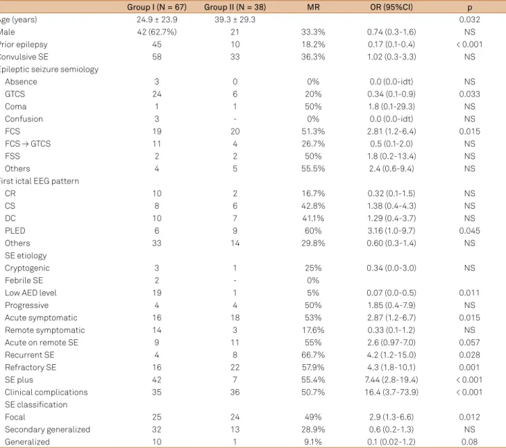
Table 1. Univariate analysis.
Group I (N = 67) Group II (N = 38) MR OR (95%CI) p
Age (years) 24.9 ± 23.9 39.3 ± 29.3 0.032
Male 42 (62.7%) 21 33.3% 0.74 (0.3-1.6) NS
Prior epilepsy 45 10 18.2% 0.17 (0.1-0.4) < 0.001
Convulsive SE 58 33 36.3% 1.02 (0.3-3.3) NS
Epileptic seizure semiology
Absence 3 0 0% 0.0 (0.0-idt) NS
GTCS 24 6 20% 0.34 (0.1-0.9) 0.033
Coma 1 1 50% 1.8 (0.1-29.3) NS
Confusion 3 - 0% 0.0 (0.0-idt) NS
FCS 19 20 51.3% 2.81 (1.2-6.4) 0.015
FCS → GTCS 11 4 26.7% 0.5 (0.1-2.0) NS
FSS 2 2 50% 1.8 (0.2-13.4) NS
Others 4 5 55.5% 2.4 (0.6-9.4) NS
First ictal EEG pattern
CR 10 2 16.7% 0.32 (0.1-1.5) NS
CS 8 6 42.8% 1.38 (0.4-4.3) NS
DC 10 7 41.1% 1.29 (0.4-3.7) NS
PLED 6 9 60% 3.16 (1.0-9.7) 0.045
Others 33 14 29.8% 0.60 (0.3-1.4) NS
SE etiology
Cryptogenic 3 1 25% 0.34 (0.0-3.0) NS
Febrile SE 2 - 0%
Low AED level 19 1 5% 0.07 (0.0-0.5) 0.011
Progressive 4 4 50% 1.85 (0.4-7.9) NS
Acute symptomatic 16 18 53% 2.87 (1.2-6.7) 0.015
Remote symptomatic 14 3 17.6% 0.33 (0.1-1.2) NS
Acute on remote SE 9 11 55% 2.6 (0.97-7.0) 0.057
Recurrent SE 4 8 66.7% 4.2 (1.2-15.0) 0.028
Refractory SE 16 22 57.9% 4.3 (1.8-10.1) 0.001
SE plus 42 7 55.4% 7.44 (2.8-19.4) < 0.001
Clinical complications 35 36 50.7% 16.4 (3.7-73.9) < 0.001
SE classiication
Focal 25 24 49% 2.9 (1.3-6.6) 0.012
Secondary generalized 32 13 28.9% 0.6 (0.2-1.3) NS
Generalized 10 1 9.1% 0.1 (0.02-1.2) 0.08
of patients, with PLED having a signiicantly higher mortality rate (60%; OR = 3.16; p = 0.045). As PLED predominated among elderly patients, there was no diference in ictal pattern-related
mortality when the analysis was controlled for age.
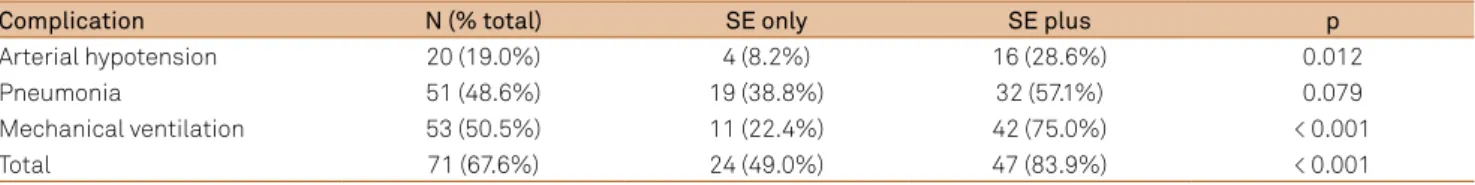
Severe clinical complications occurred in 67.6%, of pa-tients, and 48 (45.7%) had more than one complication
(Table 2). he most common complications were respira -tory failure with mechanical ventilation, arterial hypoten-sion, and pneumonia. Other complications, such as cardiac arrhythmias, and urinary tract infections, were rare in this sample. Clinical complications were more frequent among
refractory SE individuals (97.2 versus 52.2%; p < 0.0001) and in the SE plus group (49.0% versus 83.9%; p < 0.0001; Table
3). Mortality was higher in individuals with clinical
complica-tions (50.7 versus 5.9%; p < 0.001).
Multivariate analysis
In multivariate analysis, only the presence of medi-cal complications was related to a higher mortality rate
(p = 0.013; OR = 11.8; CI = 1.7-82.1).
DISCUSSION
Comparing mortality related to SE in diferent study populations is diicult because of diferences in method
-ology, deinition (seizure duration of 5 versus 30 minutes),
study design (prospective versus retrospective), population (pediatric versus adults, community versus university hos-pital), treatment (lack of medication or medical facilities
in diferent countries), and follow-up period (short- versus
long-term follow-up)2.
he short-term mortality rate in our sample (36.2%) was higher than the 7.6 to 33% reported in the ive most impor -tant previous SE epidemiology studies4,13,14,15,16. In addition, the
mortality rate in this population was higher than those in most recent studies in developing countries (7.3% in Peru17, 10.5%
in India2, 12.1% in Singapore2, 14.8% in Honduras2, 15.9% in
China2, 19.8% in Brazil18, 24.8% in Senegal2, 29% in India2, and
26.7% in hailand2), but not in all (55.4% in Taiwan19).
Our study had important biases that might explain the higher mortality rate we observed. HCFMRP-USP is the larg-est hospital in northern São Paulo state, serving as a tertiary referral facility for an area comprising more than 1.5 million
inhabitants. his may have contributed to inclusion of fewer
patients with SE etiologies having a low mortality risk, such as febrile SE (around 50% of cases in pediatric populations under 5 years of age)9,10,11 idiopathic or cryptogenic SE
(repre-senting 5 to 18% of SE cases and with a mortality approach-ing 0%)10,14,15,20 and alcohol-related SE (2.8% in our sample
compared with 8.1 to 25% in other adult studies), which has reported mortality rates of 0 to 10%)2.We employed
the 30-minute criterion for SE diagnosis, in contrast to the 5-minute criterion used by some of the recent studies men-tioned above. SE mortality has been related to longer dura-tion10,20,21. SE duration was longer in our sample due to
meth-odological issues (i.e, use of serial EEG instead of continuous EEG monitoring) and to a higher incidence of refractory SE. Also the higher mortality might be related to treatment is-sues; for example, delay in initiating medical treatment, as the majority of patients had been transferred from other
medical facilities, or the lack of medication more efective in
SE treatment, such as IV lorazepam.
Excluding the neonatal period, there is evidence that SE mortality increases with age, independent of etiology, rang-ing from 0 to 8% in children, 14 to 25% in adults, and over 35% in the elderly. In our sample, we observed a similar distribu-tion, but with higher mortality rates at every age: 25.7% in children, 34.7% in adults, and 64.7% in the elderly6,10,13.
In other hospital samples, 30 to 44% of SE patients had a history of previous epilepsy2. In our sample, 52.4% had a
history of epilepsy. We believe that this is related to a se-lection bias that may be unique to our Epilepsy Center in Ribeirão Preto.
Table 2. Clinical complications and mortality rate.
Complication N (%) MR OR p
Arterial hypotension 20 (19.0%) 70% 5.72 0.002
Pneumonia 51 (48.6%) 50.9% 3.37 0.006
Mechanical ventilation 53 (50.5%) 58.5% 8.41 < 0.001
Total 71 (67.6%) 67.6% 16.45 < 0.001
MR: mortality rate; OR: odds ratio.
Table 3. Clinical complications and SE plus.
Complication N (% total) SE only SE plus p
Arterial hypotension 20 (19.0%) 4 (8.2%) 16 (28.6%) 0.012
Pneumonia 51 (48.6%) 19 (38.8%) 32 (57.1%) 0.079
Mechanical ventilation 53 (50.5%) 11 (22.4%) 42 (75.0%) < 0.001
Total 71 (67.6%) 24 (49.0%) 47 (83.9%) < 0.001
One of the best prognostic factors of SE is etiology, and the highest mortality rates are observed in patients with acute symptomatic or progressive symptomatic etiologies. Mortality rates in acute symptomatic SE range from 27 to 34%2,4,10,11,22, which are lower than our indings (53%). Other
etiologies, such as febrile SE, are associated with lower mor-tality rates, from 0 to 3%9,10,11,14. In our sample, febrile SE also
had a 0% mortality rate.
In previously epileptic individuals, the predominant causes of SE are noncompliance to treatment or change AED therapy, associated with 20 to 55% of cases. Mortality rates in these pa-tients are low, ranging from 0 to 6%6,14,18,20. In our sample, AED
noncompliance, withdrawal, or reduction was observed in 19.6% (43.6% of previously epileptic individuals), and the
mor-tality rate in this group was 11.5% (n = 3). Two deaths were re -lated to cardiorespiratory arrest during SE treatment.
Seizure duration was associated with a increased mortal-ity in several studies6,20,23, but there is no consensus in this
inding10,18. We were not able to correlate SE duration with
mortality. SE duration was longer in our sample than in oth-ers, mainly due to a high incidence of FCS with electroen-cephalographic SE after control of clinical seizures (subtle
SE). As continuous EEG was not available, it was diicult to
determine when seizures were controlled.
he reported incidence of refractory SE is 9 to 38%, and
mortality in that group is generally higher than in other SE patients, from 16 to 100%24. he incidence (36.2%) and mor
-tality rate (57.9%) of refractory SE in our sample were similar to those in other studies.
The presence and severity of medical comorbidities is associated with a poor prognosis in SE, and can be related
to seizure duration, SE etiology, and medical treatment. As medical complications can be related to longer SE du-ration, more aggressive and prompt medical treatment may be the most effective way to prevent the development of complications. In this sample, severe medical compli-cations were described in 67.6% of patients, a higher in-cidence than other studies20,25. This was the only
inde-pendent prognostic factor in multivariate analysis. Yaffe and Lowenstein26 reported a higher survival rate among
refractory SE patients without medical complications. Respiratory insufficiency with orotracheal intubation was associated with a high mortality rate in SE27. Occurrence
of medical complications, such as arterial hypotension, multiple organ failure, and cardiac arrhythmias, was asso-ciated with high mortality in univariate analysis, but not in multivariate analysis22.
EEG is essential for SE diagnosis, especially in nonconvul-sive or subtle SE, and to guide its treatment, but its relevance for SE prognosis is not clear2. Of the diferent ictal EEG pat
-terns, only PLED has been associated with increased mortal-ity28,29, but a number of studies failed to ind an association of
EEG patterns with prognosis and mortality2. Our data found
no association between EEG patterns and mortality. In our sample, PLED was related to a higher mortality rate, but this
inding was associated with a higher prevalence of PLED in
elderly patients.
In conclusion, this prospective study, conducted in a Brazilian University Hospital, found an SE-related mortality rate (36.2%), which is higher than that reported by most pre-vious studies. Mortality was independently associated with medical complications that occurred during hospitalization.
References
1. Guidelines on epidemiologic studies on epilepsy. Commission on Epidemiology and Prognosis, International League Against Epilepsy. Epilepsia. 1993;34(4):592-6. 10.1111/j.1528-1157.1993.tb00433.x 2. Neligan A, Shorvon SD. Frequency and prognosis of convulsive status
epilepticus of different causes: a systematic review. Arch Neurol. 2010;67(8):931-40. http://dx.doi.org/10.1001/archneurol.2010.169 3. Chin RF, Neville BG, Scott RC. A systematic review of the
epidemiology of status epilepticus. Eur J Neurol. 2004;11(12):800-10. http://dx.doi.org/10.1111/j.1468-1331.2004.00943.x
4. Logroscino G, Hesdorffer DC, Cascino GD, Annegers J, Bagiella E, Hauser WA. Long-term mortality after a irst episode of status epilepticus. Neurology. 2002;58(4):537-41. http://dx.doi.org/10.1212/WNL.58.4.537
5. Sutter R, Kaplan PW, Rüegg S. Outcome predictors for status epilepticus-what really counts. Nat Rev Neurol. 2013;9(9):525-34. http://dx.doi.org/10.1038/nrneurol.2013.154
6. Logroscino G; Hesdorffer DC, Cascino G, Hauser WA, Coeytaux A, Galobardes B et al. Mortality after a irst episode of status epilepticus in the United States and Europe. Epilepsia. 2005;46(suppl s11):46-8. http://dx.doi.org/10.1111/j.1528-1167.2005.00409.x
7. Proposal for revised clinical and electroencephalographic classiication of epileptic seizures. Epilepsia. 1981;22(4):489-501. http://dx.doi.org/10.1111/j.1528-1157.1981.tb06159.x
8. Hauser WA. Status epilepticus: epidemiologic considerations. Neurology. 1990;40(5 Suppl 2):9-13.
9. DeLorenzo RJ, Towne AR, Pellock JM, Ko D. Status epilepticus in children, adults, and elderly. Epilepsia.1992;33(suppl 4):S15-25. 10. Logroscino G, Hesdorffer DC, Cascino G, Annegers JF,
Hauser WA. Short-term mortality after a irst episode of status epilepticus. Epilepsia. 1997;38(12):1344-9. http://dx.doi.org/10.1111/j.1528-1157.1997.tb00073.x 11. Shinnar S, Pellock JM, Moshé SL, Maytal J, O’Dell C, Driscoll
SM et al. In whom does status epilepticus occur: age-related differences in children. Epilepsia. 1997;38(8):907-14. http://dx.doi.org/10.1111/j.1528-1157.1997.tb01256.x 12. Treiman DM. Electroclinical features of status
epilepticus. J Clin Neurophysiol. 1995;12(4):343-62. http://dx.doi.org/10.1097/00004691-199512040-00005 13. Hesdorffer DC, Logroscino G, Cascino G, Annegers JF, Hauser WA.
14. DeLorenzo RJ, House WA, Towne AR, Boggs JG, Pellock JM, Penberthy L et al. A prospective, population-based epidemiologic study of status epilepticus in Richmond, Virginia. Neurology. 1996;46(4):1029-35. http://dx.doi.org/10.1212/WNL.46.4.1029 15. Coeytaux A, Jallon P, Galobardes B, Morabia A. Incidence of status
epilepticus in French-speaking Switzerland (EPISTAR). Neurology. 2000;55(5):693-7. http://dx.doi.org/10.1212/WNL.55.5.693 16. Knake S, Rosenow F, Vescovi M, Oertel WH, Mueller HH, Wirbatz
A et al. Incidence of status epilepticus in adults in Germany: a prospective, population-based study. Epilepsia. 2001;42(6):714-8. http://dx.doi.org/10.1046/j.1528-1157.2001.01101.x
17. Maldonado A, Ramos W, Pérez J, Huamán LA, Gutiérrez EL. [Convulsive status epilepticus: clinico-epidemiologic characteristics and risk factors in Peru]. Neurologia. 2010;25(8):478-84.
http://dx.doi.org/10.1016/j.nrl.2010.07.010
18. Garzon E, Fernandes RMf, Sakamoto AC. Analysis of clinical characteristics and risk factors for mortality in human status epilepticus. Seizure. 2003;12(6):337-45. http://dx.doi.org/10.1016/S1059-1311(02)00324-2 19. Tsai MH, Chuang YC, Chang HW, Chang WN, Lai SL, Huang CR et al.
Factors predictive of outcome in patients with de novo status epilepticus. QJM. 2009;102(1):57-62. http://dx.doi.org/10.1093/qjmed/hcn149 20. Scholtes FB, Renier WO, Meinardi H. Generalized
convulsive status epilepticus: causes, therapy, and outcome in 346 patients. Epilepsia. 1994;35(5):1104-12. http://dx.doi.org/10.1111/j.1528-1157.1994.tb02562.x 21. Towne AR, Pellock JM, Ko D, DeLorenzo RJ. Determinants of
mortality in status epilepticus. Epilepsia. 1994;35(1):27-34. http://dx.doi.org/10.1111/j.1528-1157.1994.tb02908.x
22. Claassen J, Lokin JK, Fitzsimmons BF, Mendelsohn FA, Mayer SA. Predictors of functional disability and mortality after status epilepticus. Neurology. 2002;58(1):139-42. http://dx.doi.org/10.1212/WNL.58.1.139
23. Neligan A, Shorvon SD. Prognostic factors, morbidity and mortality in tonic-clonic status epilepticus: a review. Epilepsy Res. 2011;93(1):1-10. http://dx.doi.org/10.1016/j.eplepsyres.2010.09.003 24. Claassen J, Hirsch LJ, Emerson RG, Mayer SA. Treatment of
refractory status epilepticus with pentobarbital, propofol, or midazolam: a systematic review. Epilepsia. 2002;43(2):146-53. http://dx.doi.org/10.1046/j.1528-1157.2002.28501.x
25. Alldredge BK, Gelb AM, Isaacs SM, Corry MD, Allen F, Ulrich S et al. A comparison of lorazepam, diazepam, and placebo for the treatment of out-of-hospital status epilepticus. N Engl J Med. 2001;345(9):631-7. http://dx.doi.org/10.1056/NEJMoa002141 26. Yaffe K, Lowenstein DH. Prognostic factors of pentobarbital
therapy for refractory generalized status epilepticus. Neurology. 1993;43(5):895-900. http://dx.doi.org/10.1212/WNL.43.5.895 27. Sagduyu A, Tarlaci S, Sirin H. Generalized tonic-clonic
status epilepticus: causes, treatment, complications and predictors of case fatality. J Neurol. 1998;245(10):640-6. http://dx.doi.org/10.1007/s004150050260
28. Garzon E, Fernandes RM, Sakamoto AS. Serial EEG during human status epilepticus: evidence for PLED as an ictal pattern. Neurology. 2001;57(7):1175-83. http://dx.doi.org/10.1212/WNL.57.7.1175 29. Nei M, Lee JM, Shanker VL, Sperling MR. The EEG and