O
r i g i n a la
rt i c l e2 1 Arq Bras Oftalmol. 2017;80(1):21-4 http://dx.doi.org/10.5935/0004-2749.20170007
INTRODUCTION
Several studies report changes in ocular flora under special cir-cumstances such as in newborns, patients with acquired immune de ficiency, people who wear contact lenses, and patients with diabe-tes(1-11). However, there is a paucity of data regarding these changes for patients in intensive care units (ICUs)(9). Patients hospitalized in ICUs are subject to numerous risk factors that predispose them to no-socomial infections(12-18). For example, the conjunctivae and corneas of these patients are predisposed to infection because such patients are motionless and sedated and lack a blink reflex. In contrast, most
patients are treated using invasive procedures involving mechanical ventilators, catheters, and other devices, which may predispose them to contamination by nasopharyngeal secretions(19-21).
The potential changes in ocular flora of ICU patients gain parti-cular importance in the era of cornea transplantation. In Turkey, most corneas are collected from ICU patients, and this issue is important to prevent the risk of post keratoplasty infections(22,23), particularly be-cause ocular flora confer a risk for these corneal infections. Therefore, ophthalmologists, working in the field of corneal transplantation, must take into account the conjunctival flora of the donors.
Changes in the conjunctival bacterial flora of patients hospitalized
in an intensive care unit
Mudanças na lora bacteriana conjuntival de pacientes internados em unidade de terapia intensiva
Afsun sAhin1, nilgun Yildirim1, sAAdet gultekin1, YurdAnur Akgun2, AbdurrAhmAn kiremitci2, mArtin schicht3, friedrich PAulsen3
Submitted for publication: January 28, 2016 Accepted for publication: October 7, 2016
1 Department of Ophthalmology, Eskisehir Osmangazi University Hospital, Eskisehir, Turkey. 2 Department of Microbiology, Eskisehir Osmangazi University Hospital, Eskisehir, Turkey. 3 Institut für Anatomie II Universität Erlangen-Nürnberg, Germany.
Funding: No specific financial support was available for this study.
Disclosure of potential conflicts of interest: None of the authors have any potential conflict of interest to disclose.
Corresponding author: Afsun Sahin. Department of Ophthalmology. Eskisehir Osmangazi University Hospital, Eskisehir, Turkey - E-mail: afsunsahin@gmail.com
Approved by the following research ethics committee: Eskisehir Osmangazi University Local Medical (#PR-12-08-03-07).
ABSTRACT
Purpose: To identify the changes in aerobic conjunctival bacterial flora and to correlate culture results with physical health and the duration of patients’ hospita-lization in an intensive care unit (ICU).
Methods: Patients hospitalized in the ICU were included in this study. Conjunctival cultures from all patients were obtained using a standard technique on days 1, 3, 7, and 14. Swabs were plated on nonselective (blood agar) and enriched (cho-colate agar) media within one hour. Visible colonies were isolated, and standard microbiological techniques were used to identify the bacteria. The frequency, identity, and correlation of culture results with patients’ physical findings and the duration of hospitalization were determined.
Results: We obtained 478 cultures (day 1, 270; day 3, 156; day 7, 36; and day 14, 16) from 135 patients; 288 (60.2%) cultures were positive, and 331 microorganisms were isolated. The most frequently isolated microorganism from the cultures was coagulase-negative Staphylococcus species (n=210/331, 63.5%), and the others were Corynebacterium diphtheriae (n=52/331, 15.7%), S. aureus (n=26/331, 7.9%), gram-negative bacilli other than Pseudomonas (n=14/331, 4.2%), Neisseria species (n=8/331, 2.4%), Pseudomonas aeruginosa (n=6/331, 1.8%), Haemophilus in fluenzae (n=7/331, 2.1%), Acinetobacter species (n=6/331, 1.8%), and Strepto-coccus species (n=2/331, 0.6%). The frequency of positive cultures significantly increased (p<0.03) with time.
Conclusions: Prolonged hospitalization significantly predisposes to bacterial co lonization. The colonization rate of S. aureus and Neisseria spp. increased sig-ni ficantly after one week.
Keywords: Conjunctiva/microbiology; Eye banks; Intensive care units; Bacterial flora
RESUMO
Objetivo: Identificar as mudanças na flora bacteriana aeróbia da conjuntiva e correlacionar os resultados da cultura com o estado de saúde física e a duração da hospitalização em pacientes em uma unidade de terapia intensiva (UTI).
Método: Pacientes que estavam na UTI foram incluídos neste estudo. Culturas con-juntivais foram obtidas nos dias 1, 3, 7 e 14 de todos os pacientes com uma técnica normalizada. Zaragatoas foram semeadas em placas não seletivas (ágar sangue) e enriquecidas (ágar chocolate) dentro de uma hora. Colônias visíveis foram separadas, isoladas, e identificadas utilizando técnicas microbiológicas convencionais. A fre-quência, identificação e correlação da cultura resulta com achados físicos e a duração da hospitalização foram determinados.
Resultados: Um total de 478 culturas (no primeiro dia 270, terceiro dia 156, sétimo dia 36 e dia catorze 16 culturas) foram obtidas de 135 pacientes hospitalizados du-rante o estudo. Duzentos e oitenta e oito (60,2% de todas as culturas obtidas) culturas foram positivas. Trezentos e trinta e um microrganismos foram isolados a partir dessas culturas. Em todos os grupos, o microrganismo mais frequentemente isolado foi o
Staphylococcus species coagulasenegativo (n=210/331, 63,5% de todos os micror-ganismos isolados). Outras bactérias isoladas foram Corynebacterium diphteriae
(n=52/331, 15,7%), Staphylococcus aureus (n=26/331, 7,9%), bacilos Gram-negativos que não sejam Pseudomonas (n=14/331, 4,2%), Neisseria species (n=8/331, 2,4%),
Pseudomonas aeruginosa (n=6/331, 1,8%), Haemophilus influenzae (n=7/331, 2,1%),
Acinetobacter species (n=6/331, 1,8%), e Streptococcus species (n=2/331, 0,6%). Como o tempo de hospitalização prolongada, a positividade em culturas aumentou significativamente (p<0,03).
Conclusões: hospitalização prolongada predispõe significativamente a frequência de colonização bacteriana. A taxa de colonização de S.aureus e Neisseria spp. au -mentou significativamente depois de uma semana.
Ch a n g e si nt h eC o n j u n C t i va lb a C t e r i a lf l o r ao fpat i e n t sh o s p i ta l i z e di na ni n t e n s i v eC a r eu n i t
2 2 Arq Bras Oftalmol. 2017;80(1):21-4
In the present study, we investigated the aerobic, conjunctival bac-terial flora and correlated culture results with physical findings and the duration of patients’ hospitalization in the ICU.
METHODS
This was a prospective observational study that included 135 patients (57 females and 78 males) who were treated in the ICU.
Inclusion criteria were as follows:
- All consecutive adult patients who were admitted to the ICU during the study period because of certain systemic diseases such as diabetic ketoacidosis, cerebrovascular events, and pneu-monia.
- No ocular history of infection. Exclusion criteria were as follows:
- Patients with hematological malignancies (leukemia, lymphoma, and myelodysplasia).
- Patients with documented sepsis (culture-positive).
- Patients receiving systemic steroids before the study commenced. - Patients who wore contact lenses.
Conjunctival cultures from both eyes were obtained on days 1, 3, 7, and 14 of hospitalization. A patient with a positive culture from one eye was not counted differently, and peripheral blood cultures were simultaneously initiated. Samples for conjunctival cultures were obtained using the Mini-tip supplied with Amies Sterile Transport Medium (brain-heart agar) wetted with distilled water. Swabs were plated on nonselective (blood agar) and enriched (chocolate agar) within 1 h. Plates were then incubated at 37°C in an atmosphere con taining 5% CO2 and were examined after 24 and 48 h. Visible colonies were separated, isolated, and identified using standard microbiologi-cal techniques such as the Gram stain, catalase assay, visual analysis of pigmentation, and oxidase assay. The patients were divided into two groups as immunocompetent and immunocompromised according to their serum IgG levels and critical care scores(24). The Acute Physio-logy and Chronic Health Evaluation scoring system was used. Diabetic patients, steroid users, and patients >80 years of age were considered to be immunocompromised. Further, patients were separated into a group that was administered systemic antibiotics (SA-group) and a group that was not administered antibiotics (NA-group).
All statistical analyses were performed using SPSS for Windows, Version 11.0 (SPSS Inc, Chicago, IL, USA). The McNemar, Student’s t, and chi-square tests were used, and p<0.05 was considered to be statistically significant.
RESULTS
The 135 patients enrolled in this study comprised 58% (n=78) males and 42% (n=57) females, mean age of 57.07 ± 17.22 years (range, 18-85 years), and 17 patients were diabetic. The age difference between both groups was not significant (p>0.05, Student’s t-test).
We acquired 478 cultures during two weeks (day 1, 270; day 3, 156; day 7, 36; and day 14, 16).Analysis of the 478 cultures revealed that 288 (60.2%) were positive, from which 331 microorganisms were iso-lated. Of the 288 positive cultures, 236 (82%) were unimicrobial, and 52 (18%) were polymicrobial. The colonization frequencies on days 1, 3, 7, and 14 are shown in table 1.
Coagulase-negative Staphylococcus species (CNS) represented 63.5% (n=210/331) of all isolates. Other isolates were Corynebacte-rium diphtheriae (n=52/331, 15.7%), S. aureus (n=26/331, 7.9%), gram -negative bacilli other than Pseudomonas species (n=14/331, 4.2%), Neisseria species (n=8/331, 2.4%), Pseudomonas aeruginosa (n=6/ 331, 1.8%), Haemophilus influenzae (n=7/331, 2.1%), Acinetobacter species (n=6/331, 1.8%), and Streptococcus species (n=2/331, 0.6%). CNS represented the majority (63%, day 1; 68%, day 3; 55%, day 7; and 45%, day 14). Although the colonization rate of CNS decreased on days 7 and 14, the change was not statistically significant and CNS
were most commonly isolated. The colonization rate of S. aureus was statistically significantly and increased after 1 week of hospitalization (2.5%, day 3 and 22%, day 7) and remained high on day 14 (25%). The identities of the isolates on days 1, 3, 7, and 14 are shown in table 2. The numbers of positive cultures increased as a function of time of hospitalization. The overall colonization rate increased from 51.1% to 86.1% after a one week in the ICU (p<0.05).
Cultures were positive for 85% of immunocompromised and 24% of immunocompetent patients. The number of positive cultu-res on the first day was significantly higher for immunocompromi-sed patients compared with that of immunocompetent patients (p<0.001, chi-square test) (Table 3). There were no differences in the numbers of positive cultures between the immunocompromised and immu nocompetent groups on days 3, 7, and 14 (Table 3). Systemic
Table 1. The duration of hospitalization and colonization frequencies in the conjunctival bacterial lora of patients hospitalized in an inten-sive care unit
Hospital stay and colonization frequency
Days Number of posıtıve cultures Number of total cultures % p value
01 138 270 51.1 NS
03 106 156 67.9 NS
07 031 036 86.1 >0.05*
14 013 016 81.2 >0.05*
*= days 1-7 and 1-14.
Table 2. Colonization frequencies on days 1, 3, 7 and 14 from the conjunctival bacterial lora of patients hospitalized in an intensive care unit
Percentage of isolated organisms p value Isolated microorganisms Day 1 Day 3 Day 7 Day 14
Coagulase negative Staphylococci (CNS)
63.0% 68.0% 55.0% 45.0% >0.05**
S. aureus 06.4% 02.5% 22.0% 25% <0.05**
C. diphtheria 19.2% 14.2% 05.5% 15% >0.05**
Neisseria 01.9% 01.6% 02.7% 10% <0.05**
Pseudomonas aeruginosa 02.5% 01.6% - - >0.05** Gram negative bacilli other than
Pseudomonas
03.2% 05.8% 05.5% - >0.05**
Streptococci 01.2% - - - >0.05**
Haemophilus influenzae 01.9% 01.6% 02.7% 05% >0.05**
Acinetobacter - 03.3% 05.5% - >0.05** *= days 1, 3 and day 7; days= 1, 3 and day 14.
**= days 1, 3, 7 and day 14.
Table 3. Colonization rates of the conjunctival bacterial lora from immunodeicient and immunocompetent patients hospitalized in an intensive care unit
Days
Immunocompromised patients
Immune competent patients
Number of positive/ total cultures %
Number of positive/
total cultures % p value
01 102/120 085.0 36/150 24.0 <0.001*
03 58/72 080.5 48/84 57.1 NS
07 18/20 090.0 13/16 81.0 NS
14 16/16 100.0 - - NS
Sa h i n a, e t a l.
2 3
Arq Bras Oftalmol. 2017;80(1):21-4
antibiotic treatment decreased the rate of positive cultures for the immunocompromised and immunocompetent groups of patients.
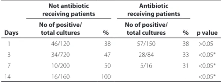
There were no statistically significant differences in the numbers of positive cultures between systemic antibiotic-receiving and no systemic antibiotic-receiving groups on day 1 (p>0.05, chi-square test). As the study progressed, the rate of positive cultures of the systemic antibiotic-receiving group decreased significantly (Table 4).
Of the 156 peripheral blood cultures, 25 (16%) were positive. Of the 25 patients with positive blood cultures, 22 had positive con-junctival cultures and, in 9 (six CNS, one C. diphtheriae, one S. aureus, one Acinetobacter species), the same microorganism was isolated simultaneously from blood and conjunctival specimens.
DISCUSSION
Data are available for ocular flora in healthy subjects, newborns, patients with acquired immune deficiency, those who wear contact lens, and patients with diabetes(1-9,11). However, there is only one stu-dy (see below) about the changes in the ocular flora of newborns hospitalized in an ICU(9). To the best of our knowledge, there is no published report concerning the effect of the duration of hospita-li zation, patients’ immune status, and administration of systemic antibiotic therapy to adult patients hospitalized in the ICU. In Turkey, most cornea donors are ICU patients. Therefore, changes in ocular flora during hospitalization are particularly important to avoid post keratoplasty infections.
Here we investigated the ocular flora of ICU patients and the effects of prolonged hospitalization, physical status, and systemic antibiotic treatment. We found that 288/478 (60.2%) cultures were po-sitive. Of interest, the highest numberof positive cultures was acqui red on day 1. Our results indicate that these cultures were already posi-tive before patients were admitted to the ICU. There are some likely explanations for this finding. First, most patients admitted to the ICU were already ill, which may have changed the flora before admission to the ICU. This explanation is partly supported by the colonization frequency, which significantly increased towards day 14 in the patient group that did not receive systemic antibiotic therapy, independent of immunocompetence.
Another possible explanation is that the ocular flora is altered in patients of advanced age, those with diabetes, and those that use steroids. The present study included 17 patients with diabetes, five patients >80 years of age, and six users of steroids. Finally, changes of the conjunctival flora mainly depend on the cause of admission to the ICU. Patients admitted to the ICU because of acute events such as trauma did not show changes in the normal flora on day 1. The limited number of patients in each of these groups did not allow per-forming statistical analysis. However, we believe that these patients may have been colonized by different ocular flora and this should be considered while analyzing the results. A study of newborns found that the frequency of colonization significantly increased from 37% to 47% after 10 weeks of hospitalization in the ICU, consistent with the results of our present study(9).
On the first day, immunocompromised patients had significantly higher colonization rates, but after receiving systemic antibiotics there was no significant difference on day 14 between the immuno-compromised and immunocompetent patients. This might be explai-ned by the effect of systemic antibiotic treatment.
The most common microorganisms isolated in our study were CNS, which represented 63.5% of all isolates, as well as representing the major pathogen. The frequency of CNS isolates increased towards day 14. The immune status of the patients was significantly affected the rate of colonization of CNS. In 9 patients, the same microorganism was isolated in the peripheral blood culture. These results further confirm the effect of nosocomial infections of these patients.
Changes in ocular flora depend on seasonal variations, tempera-ture, the host’s age, and environmental exposure. Further, traumatic ocular surgical procedures and local or systemic immune responses can modify the ocular flora(25). CNS, S. aureus, and Corynebacterium species are the most commonly isolated ocular flora present in the eyelid and conjunctiva(10,26). Our results are consistent with those of a study of newborns in the ICU showing that CNS was the most fre-quent isolate(9). Moreover, administration of anesthetics and intensive care confer a high risk of nosocomial infections with Pseudomonas and Acinetobacter species, and there are numerous reports of these bacteria causing keratitis among critically ill patients. However, we isolated Pseudomonas and Acinetobacter species in 4% of the cultu-res, which is consistent with the results of systemic cultures of these patients.
Postoperative endophthalmitis is associated with infection of donor tissues(23,27,28). Therefore, donor screening, microbiological screening, and decontamination of donor tissues are priorities of eye banking; 5% of all donor corneas are discarded because of bio logical contamina-tion(29). Our results and those of others cited above show the impor-tance of microbiological screening of donor corneas, particularly for critically ill patients hospitalized in the ICU. This is more important for ophthalmologists who collect corneas mainly from ICU patients, as is the case in Turkey.
In conclusion, patients hospitalized in the ICU are more suscepti-ble to bacterial colonization. However, we were unasuscepti-ble to generalize these results to post keratoplasty infections. Eye banks that collect corneas from ICU patients must regularly and closely follow potential donor candidates to determine bacterial colonization. Further stu-dies, particularly those that include multiple centers, are required to determine the effects of changes in ocular flora on post keratoplasty infections.
REFERENCES
1. Chaidaroon W, Ausayakhun S, Pruksakorn S, Jewsakul SO, Kanjanaratanakorn K. Ocular bacterial flora in HIV-positive patients and their sensitivity to gentamicin. Jpn J Ophthal-mol. 2006;50(1):72-3.
2. Eder M, Farina N, Sanabria RR, Ta CN, Koss M, Samudio M, et al. Normal ocular flora in newborns delivered in two hospital centers in Argentina and Paraguay. Graefes Arch Clin Exp Ophthalmol. 2005;243(11):1098-107.
3. Erdogan H, Kemal M, Toker MI, Topalkara A, Bakici Z. Effect of frequent-replacement contact lenses on normal conjunctival flora. Clao J. 2002;28(2):94-5.
4. Fleiszig SM, Efron N. Conjunctival flora in extended wear of rigid gas permeable contact lenses. Optom Vis Sci. 1992;69(5):354-7.
5. Fleiszig SM, Efron N. Microbial flora in eyes of current and former contact lens wearers. J Clin Microbiol. 1992;30(5):1156-61.
6. Gritz DC, Scott TJ, Sedo SF, Cevallos AV, Margolis TP, Whitcher JP. Ocular flora of pa-tients with AIDS compared with those of HIV-negative papa-tients. Cornea. 1997;16(4): 400-5.
7. Iskeleli G, Bahar H, Eroglu E, Torun MM, Ozkan S. Microbial changes in conjunctival flora with 30-day continuous-wear silicone hydrogel contact lenses. Eye Contact Lens. 2005;31(3):124-6.
8. Martins EN, Alvarenga LS, Hofling-Lima AL, Freitas D, Zorat-Yu MC, Farah ME, et al. Aerobic bacterial conjunctival flora in diabetic patients. Cornea. 2004;23(2):136-42. 9. Raskind CH, Sabo BE, Callan DA, Farrel PA, Dembry LM, Gallagher PG. Conjunctival
colonization of infants hospitalized in a neonatal intensive care unit: a longitudinal analysis. Infect Control Hosp Epidemiol. 2004;25(3):216-20.
Table 4. Table showing the colonization frequency in patients not receiving antibiotics and receiving antibiotics. Antibiotic receiving patients had a lower frequency of colonization
Days
Not antibiotic receiving patients
Antibiotic receiving patients
No of positive/ total cultures %
No of positive/
total cultures % p value
01 46/120 038 57/150 38 >0.05*
03 34/720 047 28/84 33 <0.05*
07 10/200 050 5/16 31 <0.05*
14 16/160 100 - - <0.05*
Ch a n g e si nt h eC o n j u n C t i va lb a C t e r i a lf l o r ao fpat i e n t sh o s p i ta l i z e di na ni n t e n s i v eC a r eu n i t
2 4 Arq Bras Oftalmol. 2017;80(1):21-4
10. Thiel HJ, Schumacher U. [Normal flora of the human conjunctiva: examination of 135 persons of various ages]. Klin Monatsbl Augenheilkd. 1994;205(6):348-57.German. 11. Yamauchi Y, Minoda H, Yokoi K, Maruyama K, Kumakura S, Usui M, et al. Conjunctival flora
in patients with human immunodeficiency virus infection. Ocul Immunol Inflamm. 2005;13(4):301-4.
12. Arunodaya GR. Infections in neurology and neurosurgery intensive care units. Neurol India. 2001;49 Suppl 1:S51-59.
13. Eggimann P, Pittet D. Infection control in the ICU. Chest. 2001;120(6):2059-93. 14. Fridkin SK, Welbel SF, Weinstein RA. Magnitude and prevention of nosocomial infections
in the intensive care unit. Infect Dis Clin North Am. 1997;11(2):479-96.
15. Girou E, Oppein F. Infection control in the ICU. Intensive Care Med. 2000;26(1):131-2. 16. Shulman L, Ost D. Managing infection in the critical care unit: how can infection control
make the ICU safe? Crit Care Clin. 2005;21(1):111-28, ix.
17. Widmer AF. Infection control and prevention strategies in the ICU. Intensive Care Med. 1994;20 Suppl 4:S7-11.
18. Wilks M, Wilson A, Warwick S, Price E, Kennedy D, Ely A, et al. Control of an outbreak of multidrug-resistant Acinetobacter baumannii-calcoaceticus colonization and infection in an intensive care unit (ICU) without closing the ICU or placing patients in isolation. Infect Control Hosp Epidemiol. 2006;27(7):654-8.
19. Ommeslag D, Colardyn F, De Laey JJ. Eye infections caused by respiratory pathogens in mechanically ventilated patients. Crit Care Med. 1987;15(1):80-1.
20. Dua HS. The conjunctiva in corneal epithelial wound healing. Br J Ophthalmol. 1998; 82(12):1407-11.
21. Dua HS. Bacterial keratitis in the critically ill and comatose patient. Lancet. 1998;351 (9100):387-88.
22. Leveille AS, McMullan FD, Cavanagh HD. Endophthalmitis following penetrating ke-ratoplasty. Ophthalmology. 1983;90(1):38-9.
23. Antonios SR, Cameron JA, Badr IA, Habash NR, Cotter JB. Contamination of donor cornea: postpenetrating keratoplasty endophthalmitis. Cornea. 1991;10(3):217-20. 24. Sue DY, Bongard FS. Philosophy and Principles of Critical Care. In: Bongard FS, Sue
DY, editors. Current Critical Care Diagnosis&Treatment. New York: McGraw Hill; 2002. pp 10-12.
25. Campos MS, Campos e Silva Lde Q, Rehder JR, Lee MB, O’Brien T, McDonnell PJ. Anae robic flora of the conjunctival sac in patients with AIDS and with anophthalmia com pared with normal eyes. Acta Ophthalmol (Copenh). 1994;72(2):241-5.
26. Grasbon T, Mino de Kaspar H, Klauss V. [Coagulase-negative staphylococci in normal and chronically inflamed conjunctiva]. Ophthalmologe. 1995;92(6):793-801. German. 27. Cameron JA, Antonios SR, Cotter JB, Habash NR. Endophthalmitis from contaminated donor corneas following penetrating keratoplasty. Arch Ophthalmol. 1991;109(1):54-9. 28. Cameron JA, Badr IA, Miguel Risco J, Abboud E, Gonnah el-S. Endophthalmitis cluster
from contaminated donor corneas following penetrating keratoplasty. Can J Ophthalmol. 1998;33(1):8-13.
29. Patel HY, Brookes NH, Moffatt L, Sherwin T, Ormonde S, Clover GM, et al. The New Zealand National Eye Bank study 1991-2003: a review of the source and management of corneal tissue. Cornea. 2005;24(5):576-82.