Application of Noninvasive Ventilation in Acute Respiratory Failure
after Cardiovascular Surgery
Vera Regina de Morais Coimbra, Rodrigo de Almeida Lara, Ériko Gonçalves Flores, Emília Nozawa, José Octávio Costa
Auler Júnior, Maria Ignez Zanetti Feltrim
Hospital das Clínicas da Faculdade de Medicina da Universidade de São Paulo – São Paulo, SP – Brazil
Summary
Objective: To examine ventilatory response, oxygenation-related, and hemodynamics of patients with hypoxemic acute respiratory failure (ARF) submitted to noninvasive mechanical ventilation (NIV) during the postoperative phase of cardiovascular surgery in order to identify predictive variables of success, and to compare the different types of NIV.
Methods: Seventy patients with hypoxemic ARF were randomized to one of three modalities of NIV - continuous positive airway pressure (CPAP) and ventilation with two pressure levels (PEEP + SP and BiPAP®). Ventilation, oxygenation-related, and hemodynamics variables were analyzed at pre-application, and 3, 6, and 12 hours after the protocol began.
Results: Thirteen patients were excluded. Thirty-one patients progressed to independence from ventilatory support and comprised the success group, and 26 required orotracheal intubation and were considered the nonsuccess group. Age, initial heart rate (HR), and respiratory rate (RR) showed elevated levels in the nonsuccess group (p=0.042, 0.029, and 0.002, respectively). A greater number of intraoperative complications were seen in the nonsuccess group (p=0.025). Oxygenation variables increased only in the success group. Among the NIV types, 57.9% of patients in the ventilator group, 57.9% in the two-pressure levels group, and 47.3% in the CPAP group progressed with success. Oxygenation and RR variables showed improvement only in the groups with two pressure levels.
Conclusion: Patients with hypoxemic ARF in the postoperative stage after cardiovascular surgery showed better oxygenation, RR, and HR during NIV application. In older patients and those with higher baseline RR and HR values, NIV was not sufficient to reverse ARF. The two-pressure level modes showed better results. (Arq Bras Cardiol 2007;88(5):270-276)
Key words: Respiration, artificial; anoxemia; respiratory insufficiency; thoracic surgery.
Mailing address: Maria Ignez Zanetti Feltrim •
Av. Dr. Enéas Carvalho de Aguiar, 44 – 2o andar – 05403-900 – São Paulo, SP – Brasil E-mail: [email protected]
Manuscript received December 28, 2006; revised manuscript received April 13, 2007; accepted May 3rd, 2007.
Introduction
Cardiac operations are associated with modifications in gas exchange and respiratory mechanics that can progress to acute respiratory failure (ARF), responsible for high postoperative morbidity and mortality rates1,2.
Among treatment alternatives, noninvasive ventilatory assistance has been used and it aims to improve alveolar ventilation and gas exchange, increase lung volumes, decrease respiratory work, and decrease mechanical ventilation time, thus avoiding reintubation and, consequently, cutting duration of stay in the intensive care unit1-8. There are also benefits
in hemodynamics, such as a decreased preload because of a reduction in venous return and decreased left ventricle postload due to reduced transmural pressure, leading to improved cardiac performance and increased cardiac output9,10.
There are two methods for noninvasive mechanical ventilation (NIV) application with positive pressure: continuous
positive airway pressure (CPAP) and ventilation with two levels of pressure (two-pressure level). NIV is well established in the treatment of respiratory failure secondary to exacerbations of chronic obstructive pulmonary disease (CPOD), in obstructive sleep apnea, and in cardiogenic pulmonary edema11-13.
However, in postoperative hypoxemic respiratory failure, the clinical response to the use of NIV has not been totally established and needs further investigation14.
This study was developed with the objective of: 1) examining the ventilatory response, oxygenation-related, and hemodynamics of patients with hypoxemic acute respiratory failure submitted to NIV application in the postoperative period of cardiovascular surgery, seeking possible predictive variables of success for this treatment modality; 2) comparing these results to those obtained with the application of different NIV modalities and equipment.
Methods
level of choice was the one that maintained SpO2 ≥ 95% for a FiO2 of 0.6. In the BiPAP Group, the minimal positive inhaled pressure (IPAP) used was 10 cmH2O, which could
be increased in 1 by 1 cmH2O increments with the objective of maintaining the exhaled tidal volume between 5 and 7 ml/kg. Initial positive exhaled pressure (EPAP) was 5 cmH2O, which could be increased in 1 by 1 cmH2O increments up to 10 cmH2O with the objective of maintaining SpO2≥ 95% for
a FiO2 of 0.6. [The FiO2 was checked by a Mini Oxi III line oximeter (MAS Medical Products®)]
Weaning from the NIV was carried according to the observation of improvement in signs and symptoms of acute respiratory failure, and was performed by progressively increasing the intervals between applications and decreasing the application time. NIV treatment was considered successful when the patient presented normalization of clinical signs and symptoms of acute respiratory failure in a maximal period of 72 hours. Nonsuccess was defined as worsening of signs and symptoms requiring orotracheal reintubation at any time, or continuation of NIV for more than 72 hours.
The ventilator-patient interface was produced by using a facial mask; with the mechanical ventilator Gibeck model (Gibeck Dryden®), masks were used, whereas for BiPAP S/TD
30 and Solo/Sullivan devices, Respironics® facial or nasal
masks were used.
Statistical analysis - The variables of heart rate and respiratory rate (with NIV measurements made during preapplication, 6 hours, and 12 hours after study began), pH, PaCO2, PaO2, and SaO2 (with NIV measurements at preapplication, 3 hours, and 12hours after study began) were analyzed in each group and comparatively among the groups, using variance analysis testing (ANOVA with Kruskal – Walls One-Way Analysis of Variance on Ranks, Tukey test, and the Two-Way Repeated Measures Analysis of Variance). For the other variables analyzed, non-paired t test and chi-squared test were used to examine occurrences of events.
Results
Seventy patients were enrolled in the study, and 13 of them were excluded for different reasons, including lack of arterial gasometry, noncompliance with the protocol, presence of pneumothorax, and modification of the ventilatory modality.
Of the 57 patients included, 22 underwent only myocardial revascularization (MR), 13 underwent valvar operations (combined or not), 5 underwent correction of an aneurism or dissection of the aorta, 3 underwent thromboendarterectomy for chronic pulmonary embolism, and 15 patients underwent combination operations.
Success X Nonsuccess - Of the 57 patients who completed the study, 31 (54.4%) progressed to independence from ventilatory support and comprised the success group, and 26 (45.6%) progressed with a need for orotracheal intubation and were considered the nonsuccess group for NIV strategy. Causes for orotracheal intubation were: a) worsening of respiratory insufficiency (n=11); b) worsening of the ARF associated with hemodynamic instability (n=7); c) lowered level of consciousness (n=6); and d) hemodynamic instability Institute] of the Hospital dasClínicas da Faculdade de Medicina
da Universidade de São Paulo (InCorHC – FMUSP) during the period from July 1999 to December 2002. The sample consisted of 70 patients undergoing cardiovascular operations. Patients were admitted to the immediate postoperative recovery unit and were extubated as per institute protocol. In the presence of acute respiratory failure, according to previously defined criteria, patients were randomized to one of three study groups: ventilator group, CPAP group, or two-pressure level group.
For inclusion, patients had to present with at least three of the five proposed signs and symptoms: dyspnea, tachypnea (respiratory rate > 25 rpm), use of accessory muscles, and hypoxemia with arterial blood hemoglobin saturation measured by pulse oxymetry (SpO2) < 95% with O2 catheter at 5l/min and chest X-ray showing abnormalities in at least two quadrants.
Patients showing agitation or non-cooperation, swallowing reflex changes, hemodynamic instability,difficult to manage secretions, acute cardiac arrhythmias, active gastric or upper airway bleeding, or who had used some form of noninvasive ventilation after the last extubation were excluded from the study.
Once included, ventilatory, oxygenation-related, and hemodynamics data were collected with the patients in elevated dorsal decubitus, before NIV installation. Respiratory rate was counted by observation of thoracoabdominal movements; heart rate (HR) and mean arterial pressure (MAP) readings were obtained by a monitor (Hewlet Packard®).
These data were collected every 6 hours. A sample of arterial blood was collected with the patient breathing spontaneously while receiving 5L/min O2 by catheter for at least 15 minutes (1st measurement). This collection was repeated 3 hours (2nd
measurement) and 12 hours (3rd measurement) after beginning
NIV, with the patient in the same oxygen therapy conditions as for the 1st measurement.
NIV was applied using three different methods: 1) Ventilator Group (Group A) – use of two pressure levels by means of a HamiltonVeolar or Amadeus Mechanical Ventilator in the spontaneous mode, using PEEP and supporting pressure (SP); 2) CPAP Group (Group B) – use of continuous positive airway pressure with the Sullivan or Solo (Respironics®) apparatus; 3)
BiPAP Group (Group C) – use of two pressure levels in BiPAP®
S/TD 30 (Respironics®).
All groups received an initial NIV application for 3 hours, with a 30-minute interval if needed. Posterior applications were at least one hour long during the first 12 hours. The interval between applications was always less than three hours.
In the Ventilator Group, a minimal 10 cmH2O SP was
used, which could be increased by 1 cm H2O increments with the objective of reaching an exhaled tidal volume of 5 to 7 ml/kg. The initial PEEP value was 5 cmH2O, which could also be increased by 1 cmH2O increments up to 10 cmH2O, seeking to maintain SpO2≥ 95% for an inhaled oxygen fraction
(n=2).
In analysing the clinical-surgical data, we observed that the nonsuccess group showed a larger number of patients who smoked, had undergone previous cardiovascular operations, and had experienced a greater number of intraoperative surgical complications, as is displayed on Table 1. The mean values for the preceding left ventricular ejection fraction (LVEF) were 58% in the success group and 56% in the nonsuccess group, with no significant difference between the groups.
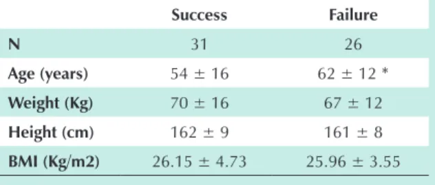
The anthropometric data from both groups are shown on Table 2. Patients in the nonsuccess group were older than those in the success group.
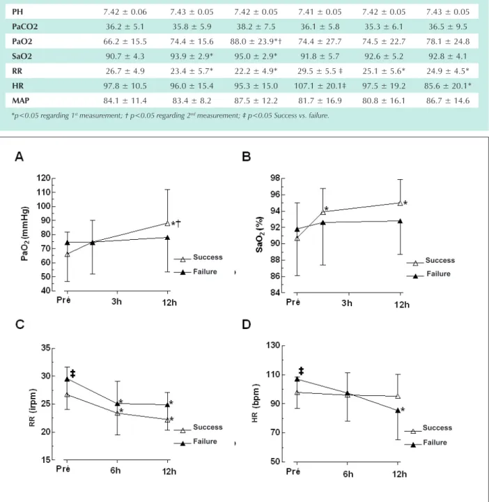
Oxygenation-related, ventilation, and hemodynamics data are shown for both the groups on Table 3 in mean ± standard deviation, and in Figure 1 for the three measurements obtained over time. The success group presented blood elevations of PaO2 and SaO2, reaching higher levels at the 12th hour of the
study. Oxygenation in the nonsuccess group did not improve, even with installation of ventilatory support.
Respiratory rate was high in both groups at the beginning of the study, but proved significantly higher in the nonsuccess group (p= 0.002). After 6 hours and 12 hours, mean values were significantly lower than in the pre-study situation, although with no difference between the groups.
Mean pre-study values of HR were statistically higher in the nonsuccess group, and were significantly lower at the 12th hour. For the success group, HR behavior was similar
over time.
Two-pressure level, CPAP, and Ventilator Treatment Modalities
Patients were regrouped according to the modality of NIV applied, producing three groups of 19 patients each: two-pressure level, CPAP, and ventilator. Eleven (57.9%) patients progressed to suppression of the noninvasive ventilatory support in the ventilator group, 11 (57.9%) in the two-pressure level group, and 9 in the CPAP group (47.3%).
Groups were homogenous as to age, weight, height, and BMI, as is displayed on Table 4.
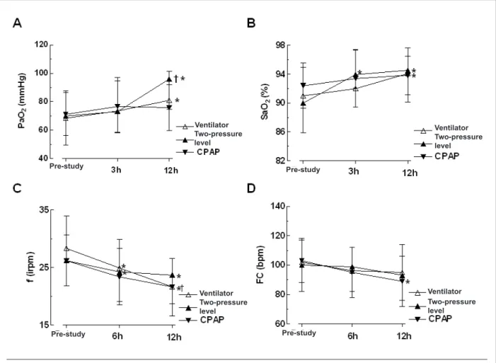
Oxygenation-related, ventilation, and hemodynamics data are shown on Table 5 and illustrated in Figure 2. As to oxygenation values (PaO2 and SaO2), statistically significant increases were observed only in the ventilator and two-pressure level groups over time, although with no difference between these two modalities.
Respiratory rate did not show a statistically significant difference among the groups, but there was a significant reduction of its values in the two-pressure level and ventilator groups over time when compared to the initial levels.
The HR did not differ among the groups, but there was a statistically significant reduction in the 3rd measurement when
compared to the pre-values in the CPAP group. MAP did not show any significant changes among the groups over time.
Discussion
Our results show that the application of NIV in patients with hypoxemic ARF post cardiovascular surgery avoided
reintubation in 54.4% of the cases. Several studies have already demonstrated the efficacy of NIV in preventing or avoiding orotracheal intubation in situations of hypoxemic acute respiratory failure. Recent randomized studies15-19 involving
a heterogeneous population of patients with hypoxemia have reported success in avoiding orotracheal intubation in 69 to 79%. The difference in percentage of success among the various studies, including ours, is because of the different
causes that lead to hypoxemic ARF. Antonelli et al20 studied
354 patients with hypoxemic ARF, observing a 51% success rate in patients with acute respiratory distress syndrome (ARDS), 50% in patients with nosocomial pneumonia, and 69% in patients with atelectasia. These numbers are similar to our results, which can be explained by the fact that most of the patients with hypoxemic ARF in the postoperative phase after cardiovascular operations have lung collapse and infiltration processes as the main causes. Although our patients had ARF, in most cases the primary basis of this involvement was correlated to cardiac dysfunction, since most unsuccessful patients showed a greater number of intraoperative events such as cardiorespiratory arrest (CRA) and hemodynamic instability requiring mechanical circulatory assistance, and bleeding.
Predictors of nonsuccess for the use of NIV are not well characterized. In the study by Meduri et al5 gasometry
improvement was associated with the NIV success, but no predictive correlation for success was found for hypoxemic ARF. Rana et al21 observed patients with acute pulmonary
injury and noted that shock, metabolic acidosis, and serious Table 1 - Clinical and surgical data of the groups that progressed to
success and failure of NIV
Success Failure P
Smoking habit 14/31 16/26 0.025
Intraoperative
events * 11/31 17/26 NS Previous surgeries 06/31 07/26 NS
Previous surgeries 06/31 07/26 NS
* Difficult ventilation and hypoxemia. Hemodynamic instability requiring mechanical circulatory assistance, bleeding and CRA.
Table 2 - Mean values and SD of anthropometric variables of the groups that progressed to success and failure of NIV
Success Failure
N 31 26
Age (years) 54 ± 16 62 ± 12 *
Weight (Kg) 70 ± 16 67 ± 12
Height (cm) 162 ± 9 161 ± 8
BMI (Kg/m2) 26.15 ± 4.73 25.96 ± 3.55
hypoxemia were predictors of nonsuccess for NIV strategy. Antonelli et al20 found the variables of age, initial respiratory
rate, and lack of oxygenation improvement within one hour of the treatment application as correlated to nonsuccess of NIV.
Our data show that before NIV application, there were statistically significant differences as to age, heart rate, and
respiratory rate, with the highest values in the nonsuccess group. It is known that aging provokes a series of cardiorespiratory changes, translated as a decrease of cardiorespiratory reserve. This condition is a predisposing factor for respiratory failure in cases of infectious, traumatic, and surgical aggressions. Our patients who progressed to nonsuccess were older, and at the beginning of the study presented higher heart rates Table 3 - Mean values and SD of oxygenation-related, ventilatory and hemodynamic variables of the groups that progressed to success and failure of
NIMV
Success Insuccess
1st 2nd 3rd 1st 2nd 3rd
PH 7.42 ± 0.06 7.43 ± 0.05 7.42 ± 0.05 7.41 ± 0.05 7.42 ± 0.05 7.43 ± 0.05
PaCO2 36.2 ± 5.1 35.8 ± 5.9 38.2 ± 7.5 36.1 ± 5.8 35.3 ± 6.1 36.5 ± 9.5
PaO2 66.2 ± 15.5 74.4 ± 15.6 88.0 ± 23.9*† 74.4 ± 27.7 74.5 ± 22.7 78.1 ± 24.8
SaO2 90.7 ± 4.3 93.9 ± 2.9* 95.0 ± 2.9* 91.8 ± 5.7 92.6 ± 5.2 92.8 ± 4.1
RR 26.7 ± 4.9 23.4 ± 5.7* 22.2 ± 4.9* 29.5 ± 5.5 ‡ 25.1 ± 5.6* 24.9 ± 4.5*
HR 97.8 ± 10.5 96.0 ± 15.4 95.3 ± 15.0 107.1 ± 20.1‡ 97.5 ± 19.2 85.6 ± 20.1*
MAP 84.1 ± 11.4 83.4 ± 8.2 87.5 ± 12.2 81.7 ± 16.9 80.8 ± 16.1 86.7 ± 14.6
*p<0.05 regarding 1st measurement; † p<0.05 regarding 2nd measurement; ‡ p<0.05 Success vs. failure.
Fig. 1 - Mean values and SD of variables in the groups success and failure, at three moments assessed. A) PaO2; B) SaO2; C) RR and D) HR, (* p<0.05 regarding 1st measurement; † p<0.05 regarding 2nd measurement; ‡ p<0.05 success vs. failure)
Success
Success
Success
Success Failure
Failure
Failure
Failure
R
and respiratory rates, which could be suggestive of a greater ventilatory and hemodynamic overload, contributing to a respiratory deterioration more difficult to reverse with NIV.
We observed that along the first 12 hours of the protocol, the variables SaO2 and PaO2 showed significant increases in the success group, whereas in the nonsuccess group there was no such change. These results are similar to those found by Antonelli et al20, and Rana et al21, suggesting that patients
who showed an improvement in oxygenation during the first hours of treatment have better results in terms of avoiding orotracheal intubation.
The respiratory rate showed a similar behavior between the two groups, with a significant reduction in the first 12 hours within each group, albeit with no difference between the groups.
Thus, NIV was an effective ventilatory strategy in most cases, avoiding installation of invasive mechanical ventilation; nevertheless, factors such as age, and previous heart rate and respiratory rate can compromise the efficacy of the ventilatory
Table 5 - Mean values and SD of oxygenation-related, ventilatory and hemodynamic variables of the groups of patients characterized by the NIMV modality received
Ventilator Two-pressure level CPAP
1st 2nd 3rd 1st 2nd 3rd 1st 2nd 3rd
pH 7,42 ±
0,05 7,42 ± 0,06 7,40 ± 0,06 7,43 ± 0,05 7,43 ± 0,05 7,42 ± 0,07 7,40 ± 0,06 7,42 ± 0,05 7,42 ± 0,07
PaCO2 36,6 ±
4,6 35,5 ± 5,2 38,0 ± 4,5 36,9 ± 6,9 37,3 ± 8,1 38,9 ± 12,0 34,9 ± 4,3 33,5 ± 4,0 35,1 ± 4,8
PaO2 68,5 ±
19,0 73,6 ± 23,6 81,1 ± 20,2* 70,0 ± 20,4 73,2 ± 15,0 96,0 ± 21,2*† 71,3 ± 15,2 76,7 ± 17,9 75,8 ± 16,1
SaO2 91,0 ±
3,9 92,0 ± 5,3 94,1 ± 3,5* 90,0 ± 4,1 93,9 ± 1,8* 94,5 ± 4,4* 92,4 ± 3,1 93,4 ± 4,0 93,8 ± 2,7
F 28,3 ±
5,6 24,9 ± 5,0* 21,6 ± 5,0*† 29,4 ± 4,3 24,2 ± 5,1* 23,7 ± 5,0* 26,2 ± 4,4 23,4 ± 4,9 21,6 ± 5,0
FC 102 ± 15 96 ± 16 95 ± 19 100 ± 18 99 ± 17 93 ± 17 103 ± 15 95 ± 17 89 ± 17*
PAM 84,6 ±
16,3 82,4 ± 11,3 86,4 ± 14,0 79,1 ± 9,9 82,2 ± 10,2 88,1 ± 13,7 85,2 ± 15,2 82,2 ± 15,6 87,0 ± 12,4
* p<0.05 regarding 1st measurement; † p<0.05 regarding 2nd measurement.
support.
Two-pressure level, CPAP, and Ventilator Treatment Modalities
Comparing the three NIV modalities as to the occurrence of success and nonsuccess, we found no statistically significant difference among them (p=0.531), although there was a tendency towards better results with the use of two-pressure level modalities, which contributed to avoid reintubation in most of the cases studied in each group (57.9%). In the CPAP modality, there was a greater number of reintubations, with 47.3% success, even though the difference between the groups was not significant.
Ventilator and two-pressure level modalities were also responsible for significant improvements over time. The ventilator and two-pressure level groups showed oxygenation rises in the first 12 hours of application, while in the CPAP group, this change was discreet. Matte et al22
studied 96 patients in the postoperative phase of myocardial revascularization surgery and compared the use of an incentive Table 4 - Mean values and SD of anthropometric variables of the groups of patients characterized by the NIMV modality received
Ventilator Two-pressure level CPAP
n 19 19 19
Age (years) 58.5 ±15.4 54.9 ± 15.4 60.3 ± 13.3
Weight (Kg) 67.2 ±15.2 68.9 ± 17.7 73.2 ± 10.8
Height (cm) 1.60 ± 0.07 1.64 ± 0.10 1.62 ± 0.07
inspirometer, CPAP, and two-pressure level in preventing deterioration of lung function and gasometry values in these patients with no signs of ARF. The study showed that both CPAP and two-pressure level afforded an improvement of PaO2 over 24 hours of application, but there was no significant
difference between the modalities13. In treating 83 patients
with cardiogenic pulmonary edema, Park et al13 compared the
use of oxygen therapy (n=27), CPAP (n=27), and two-pressure level (n=29), and noted that with 30 minutes of application, there was a significant increase of PaO2 in the two-pressure level and CPAP groups. In this study, the respiratory rates of patients from the two-pressure level and CPAP groups were lower than those of the oxygen therapy group. The behavior of the respiratory rate in our patients was similar to that seen in the study by Matte et al21, who showed a decrease in the
two-pressure level group during the first 12 hours of application, whereas in the CPAP group there was a small elevation in the same period, producing a statistically significant difference between the two methods.
Macintyre et al23, using pulmonary complacency and
airway pressure measurements in Intensive Care Unit patients with stable pulmonary and hemodynamic conditions, demonstrated a reduction in respiratory muscle work function when using two-pressure level, showing that this method
reduces inspiratory effort and respiratory work. Mechanical work is equal to the force multiplied by the dislocation. In the respiratory system, the work generated by the inspiratory muscles creates a pressure that causes dislocation of the air. Therefore, when additional positive pressure is used helping muscular function during inspiration, there is a reduction in respiratory work.
The heart rate and arterial blood pressure were not modified during application of positive pressure, and their mean values remained within normal limits during most of the protocol. A statistically significant change was noted in the HR over time only in the CPAP group. Philip-Jöet et al10,
when studying hemodynamic modifications resulting from the use of CPAP and two-pressure level methods, found no significant differences in patients with normal left ventricular function. There was a modification of the cardiac output in patients who already presented a decrease in mean pulmonary artery pressure (less than 12 mmHg). In our study, it was not possible to correlate this information with the hemodynamic condition, since the left ventricular function was not assessed in these patients.
In conclusion, patients with hypoxemic ARF in the postoperative phase after cardiovascular surgery improved
Fig. 2 - Mean values and SD of variables in different modalities. A) PaO2; B) SaO2; C) RR and D) HR.
(* p<0.05 regarding 1st measurement; † p<0.05 regarding 2nd measurement).
Ventilator Ventilator
Ventilator Ventilator
Two-pressure level Two-pressure
level Two-pressure level
Two-pressure level
Pre-study
Pre-study
References
1. Irwin S, Tecklin JS. Fisioterapia cardiopulmonar. 2a ed. São Paulo: Editora Manole; 1994.
2. Kildgen-Milles D, Buhl R, Gabriel A, Bohner H, Muller E. Nasal continuous positive airway pressure: a method to avoid endotracheal reintubation in postoperative high-risk patients with severe nonhypercapnic oxygenation failure. Chest. 2000;117(4):1106-11.
3. Aguilo R, Togores B, de la Pena A, Santos C, Agusti AG. Noninvasive ventilatory support after lung resectional surgery. Chest. 1997;112(1):117-21.
4. Kilger E, Briegel J, Haller M, Frey L, Schelling G, Stoll C, et al. Effects of noninvasive positive pressure ventilatory support in non-COPD patients with acute respiratory insufficiency after early extubation. Intens Care Med. 1999;25:1374-80.
5. Meduri GU, Turner RE, Abou-Shala N, Wunderink R, Tolley E. Noninvasive positive pressure ventilation via face mask: first-line intervention in patients with acute hypercapnic and hypoxemic respiratory failure. Chest. 1996; 109 (1): 172-92.
6. Wysocki M, Tric L, Wolff MA, Gertner J, Millet H, Herman B. Noninvasive pressure support ventilation in patients with acute respiratory failure. Chest. 1993;107:761-8.
7. Pang D, Keenan SP, Cook DJ, Sibbald WJ. The effect of positive pressure airway support on mortality and the need for intubation in cardiogenic pulmonary edema: a systematic review. Chest. 1998;114:1185-92.
8. Kramer N, Meyer TJ, Meharg J, Cece RD, Hill NS. Randomized, prospective trial of noninvasive positive pressure ventilation in acute respiratory failure. Am J Respir Crit Care Med. 1995;151(6):1799-806.
9. Meyer JJ, Hill NS. Noninvasive positive pressure ventilation to treat respiratory failure. Ann Intern Med. 1994;120:760-70.
10. Philip-Jöet FF, Paganelli FF, Dutau HL, Saadjian AY. Hemodynamic effects of bilevel nasal positive airway pressure ventilation in patients with heart failure. Respiration. 1999;66:136-43.
11. Brochard L, Mancebo J, Wysocki M, Lofaso F, Conti G, Rauss A, et al. Noninvasive ventilation for acute exacerbations of chronic obstructive pulmonary disease. N Engl J Med. 1995;333(13):817-22.
12. Sullivan CE, Berthon-Jones M, Issa FG, Eves L. Reversal of obstructive sleep apnoea by continuous positive airway pressure applied through the nares. Lancet. 1981;1(8225):862-5.
13. Park M, Sangean MC, Volpe MDS, Feltrim MIZ, Nozawa E, Leite PF, et al. Randomized, prospective trial of oxygen, continuous positive airway pressure, and bilevel positive airway pressure by face mask in acute cardiogenic pulmonary edema. Crit Care Med. 2004;32(12):2407-15.
14. International Consensus Conferences in Intensive Care Medicine: noninvasive positive pressure ventilation in acute respiratory failure. Am J Respir Crit Care Med. 2001;163(1):283-91.
15. Masip J, Betbese AJ, Paez J, Vecilla F, Canizares R, Padro J, et al. Non-invasive pressure support ventilation versus conventional oxygen therapy in acute cardiogenic pulmonary oedema: a randomised trial. Lancet. 2000;356:2126-32.
16. Confalonieri M, Potena A, Carbone G, Porta RD, Tolley EA, Meduri UG. Acute respiratory failure in patients with severe community-acquired pneumonia. A prospective randomized evaluation of noninvasive ventilation. Am Respir Crit Care Med. 1999;160:1585-91.
17. Antonelli M, Conti G, Bufi M, Costa MG, Lappa A, Rocco M, et al. Noninvasive ventilation for treatment of acute respiratory failure in patients undergoing solid organ transplantation: a randomized trial. JAMA. 2000;283(2):235-41.
18. Martin TJ, Hovis JD, Costantino JP, Bierman MI, Donahoe MP, Rogers RM, et al. A randomized, prospective evaluation of noninvasive ventilation for acute respiratory failure. Am Respir Crit Care Med. 2000;161:807-13.
19. Keenan SP, Sunuff T, Cook DJ, Hill NS. Does noninvasive positive pressure improve outcome in acute hypoxemic respiratory failure? A systematic review. Crit Care Med. 2004;32(12):2516-23.
20. Antonelli M, Conti G, Moro ML, Esquinas A, Gonzales-Diaz G, Confalonieri M, et al. Predictors of failure of noninvasive positive pressure ventilation in patients with acute hypoxemic respiratory failure: a multi-center study. Intensive Care Med. 2001;27(11):1718-28.
21. Rana S, Jenad H, Gay PC, Buck CF, Hubmayr RD, Gajic O. Failure of non-invasive ventilation in patients with acute lung injury: observational cohort study. Crit Care. 2006;10 (3): R79.
22. Matte P, Jacquet L, Van Dyck M, Goenen M. Effects of conventional physiotherapy, continuous positive airway pressure and non-invasive ventilatory support with bilevel positive airway pressure after coronary artery bypass grafting. Acta Anaesthesiol Scand. 2000;44:75-81.
23. MacIntyre NR. Respiratory function during pressure support ventilation. Chest. 1986;89(5):677-83.
oxygenation, respiratory rate, and heart rate during NIV application. In older patients and those with higher initial respiratory and heart rates, NIV may not be sufficient for reversing the hypoxemic respiratory failure condition.
The methods with two pressure levels showed results superior to those of the CPAP method.
Potential Conflict of Interest
No potential conflict of interest relevant to this article was
reported.
Sources of Funding
There were no external funding sources for this study
Study Association