Biometria ocular, estimativa matemática
e variação esférica pós-facectomia
Ocular biometry, mathematical estimation
and spherical variation after facectomy
Received for publication 20/05/2015 - Accepted for publication 04/08/2015 The authors declare no conflicts of interests.
A
BSTRACTObjective: To assess ocular biometric parameters by optical biometry and to observe a possible spherical refractive difference, as well as its variation based on estimated preoperative calculation and the spherical refraction post cataract surgery by phacoemulsification with intraocular lens implant (IOL). Methods: After reviewing 252 electronic medical records between 2013 and 2014, 117 adult patients (189 eyes) were selected. The patients underwent phacoemulsification with foldable IOL implantation by the same surgeon and were examined by IOLMaster® 500. The IOL power was calculated using the Haigis formula. The Wilcoxon test was applied to identify the existence of significant differences (p<0.05) between the spherical expected refraction (SER) and the final spherical refraction (FSR) of the eyes.
Results: There were operated 98 right eyes (OD) and 91 left (OS). A calculation of the variation between FSR and SER indicated that 55% of the OD reached results within ± 0.5 diopters (D) and 89% within ± 1D. With respect to OS, 46% achieved results within ± 0.5D and 78% within ± 1D. Conclusion: Optical biometry is a reliable, predictable and reproducible method to estimate the FSR of both eyes.
Keywords: Phacoemulsification; Cataract resection; Biometry
R
ESUMOObjetivo: Avaliar os parâmetros biométricos oculares por meio da biometria óptica e observar uma possível diferença refratométrica
esférica, assim como sua variação, baseada no cálculo pré-cirúrgico estimado e na refração esférica pós-cirurgia de catarata pela facoemulsificação com implante de lente intraocular (LIO). Métodos: Foram revisados 252 prontuários eletrônicos entre 2013 e 2014 dos quais foram selecionados 117 pacientes adultos (189 olhos) submetidos à facoemulsificação com implante de LIO dobrável pelo mesmo cirurgião e examinados através do IOLMaster® 500. O poder dióptrico da LIO foi calculado pela fórmula de Haigis. O teste de Wilcoxon foi empregado para testar a existência de diferença significativa (p<0,05) entre o grau esférico esperado (GEE) e o grau esférico final (GEF). Resultados: Foram operados 98 olhos direitos (OD) e 91 esquerdos (OE). Após calculada a variação entre o GEE e o GEF observou-se que 55% dos OD alcançaram resultados dentro de ± 0,5 dioptrias (D) e 89% resultados dentro de ± 1D. Quanto ao OE, 46% alcançaram resultados dentro de ± 0,5D e 78% dentro de ± 1D. Conclusão: A biometria óptica pode ser utilizada como um método confiável, previsível e reprodutível para que seja estimado o GEF de ambos olhos.
Descritores: Facoemulsificação; Exérese de catarata; Biometria
Francisco Wellington Rodrigues
1, Lucas Lauar Cortizo Vidal
2, Ana Luiza Rassi de Mendonça
2, Rodrigo Egidio da Silva
1I
NTRODUCTIONC
ataract is the name given to any opacity of crystalline1that, despite being a major cause of blindness and visual impairment, not necessarily affect vision compromising labor activities laborais.1,2 It is the biggest cause of reversible
blindness in Latin America3,4 and worldwide (50%)3-6, but out
of the 45 million blind existing 40% are due to cataract.7
According to the WHO, the annual incidence of the disease is estimated to be 0.3%8, which would represent in Brazil about
550,000 new cases of cataract by year8, being characterized as a
public health problem.2,9
Cataract is a disease that is curable by restoring the vision of the person operated by a surgical procedure which requires only one intervention and that does not depend on the adhesion of the patient to the use of medication, named facectomia.1,2 This
surgery to replace the opaque crystalline by a prosthesis called intraocular lens (IOL) can be performed by various techniques, the phacoemulsification being currently the most used one for being the safest and providing a fast post-operative recovery. 10-14 With the precision and predictability of this technique, the
surgical indication is made earlier, with the only problematic factor being to calculate the IOL, which explains the great importance of ocular biometry.15
The ocular biometry is the exam to measure the axial length of the eyeball and its structures, essential for defining the origin of ametropy and composes one of the bases for the calculation of intraocular lenses (IOLs).1,16,17 The biometric accuracy, coupled
to improved surgical technique and the development of IOLs, is what approaches the cataract surgery to the refractive procedure nowadays, since besides the cure it can also correct pre-existing ametropies. 11-13
Given the above, we aim to assess the ocular biometric parameters by optical biometry. It is possible to observe a possible spherical refractive difference, as well as its variation, based on the estimated pre-surgical calculation and the spherical refraction post facectomy surgery by the phacoemulsification technique with foldable intraocular lens implantation.
M
ETHODSThis is an observational cross-sectional study approved by the ethics and Research Committee (CEP) of the Pontifical Catholic University of Goiás (PUC-Goiás). We reviewed 252 electronic medical records at the Hospital Ver in the city of Goiania in the years 2013 and 2014, of which 117 adult patients (total of 189 eyes) operated for cataract were selected. The sample was randomly selected from the total volume of patients undergoing facectomy with IOL implantation by the same surgeon.
The inclusion criteria were: patients operated at Hospital Ver above 18 years of age who underwent phacoemulsification with foldable IOL implant (brand B-Lens Hanita) by the same surgeon in the years 2013 and 2014, examined by optical biometry (IOLMaster® 500, Zeiss System) by the same ophthalmologist,
and with no complications in the surgery.
Patients under 18 years of age, contact lens users, patients with eye diseases and/or previous systemic, and/or eyes which underwent ophthalmological surgery previously did not participate in this study.
Before starting the optical biometry examination with IOLMaster® 500, 1 drop of ophthalmic anesthetic solution
(Proparacaine hydrochloride 5 mg/mL) was administered in each eye of the patient, and then 1 drop of ocular lubricant (Carmellose Sodium 5 mg/mL) to regularize the tear film.
The biometric formula used for the calculation of intraocular lens of all samples in this research was Haigis (4th generation).
The post surgical evaluation was 30 days after the cataract surgery, when all subjects in the research had the degree of spherical refraction measured by the same ophthalmologist.
The data from the medical records were typed in the software Microsoft Excel® and analyzed using the software package SPSS 21.0 (Statistical Package for Social Science Inc., Chicago, Illinois, USA) for Windows®.
The normality test of Kolmogorov Smirnov was used to evaluate if the continuous variables (number of lens, spherical degree expected, final spherical degree and difference between the expected and final spherical degrees) showed normal distribution. These variables were presented as mean, standard deviation, median and 95% confidence interval.
The Wilcoxon test was used to test the existence of significant difference between the spherical and the expected degrees (estimated by optical biometry with the IOLMaster® 500) and spherical degree end in each eye after surgery.
All tests considered a 95% confidence level, meaning it was considered significant p < 0.05.
R
ESULTSThe sample was composed by a total of 117 patients with an average age of 68.02 years (SD ± 10.53), and 69 (59%) were female and 48 (41%) male. Of the 189 operated eyes of the sample, 98 (52%) were right eyes and 91 were left eyes (48%).
Of all continuous variables studied for both eyes just the final spherical degree of the left eye and the difference between the expected and final spherical degrees of the left eye showed normal distribution. The other variables did not show normal distribution, since they all had p < 0.05. (Table 1)
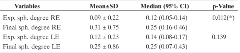
There was statistically significant difference in the comparison between the expected spherical degree and the final spherical degree in the right eye (p = 0.012), but not in the left eye (p = 0.259). (Table 2)
After calculated the variance between the expected spherical degree and the final spherical degree of the 189 eyes operated, we found that 55% of right eyes (50 eyes) achieved results within ± 0.5 D; 89% (81 eyes) achieved results within ± 1D and 97% (88 eyes) achieved results within ± 2D. As for the left eye, 46% (45 eyes) achieved results within ± 0.5D; 78% (76 eyes) achieved results within ± 1D and 96% (94 eyes) achieved results within ± 2D. (Figure 1)
D
ISCUSSIONSenile cataract has a higher incidence in the population over 50 years18. It is the most common type of cataract, and a
Variables n N.I Mean ± SD Median (95% CI) p-Value
No. lens RE 98 19 20.4±4.1 21.0 (19.6-21.2) < 0.001 No. lens LE 91 26 20.1±3.6 20.5 (19.4-20.9) < 0.001 Exp. sph. degree RE 98 19 0.09±0.22 0.12 (0.05-0.14) < 0.001 Exp. sph. degree LE 91 26 0.12±0.23 0.14 (0.08-0.17) < 0.001 Final sph. degree RE 98 19 0.31±0.75 0.25 (0.16-0.46) < 0.001 Final sph. degree LE 91 26 0.25±0.86 0.25 (0.07-0.43) 0.050 Diff. final and exp. sph. degrees RE 98 19 0.55±0.51 0.46 (0.45-0.65) < 0.001 Diff. final and exp. sph. degrees LE 91 26 -0.12±0.87 -0.16 (-0.30-0.87) 0.200
Table 1
Distribution of variables: number of lens, spherical degree expected, final degree expected and difference between the final and the expected spherical degrees of both eyes
Test: Kolmogorov Smirnov
Diff: difference, Sph: spherical, Exp: expected, N: number of the lens, RE: right eye, LE: left eye, N.I.: no information
Figure 1: Distribution of eyes as to the percentage varying between the expected spherical degree and the final spherical degree within ± 0.5 diopters (D); ± 1D and ± 2
Table 2
Comparison between the results of the expected spherical degree and the final spherical degree in the right and left eyes
Variables Mean±SD Median (95% CI) p-Value
Exp. sph. degree RE 0.09 ± 0,22 0.12 (0.05-0.14) 0.012(*) Final sph. degree RE 0.31 ± 0.75 0.25 (0.16-0.46)
Exp. sph. degree LE 0.12 ± 0.23 0.14 (0.08-0.17) 0.139 Final sph. degree LE 0.25 ± 0.86 0.25 (0.07-0.43)
Test: Wilcoxon; *significant
an estimated prevalence of 2.5% between 40 and 49 years, 6.8% between 50 and 59 years, 20% between 60 and 69 years, 42.8% between 70 and 79 years, and 68.3% in over 80 years.18
During this research we followed the recommendations for good results in cataract surgery described in the literature:19
standardization of biometric equipment used for axial length measurement and keratometry, use of optical biometry (IOLMaster® 500), seamless facectomy and with the foldable
intraocular lens implant in the capsular bag (“in the bag”), appropriate use of the formula of 4th generation for the calculation of IOL and optimization of their constants.19
After calculated the variance between the expected spherical degree and the final spherical degree of the sample, we found that 55% of right eyes achieved results within ± 0.5 D; 89% achieved results between 0 and ± 1D, and 97% achieved results between 0 and ± 2D. As for the left eye, 46% achieved results within ± 0.5D; 78% achieved results between 0 and ± 1D and 96% achieved results between 0 and ± 2D. (Figure 1) These data demonstrate good reproducibility and an acceptable index of reliability for the cataract surgery performed and for the biometry method used.
The guidelines of the Royal College of Ophtalmologists Cataract Surgery say that the most important thing in biometry is to achieve excellent results.20 They must be calculated by the
biometric error prediction, that is, the difference between the equivalent final spherical degree and the equivalent spherical calculated expected (target refraction), which may be represented in terms of percentage of eyes with 0.5 to 1.0D of target refraction alvo.20 Recent studies suggest that the target refraction is easily
reached with advent of modern optical biometry, correct choice of the IOL formula and optimization of its constant, with possible results of over 90% with ± 1D and 60% with ± 0.5D.21-25
However, in the present study, it was observed an acceptable variability for the spherical degree, demonstrating good safety and predictability post facectomy. More randomized studies are recommended in multi-centers addressing other variables such as the spherical and cylindrical equivalent to ob-serve the same correlation.
C
ONCLUSIONThe optical biometry may be used as a reliable, predictable and reproducible method, so that the final spherical degree of the patient is estimated. We need more high-impact epidemiological studies to corroborate the results found.
R
EFERENCES1. Centurion V, Figueiredo CG, Carvalho D, Trindade F, Rezende F, Almeida HG, et al. Catarata: Diagnóstico e tratamento. Projeto Diretrizes, 2003. Conselho Brasileiro de Oftalmologia. 2012; p.16-27. [Internet]. [citado 2015 Fev 8]. Disponível em: http://www. cbo.com.br/novo/medico/pdf/Diretrizes_CBO_ AMB_ CFM.pdf. 2. Kara-Junior N, Santhiago MR, Parede TR, Espindola RF, Mazurek MG, Germano R, Kara-Jose N. [Influence of cataract surgical correction on working perception]. Arq Bras Oftalmol. 2010; 73(6):491-3. Portuguese.
3. Temporini ER, Kara N Jr, Jose NK, Holzchuh N. Popular beliefs regarding the treatment of senile cataract. Rev Saude Publica. 2002;36(3):343-9.
4. Marback R, Temporini E, Kara Júnior N. Emotional factors prior to cataract surgery. Clinics (Sao Paulo). 2007;62(4):433-8.
5. Organização Mundial de Saúde (OMS). Prevention of blindness and visual impairment: Priority eye disese. [Internet]. WHO: 2105. [cited 2015 Mar 8]. Available from: http://www.who.int/blind-ness/causes/priority/en/index1.html
6. Resnikoff S, Pascolini D, Etya’ale D, Kocur I, Pararajasegaram R, Pokharel GP, Mariotti SP. Global data on visual impairment in the year 2002. Bull World Health Organ. 2004 Nov;82(11):844-51. 7. Snellingen T, Evans JR, Ravilla T, Foster A. Surgical interventions
for age-related cataract. Cochrane Database Syst Rev. 2002;(2):CD001323.
8. Taleb A, Ávila M, Moreira H. As condicões de saúde ocular no Brasil - São Paulo: International Standard Book; 2009. 9. Medina NH, Muños EH. Atenc’aÞo a sauìde ocular da pessoa
idosa. Bepa. 2011; 8(85):23-8.
10. Eleftheriadis H. IOL master biometry: refractive results of 100 consecutive cases. Br J Ophthlmol. 2003;87(8):960-3.
11. Reeves SW. Advances in cataract surgery and intraocular lenses. Minn Med. 2009;92(6):38-40.
12. The Royal College of Ophthalmologists Cataract Surgery. Comissioning Guide: Cataract Surgery. 2015. [Internet]. [cited 2015 Mar 10]. Available from: https://www.rcophth.ac.uk/wp- content/uploads/2015/03/Commissioning-Guide-Cataract-Sur-gery-Final-February-2015.pdf.
13. Vasavada AR, Vasavada V, Raj SM. Advances in cataract and IOL implant surgery. JIMSA. 2010; 23(3):127-31.
14. Minassian DC, Rosen P, Dart JK, Reidy A, Desai P, Sidhu M, Kaushal S, Wingate N. Extracapsular cataract extraction com-pared with small incision surgery by phacoemulsification: a randomised trial. Br J Ophthalmol. 2001 Jul;85(7):822-9. Erra-tum in: Br J Ophthalmol. 2001;85(12):1498.
15. Monteiro EL, Allemann N. Biometria óptica. Arq Bras Oftalmol. 2001; 64:367-70.
16. Pereira GC, Allemann N. Biometria ocular, erro refrativo e sua relação com a estatura, idade, sexo e escolaridade em adultos brasileiros. Arq Bras Oftalmol. 2007;70(3):487-93.
17. Zacharias W. Biometria: sua importância. In: Centurion V. Faco total. Rio de Janeiro: Cultura Médica; 2000. p.61-88.
18. Ávila MP, Oliveira LL, Isaac DL, Rocha MN, Mendonça LS. Análise da prevalência e epidemiologia da catarata na população atendida no centro de referência em Oftalmologia da Universidade Fe-deral de Goiás. Goiânia: Faculdade de Medicina da Universidade Federal de Goiás; 2011. [Anais/Resumos da 63ª Reunião Anual da Sociedade Brasileira para o Progresso da Ciência (SBPC). 2011. [Internet]. [citado 2014 novembro 22]. Disponiìvel em: http:/ /www.sbpcnet.org.br/livro/63ra/conpeex/pivic/trabalhos/ LAIS_LEA.PDF.
19. Aristodemou P, Knox Cartwright NE, Sparrow JM, Johnston RL. Improving refractive outcomes in cataract surgery: A global per-spective. World J Ophthalmol. 2014;4(4):140-6.
20. The Royal College of Ophthalmologists Cataract Surgery Guide-lines. 2010; p. 45. [Internet]. [cited 2014 Dec 10]. Available from: https://www.rcophth.ac.uk/wp-content/uploads/2014/12/2010- SCI-069-Cataract-Surgery-Guidelines-2010-September-2010.pdf.
21. Gale RP, Saldana M, Johnston RL, Zuberbuhler B, McKibbin M. Benchmark standards for refractive outcomes after NHS cata-ract surgery. Eye (Lond). 2009;23(1):149-52.
Corresponding author:
Francisco Wellington Rodrigues
Av. Americano do Brasil, 260, Goiânia (GO), Brazil, 74180-110 E-mail: [email protected]
23. Aristodemou P, Knox Cartwright NE, Sparrow JM, Johnston RL. Intraocular lens formula constant optimization and partial coher-ence interferometry biometry: Refractive outcomes in 8108 eyes after cataract surgery. J Cataract Refract Surg. 2011;37(1):50-62. 24. Nemeth G, Nagy A, Berta A, Modis L Jr. Comparison of intraocu-lar lens power prediction using immersion ultrasound and optical biometry with and without formula optimization. Graefes Arch Clin Exp Ophthalmol. 2012;250(9):1321-5.