AssessmentofendometriAlmorphologyAndhistologyinpostmenopAusAlwomen
711
Rev Assoc Med Bras 2010; 56(6): 711-4I
ntroductIonEndometrial cancer is the most common neoplasm of the lower genital tract In the United States and it is the fifth most diagnosed among Brazilian women. Estimates suggest an incidence of 40100 new cases in the United States in 2009, and the number of deaths was 7470. In the majority of cases, early diagnosis is achieved, still in stage I, when the 5-year survival rate is 83%. 1
The progesterone test used to be one of the most widely used of the many screening tests for endometrial cancer. When positive, it suggests estrogen stimulation in the endometrium. However, it has low sensitivity and specificity for post-meno-pausal endometrial abnormalities and cannot provide an etio-logic diagnosis for any lesions found. 2
Uterine curettage, which was the established gold standard for a long time, is no longer recommended as first choice for screening of endometrial cancer, 5 because its successor,
diag-nostic hysteroscopy with guided biopsy, 3,4 offers high sensitivity
and specificity, few contraindications or complications and lower cost, since it can be conducted in an ambulatory setting. 6
Transvaginal ultrasonography, another possibility for scree-ning and diagnosis of endometrial abnormalities, does not alone improve on the results achieved with hysteroscopy and biopsy,
but when combined with hysteroscopy and biopsy, it makes endometrial assessment much more precise. 5 Although
ultraso-nography is a fast, low-cost and noninvasive test, it suffers from certain technical limitations that are operator-dependent. 5,7,8
Indeed, even when the observers are experienced, when endometrial thickness is measured twice, by the same observer or by two different observers, variations are observed of 0.8mm and 1.0mm respectively, with precision errors of less than 0.7mm. 7 Epstein et al. (2002) observed that with women
whose endometrial thickness is close to 5mm, the maximum intraobserver difference was 2mm. 9 Another limitation is that
although ultrasound identifies abnormalities of the uterine cavity, it does not provide etiologic diagnosis. As a result, 50% of postmenopausal women subjected to transvaginal ultrasound for screening of endometrial cancer needed hysteroscopy for a full diagnosis. 5 Therefore, when compared with diagnostic
hysteroscopy, transvaginal ultrasound offers little accuracy and poor specificity for endometrial disorders. 10,11
Studies that have assessed the endometrium of postme-nopausal women using hysteroscopy investigated women on hormone therapy or with vaginal bleeding and/or endometrial thickening on ultrasound. However, it was the scarcity of studies investigating the endometrium of postmenopausal women not
*Correspondence:
CAISM - Universidade Estadual de Campinas – Unicamp
Av. Alexandre Fleming, 101 Barão Geraldo
Campinas – SP, Brazil CEP: 13083-881
AbstrACt
objectIve. To perform outpatients evaluation of endometrial morphology and histology in non- bleeding
postmenopausal women.
Methods. We conducted a descriptive study that recruited 52 menopausal women aged 50 to 60 who
had not used hormone replacement therapy in the previous 6 months and who did not present any kind of vaginal bleeding after menopause. These women underwent ultrasound examination, hysteroscopy and biopsy, and then endometrial findings were analyzed.
results. Thirty-two of the 52 women (61.5%) had normal ultrasound results and a normal uterine
cavity with atrophic endometrium according to hysteroscopy and confirmed by endometrial biopsy. Twenty (38.4%) had hysteroscopic and histological abnormalities and just five of these women had endometrial thickness of more than five millimeters on ultrasound.
conclusIon. Diagnostic Hysteroscopy combined with aspiration biopsy (Pipelle) performed in an
ambulatory setting can detect endometrial abnormalities that cannot be diagnosed by transvaginal ultrasound alone.
Key Words: Endometrium. Menopause. Endometrial neoplasms. Ultrasound. Hysteroscopy.
assessMent
of
endoMetrIal
Morphology
and
hIstology
In
postMenopausal
woMen
luIs paulo galvao wolff1*, andré aguIardo Monte2, ana carolInade souza attI3, Ilza MarIa urbano MonteIro4
Study conducted at the Universidade Estadual de Campinas – Unicamp and Centro Assistência Integral a Saude da Mulher – CAISM, Campinas, SP, Brazil
1- Mestrado em Ginecologia; médico ginecologista da SPDM- Associação Paulista Desenvolvimento da Medicina, São José dos Campos, SP 2- Coordenador médico do Serviço de Ginecologia da SPDM – Associação Paulista para o Desenvolvimento da Medicina, São José dos Campos, SP 3- Estudante do curso de Medicina pela Universidade Estadual de Campinas, Campinas, SP
4- Professora livre-docente da Universidade Estadual de Campinas; Docente do Departamento Tocoginecologia, Campinas, SP
wolff lpg etAl.
712
Rev Assoc Med Bras 2010; 56(6): 711-4on hormone therapy and with no vaginal bleeding using trans-vaginal ultrasonography, hysteroscopy and suction biopsy that motivated us to undertake this study of the subject.
M
ethodsThis study was approved by the Research Ethics Committee at the Medical Sciences Faculty at the Universidade Estadual de Campinas (FCM-Unicamp) and is in compliance with all of the principles set out in the Helsinki Declaration, as amended in Edinburgh, Scotland, in October of 2000. 12 This was a
descriptive study that recruited 52 women for endometrial evaluation. These women were being treated at the Gyneco-logy department at the Center for Integral Women’s Healthcare (CAISM-Unicamp) and had taken part in an endometrial evalu-ation study. 13 Since these women had not been on hormone
therapy in the previous 6 months and did not present with any type of genital bleeding after menopause, they were defined as asymptomatic. All of them initially underwent ultrasound, ambulatory hysteroscopy and endometrial aspiration biopsy using a Pipelle suction curette, performed by a single observer. Ultrasound scans were performed transvaginally using a Sonoace 8800 or Aloka SSD 500 machine with a 7.5MHz transducer. Endometrial thickness was measured on ultrasound with the uterus in a longitudinal slice and included measure-ment of both endometrial basal layers. Results were expressed in millimeters and were considered abnormal if the endometrial thickness was greater than 5m. 8
Diagnostic hysteroscopy was conducted in outpatients using a 4.9mm optical system. The uterine cavity was expanded using CO2 with pressure maintained at 60 mmHg to 100
mmHg. Endometrial hysteroscopy findings were classified as uterine cavity normal, endometrial thickening or others (polyps, myomas, synechiae or foreign bodies). Images of the uterine cavity were defined as normal when they were compatible with an atrophic endometrium, characterized by pale and thin mucosa, few glands and visible capillary vascularization. Histologically, the endometrium specimens obtained via Pipelle curette were classified as inactive, when atrophic, or active, when there were proliferative abnormalities, typical or atypical hyperplasia, polyps, mixed findings or malignant neoplasms. 14
r
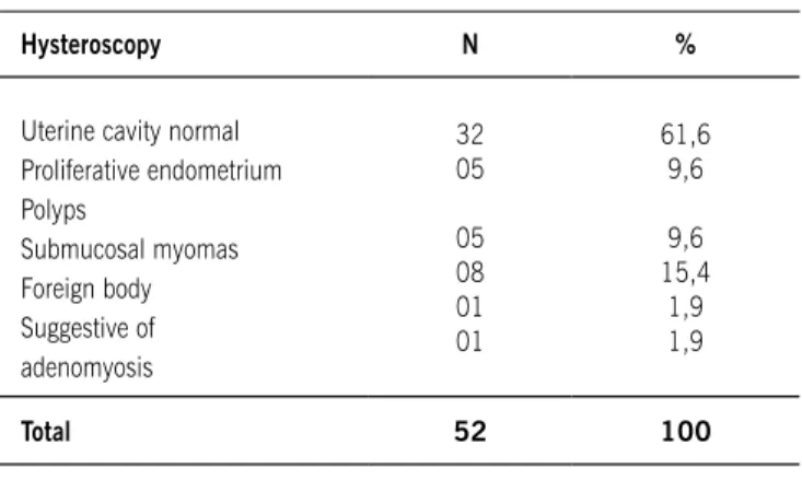
esultsThirty-two of the 52 women recruited (61.5%) had a normal uterine cavity with an endometrium that appeared atrophic on hysteroscopy and biopsy. All of them had an endometrial thickness of less than 5mm on ultrasound, which demonstrates that the transvaginal ultrasound had good specificity (100%). Twenty (38.4%) women had abnormal hysteroscopy findings (Table 1).
Ten of the twenty women with abnormal hysteroscopy findings (50%) had an active endometrium on histological
assessment. Inactive endometrium findings included submu-cosal myomas, foreign bodies and adenomyosis. Fifteen of the women who had abnormal hysteroscopy findings had ultrasound thicknesses of less than 5mm. Just five of these women had an endometrium thickness greater than five milli-meters (Table 2).
Diagnostic hysteroscopy was conducted for all of the women, irrespective of endometrial thickness, resulting in an increase in sensitivity of 15 cases over ultrasound alone (75%). In these fifteen extra cases, hysteroscopy identified polyps and proliferative abnormalities which were then confirmed by anatomopathological tests. No cases of endometrial neoplasm or hyperplasia were detected.
d
IscussIonThis study has shown that just five out of 20 women who had endometrial abnormalities on hysteroscopy and biopsy (25%) also had an endometrial thickness that was greater than 5mm according to ultrasound. Three of the five detected by ultrasound had a proliferative endometrium and two had endometrial polyps. Ultrasound did not detect abnormalities in the other 15 women, demonstrating low diagnostic sensitivity. This is probably because of the clinical conditions of the patients, who were asymptomatic, which could lead the ultrasound operators to underestimate endometrial thickness measurements.
This has also been observed by Goldstein (2009) who
Hysteroscopy N %
Uterine cavity normal Proliferative endometrium Polyps
Submucosal myomas Foreign body Suggestive of adenomyosis
32 05
05 08 01 01
61,6 9,6
9,6 15,4
1,9 1,9
total 52 100
table 1 – Classification of hysteroscopy findings
Hysteroscopy Endometrial thickness (mm)
Proliferative endometrium Polyps
Submucosal myomas Foreign bodies Adenomyosis
< 5mm 2 3 8 1 1
>5mm 3 2
AssessmentofendometriAlmorphologyAndhistologyinpostmenopAusAlwomen
713
Rev Assoc Med Bras 2010; 56(6): 711-4investigated postmenopausal women given transvaginal ultra-sound and found variations in endometrial thickness measu-rements, identifying measurement errors that were dependent on the presence of vaginal bleeding. 15 When menopausal
women with bleeding and with endometrial thickness close to 5 mm were assessed by different observers using transvaginal ultrasound, there were measurement variations of up to 2 mm, which were explained by the fact that endometrial thickness close to 5 mm is considered a cutoff point for the presence or absence of postmenopausal endometrial disorders. This may result in a tendency towards overestimating the endometrial thickness measurement. 16
Indeed, the negative predictive value for postmenopausal ultrasound endometrial thickness measurement varies depen-ding upon whether the women being assessed present with or without genital bleeding. 16
Endometrial thickness measured using transvaginal ultra-sound was also ineffective for diagnosing endometrial polyps in women with bleeding after the menopause.17 Along the same
lines, Dreisler et al. (2009) assessed the uterine cavity using hysteroscopy and observed that 82% of women with endome-trial polyps were asymptomatic. 18
With relation to endometrial carcinoma, a recent meta-anal-ysis suggested reducing the cutoff point for endometrial thick-ness measured by transvaginal ultrasonography to 3mm with the objective of increasing the method’s sensitivity for ruling out neoplasms in postmenopausal women with bleeding. 19
In our study, 15 women who did have abnormal hyste-roscopic findings were not identified by ultrasound screening, taking normal endometrial thickness as less than 5 mm. Notwi-thstanding, just six of these 15 women had histologically active endometrial lesions and no cases of hyperplasia or neoplasm were observed.
The most common abnormal hysteroscopy finding was submucosal myoma (15.4%), which is in contrast to reports in the literature where proliferative findings such as polyps and hyperplasia predominate. 3,20 It is probable that our results have
been affected by the small number of women studied. Diagnostic hysteroscopy of postmenopausal women is often associated with the finding of an atrophied endometrium and this was confirmed by our study, since 66% of the hysteroscopy test results were atrophic endometrium. In such cases, where the endometrium has been defined as inactive, the histological biopsy sample adds very little to diagnosis. Despite this, some authors recommend performing the biopsy even when hyste-roscopy findings are of atrophied endometrium. 4,20 We did not
however detect any discordance between hysteroscopy findings and the histological results from the endometrial samples. All of our endometrial samples, which were obtained by Pipelle suction curette, agreed with the hysteroscopy findings.
Hysteroscopy associated with endometrial histology obtained via guided biopsy is considered the gold standard
for identification of endometrial disorders. Hysteroscopy alone offers sensitivity of 93% for detection of malignant lesions, with 98.4% specificity, 21 and when combined with guided suction
biopsy with a Pipelle curette, the rates of endometrial cancer detection reach 99.6%. 21 However, certain focal lesions may
not provide a suitable sample if the biopsy is guided. In such cases it is necessary to take the endometrial sample while viewing directly via hysteroscope. Preparatory hysteroscopy is recommended for uterine curettage in order to increase diag-nostic efficacy. 3
To date, no satisfactory screening method has been deve-loped for diagnosis of endometrial carcinoma. 3 After the
menopause, when vaginal bleeding is present, transvaginal ultrasound is simple, minimally invasive and well-tolerated for screening patients, 15 and should be used before
hysteros-copy. However, in these cases a need for hysteroscopy and endometrial histological assessment should not be ruled out even for women with normal endometrial thickness. Dreisler et al. (2009) suggest that for women without abnormal vaginal bleeding, endometrial thickness measured via transvaginal ultrasound is enough to exclude focal endometrial disorders, but that when vaginal bleeding is present, the value of 4-5mm that is generally used after the menopause should not be relied on for excluding intrauterine lesions. 22
Where women are asymptomatic but endometrial thickening has been diagnosed by ultrasound, hysteroscopy and histology are indispensable for arriving at the correct diagnosis. Hyste-roscopy is therefore the correct test to use in any circumstance in which observation of the uterine cavity may provide a basis for a precise diagnosis and the most appropriate treatment, particularly for postmenopausal assessments. 20,23 Combining
these methods not only allows early diagnosis of endometrial lesions, but also reduces unnecessary curettage.
c
onclusIonsAmbulatory diagnostic hysteroscopy combined with suction biopsy (Pipelle) offers high efficacy and low rates of compli-cations for diagnosis of endometrial disorders. In this study we observed abnormal hysteroscopy findings in asymptomatic women that were not detected by transvaginal ultrasound. Hysteroscopy should therefore be indicated for postmenopausal women with endometrial thickening on ultrasound and for postmenopausal women with bleeding, irrespective of endo-metrial thickness.
Conflict of interest: none
r
eferences1. American Cancer Society, Surveillance Research; 2009. [cited 2010 feb 8]> Available from: http://cancer.org/downloads/STT/2008CAFfinalsecured.pdf. 2. Surita R, Barbosa CP, Hernendes PS, Napolitano AC, Martins SV. Achados
wolff lpg etAl.
714
Rev Assoc Med Bras 2010; 56(6): 711-43. Schmidt T, Breidenbach M, Nawroth F, Mallmann P, Beyer IM, Fleisch C, et al. Hysteroscopy for asymptomatic postmenopausal women with sonographically thickened endometrium. Maturitas. 2009;62:176-8.
4. Loffer ED. Hysteroscopy with selective endometrial sampling compared with D&C for abnormal uterine bleeding: the value of a negative hysteroscopic view. Obstet Gynecol. 1989;73:16-20.
5. Symonds I. Ultrasound, hysteroscopy and endometrial biopsy in the inves-tigation of endometrial cancer. Best Pract Res Clin Obstet Gynaecol. 2001;15:381-91.
6. Goldrath MH, Sherman AI. Office hysteroscopy and suction curettage: can we eliminate the hospital diagnostic dilatation and curettage ? Am J Obstet Gynecol. 1985;152:220-9.
7. Warming L, Ravn P, Skouby S, Christiansen C. Measurement precision and normal range of endometrial thickness in a postmenopausal population by transvaginal ultrasound. Ultrasound Obstet Gynecol 2002; 20 (5): 492-95. 8. Bourne T, Hamberger L, Hahlin M, Granber S. Ultrasound in gynecology:
endometrium. Int J Gynecol Obstet. 1997;56:115-27.
9. Epstein E, Valentin L. Intraobserver and interobserver reproducibility of ultra-sound measurements of endometrial thickness in postmenopausal women. Ultrasound Obstet Gynecol. 2002;20:486-91.
10. Yela DA, Ravacci SH, Monteiro IM, Pereira KC, Gabiatti JR. Comparative study of transvaginal sonography and outpatient hysteroscopy for detection of pathologic endometrial lesions in postmenopausal women. Rev Assoc Med Bras. 2009;55:553-6.
11. Giusa-Chiferi MG, Gonçalves WJ, Baracat EC, Albuquerque Neto LC, Bortoletto CCR, Lima GR. Transvaginal ultrasound, uterine biopsy and hysteroscopy for postmenopausal bleeding. Int J Obstet Gynecol. 1996;55:39-44
12. World Medical Association. Declaration of. Helsinki: Recommendations Guiding Physicians in Biomedical Research Involving Human Subjects. Somerset West. Republic of South África, 2000.
13. Wolff LPG, Martins MR, Bedone AJ, Monteiro IMU. Avaliação do endométrio em mulheres menopausadas após seis meses de isoflavona. Rev Assoc Med Bras. 2006;52:419-23
14. Moura AP, Pastore AR, Cerri GG. Endométrio. In: Pastore AR, Cerri GG. Ultra-sonografia obstetrícia ginecologia. São Paulo: Sarvier; 1997. p.555-71.
Artigo recebido: 24/08/10 Aceito para publicação: 14/09/10
15. Goldstein SR. The role of transvaginal ultrasound or endometrial biopsy in the evaluation of the menopausal endometrium. Am J Obstet Gynecol. 2009;201:5-11.
16. Epstein E, Valentin L. Intraobserver and interobserver reproducibility of ultra-sound measurements of endometrial thickness in postmenopausal women. Ultrasound Obstet Gynecol. 2002;20:486-91.
17. Timmermans A, Gerritse MB, Opmeer BC, Jansen FW, Mol BW, Veersema S. Diagnostic accuracy of endometrial thickness to exclude polyps in women with postmenopausal bleeding. J Clin Ultrasound. 2008;36:286-90. 18. Dreisler E, Stampe Sorensen S, Ibsen PH, Lose G. Prevalence of endometrial
polyps and abnormal uterine bleeding in a Danish population aged 20-74 years. Ultrasound Obstet Gynecol. 2009;33:102-8.
19. Timmermans A, Opmeer BC, Khan KS, Bachmann LM, Epstein E, Clark TJ, et al. Endometrial thickness measurement for detecting endometrial cancer in women with postmenopausal bleeding: a systematic review and meta-analysis. Obstet Gynecol. 2010;116:160-7.
20. Metello J, Relva A, Milheras E, Colaço J, Retto H. Eficácia diagnóstica da histe-roscopia nas metrorragias pós-menopausa. Acta Med Port. 2008;21:483-8. 21. Elliott J, Connor J, Lashen H. The value of outpatient hysteroscopy in diagnosing
endometrial pathology in postmenopausal women with and without hormone replacement therapy. Acta Obstet Gynecol Scand. 2003;82:1112-9. 22. Dreisler E, Stampe Sorensen S, Ibsen PH, Lose G. Value of endometrial thickness
measurement for diagnosing focal intrauterine pathology in women without abnormal uterine bleeding. Ultrasound Obstet Gynecol. 2009;33:344-8. 23. Machado SB, Pina H, Esteve M, Neves NA, Schidler SS, Borges AS. Indicações,