Major Article
INTRODUCTION
Address to: Dr. Selwyn Arlington Headley. Depto de Medicina Veterinária
Preventiva/UEL. Rodovia Celso Garcia Cid, PR 445, km 380, Campus Universitário, Caixa Postal 10.001, 86057-970 Londrina, PR, Brasil. Phone/Fax: 55 43 3371-4485
e-mail: [email protected] Received 18 April 2014 Accepted 30 June 2014
Serological evidence for Saint Louis encephalitis virus
in free-ranging New World monkeys and horses within
the upper Paraná River basin region, Southern Brazil
Walfrido Kühl Svoboda
[1],
Lívia Carício Martins
[2],
Luciano de Souza Malanski
[3],
Marcos Massaaki Shiozawa
[4],
Kledir Anderson Hofstaetter Spohr
[5],
Carmen Lúcia Scortecci Hilst
[6],
Lucas M. Aguiar
[7],
Gabriela Ludwig
[8],
Fernando de Camargo Passos
[9],
Lineu Roberto da Silva
[10],
Selwyn Arlington Headley
[11]and Italmar Teodorico Navarro
[11][1]. Universidade Federal da Integração Latino-Americana (UNILA), Instituto Latino-Americano de Ciências da Vida e da Natureza (ILACVN), Foz do Iguaçu, PR, Brasil [2]. Departamento de Arbovirologia e Febres Hemorrágicas, Instituto Evandro Chagas, Ananindeua, PA. [3]. Instituto Chico Mendes de Conservação da Biodiversidade, Porto Velho, RO. [4]. Escola de Medicina Veterinária, Universidade Norte do Paraná, Arapongas, PR. [5]. Escola de Medicina Veterinária, Universidade de Cuiabá, Cuiabá, MT. [6]. Departamento de Clínica Veterinária, Universidade Estadual de Londrina, Londrina, PR. [7].Universidade Federal da Integração Latino-Americana, Foz do Iguaçu, PR. [8].Centro Nacional de Pesquisa e Conservação de Primatas Brasileiros, Instituto Chico Mendes de Conservação da Biodiversidade, João Pessoa, PB. [9]. Departamento de Zoologia, Universidade Federal do Paraná, Curitiba, PR. [10]. Secretaria da Saúde do Paraná, Curitiba, PR. [11]. Departamento de Medicina Veterinária Preventiva, Universidade Estadual de Londrina, Londrina, PR.
ABSTRACT
Introduction: Saint Louis encephalitis virus (SLEV) primarily occurs in the Americas and produces disease predominantly in humans. This study investigated the serological presence of SLEV in nonhuman primates and horses from southern Brazil.
Methods: From June 2004 to December 2005, sera from 133 monkeys (Alouatta caraya, n=43; Sapajus nigritus, n=64; Sapajus cay, n=26) trap-captured at the Paraná River basin region and 23 blood samples from farm horses were obtained and used for the serological detection of a panel of 19 arboviruses. All samples were analyzed in a hemagglutination inhibition (HI)
assay; positive monkey samples were confi rmed in a mouse neutralization test (MNT). Additionally, all blood samples were
inoculated into C6/36 cell culture for viral isolation. Results: Positive seroreactivity was only observed for SLEV. A prevalence of SLEV antibodies in sera was detected in Alouatta caraya (11.6%; 5/43), Sapajus nigritus (12.5%; 8/64), and S. cay (30.8%; 8/26) monkeys with the HI assay. Of the monkeys, 2.3% (1/42) of A. caraya, 6.3% 94/64) of S. nigritus, and 15.4% (4/26) of
S. cay were positive for SLEV in the MNT. Additionally, SLEV antibodies were detected by HI in 39.1% (9/23) of the horses evaluated in this study. Arboviruses were not isolated from any blood sample. Conclusions: These results confi rmed the presence of SLEV in nonhuman primates and horses from southern Brazil. These fi ndings most likely represent the fi rst detection of this
virus in nonhuman primates beyond the Amazon region. The detection of SLEV in animals within a geographical region distant from the Amazon basin suggests that there may be widespread and undiagnosed dissemination of this disease in Brazil.
Keywords: Saint Louis encephalitis. Serology. New World monkeys. Horses. Arboviruses.
Saint Louis encephalitis virus (SLEV) belongs to the genus
Flavivirus, family Flaviviridae, which consists of approximately 70 virus species and subspecies distributed worldwide1. Most
fl aviviruses are transmitted between susceptible vertebrates
by hematophagous arthropods, in particular mosquitos1,2.
Flaviviruses are the most import causes of infectious diseases
in humans from Brazil3; these viruses include Bussuquara,
Cacipacore, dengue (serotypes 1, 2, 3, and 4), Iguape, Ilhéus, Rocio, Saint Louis encephalitis, and yellow fever3,4.
SLEV may have initially originated in Central America5, but
it has now disseminated throughout the Americas, with reports of its presence in the USA6,7, Canada7, Argentina8,9, Uruguay10,
and Trinidad11. In Brazil, SLEV was likely fi rst isolated in the
1960s from a pool of Sabethes belisarioi mosquitoes captured on the Belém-Brasília Highway12. Since then, this virus has
been identifi ed predominantly in humans13-15 and horses16-18
from Brazil.
Serological confi rmation of SLEV in wildlife is very rare;
it has been described in the white-tailed deer in the USA19,
while seropositivity has been demonstrated in wild and sentinel animals and arthropods from both the Amazon region20,21 and the
State of São Paulo22, Brazil. Moreover, at least three epizootic
METHODS
including sentinel Cebus monkeys in the Brazilian Amazon
region21. Additionally, a serological survey conducted in French
Guiana detected low levels of SLEV antibodies in free-ranging primates23.
This study presents the fi ndings of a serological investigation
of SLEV in free-ranging New World monkeys and farm horses from southern Brazil.
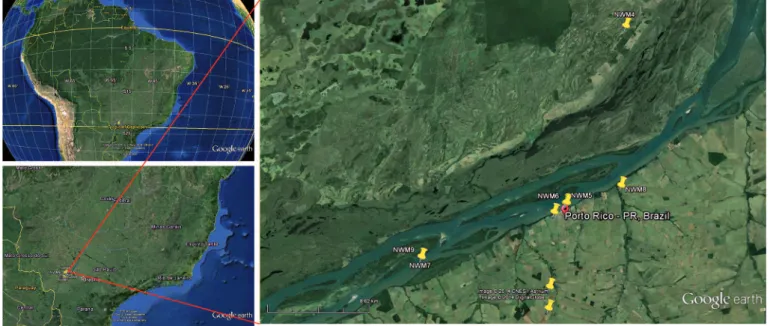
FIGURE 1 - Google earth images illustrating the region where the research was conducted. The yellow pins indicate the coordinates where monkeys seropositive for Saint Louis encephalitis virus were trap-captured.
Study location
All monkeys used in this study were trap-captured within the Porto Rico County region, located between the northwestern region of the State of Paraná and the southeast region of the State of Mato Grosso do Sul, on the upper Paraná River in Brazil
(Figure 1). This region consists of islands and sub-tropical forest reserves (where the animals were captured) that are protected by the Instituto Brasileiro do Meio Ambiente e dos Recursos Naturais Renováveis (IBAMA). The riparian forests of the islands of Mutum, Porto Rico, Gaivota, and Japonesa and the forests on the opposite shore of the Paraná River were included in this study. Porto Rico County is located in the northwestern region of the State of Paraná (22°46’20”S latitude and 53°16’01”W-GR longitude). This sub-tropical region has an average annual rainfall of 1,200-1,300mm and temperatures ranging from 16 to 29°C.
Nonhuman primates
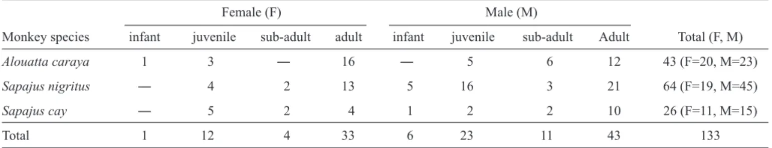
One hundred thirty-three nonhuman primates [43 Alouatta caraya (black and golden howler monkey), 64 Sapajus nigritus,
and 26 S. cay (both called capuchin monkeys)] were
trap-captured as previously described24. The biological data for the
captured nonhuman primates are given in Table 1; all primates were captured between June 2004 and December 2005 by a team of biologists and veterinarians with permission from IBAMA (license number 104/04). All primates were anesthetized25,
after which blood samples were obtained by jugular or brachial venipuncture, and sera were centrifuged (±1,000g) and stored at -196°C until used. All trapped animals were released after complete recovery at the capture location.
All captured primates were strict forest inhabitants. However, a number of S. nigritus specimens (n=13) were trapped in a fo rest reserve (within the Paraná River basin) close to a farm whose owner reportedly had frequent contact with monkeys.
H orses
Blood samples were obtained by jugular venipuncture from 23 mixed-breed adult horses (16 males and 7 females) located in proximity to where the S. cay monkeys were captured. All sampled horses were located on the same property and were representative of the total horse population maintained at this holding. Serum samples were obtained and stored at -196°C until they were used.
Serological assays
All serological assays were performed at the Evandro Chagas Institute (IEC-PA), Department of Arbovirology and Hemorrhagic Fevers, Belém, PA, Brazil. All samples were stored on dry ice and then air-shipped to Belém.
Hemagglutination inhibition test
All samples were initially subjected to a microplate hemagglutination inhibition (HI) test26 against a panel of
standardized antigens for 19 arboviruses, including four from
the genus Alphavirus (eastern equine encephalomyelitis,
RESULTS
TABLE 1 - The species, sex, and age distributions of free-ranging New World monkeys captured within the Porto Rico County region, Southern Brazil.
Female (F) Male (M)
Monkey species infant juvenile sub-adult adult infant juvenile sub-adult Adult Total (F, M)
Alouatta caraya 1 3 ― 16 ― 5 6 12 43 (F=20, M=23)
Sapajus nigritus ― 4 2 13 5 16 3 21 64 (F=19, M=45)
Sapajus cay ― 5 2 4 1 2 2 10 26 (F=11, M=15)
Total 1 12 4 33 6 23 11 43 133
TABLE 2 - Results of hemagglutination inhibition assays and mouse neutralization tests for free-ranging New World monkeys captured within the Porto Rico County region, Southern Brazil.
Alouatta caraya Sapajus nigritus Sapajus cay Total
HI result*(antibody titer) male female male female male female (male/female)
Negative 21 17 40 16 10 8 112 (71/41)
20 ― 2 2 1 2 2 9 (4/5)
40 2 1(2.7) 1 ― 2(1.8/2.7) ― 6 (5/1)
80 ― ― 1(1.9) 1(2.1) ― 1(2.7) 3 (1/2)
160 ― ― ― 1(2.8) ― ― 1 (―/1)
320 ― ― ― ― ― ― ―
640 ― ― 1(3.5) ― 1(3.1) ― 2 (2/―)
Total 23 20 45 19 15 11 133 (83/50)
HI: hemagglutination inhibition. *HI test results: positive HI ≥ 20; (n)neutralization logarithmic index (NLI): positive NLI ≥ 1.8.
Serological assay and virus isolation from nonhuman primates
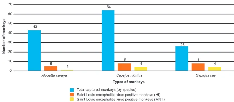
According to the HI assay, SLEV antibodies were present in
primates, specifi cally in 11.6% (5/43) of A. caraya, 12.5% (8/64) of S. nigritus, and 30.8% (8/26) of S. cay animals (Table 2).
However, confi rmation of SLEV infection by MNT was obtained
for only 2.3% (1/43) of A. caraya, 6.3% (4/64) of S. nigritus
and 15.4% (4/26) of S. cay primates (Table 2). Arboviruses were not isolated from any of the 133 primate-derived six Flavivirus (yellow fever, Saint Louis encephalitis, Rocio,
Ilhéus, Cacipacoré and Bussuquara), eight Orthobunyavirus
(Oropouche, Caraparu, Catú, Guaroa, Maguari, Tacaiuma,
Utinga and Belém) and one from the genus Phlebovirus
(Icoaraci). Animals were considered seropositive when a titer
of ≥ 20 was observed with the HI assay. Mouse neutralization test
All positive monkey sera with HI titers >20 were subjected
to a mouse neutralization test (MNT), a confi rmatory assay that
was used to characterize the virus associated with the infection and performed according to a previously described protocol26.
The results were calculated using the neutralization logarithmic index (NLI)27. Sera from horses were not evaluated by MNT.
Virus isolation
All blood samples were inoculated into Aedes albopictus
cell culture (clone C6/36); immunofl uorescence was used for viral identifi cation28.
Statistical analysis
Statistical signifi cance was analyzed using the Chi-square
test (Yates corrected) to establish differences between the
characteristics evaluated (species, gender, age, and the presence of horses). Associations between variables and positivity were
determined by odds ratios (ORs) with 95% confi dence levels. T he results were considered statistically signifi cant when the ρ-value was 5%.
Ethical considerations
This study was approved by the Animal Experimental
Ethics Committee, Universidade Estadual de Londrina
blood samples, although SLEV antibodies were detected by HI in 15.8% (21/133) of serum samples, with HI titers to SLEV ranging from 20 to 640 (Table 2 and Figure 2). Negative seroreactivity was observed for all other arboviruses.
The association of the results obtained based on NMT analysis of characteristics evaluated (species, sex, age, and the presence of horses within the same habitat) with the neutralization logarithm index (NLI) is summarized in Table 3. The prevalence of anti-SLEV antibodies was elevated in S. cay
(15.4%; 4/26) relative to S. nigritus (6.3%; 4/64) and A. caraya
(2.3%; 1/43) primates. Interestingly, negative seroreactivity of HI to SLEV antibodies was observed in infant and juvenile
primates, while SLEV antibody serum prevalence was identifi ed
in sub-adult (13.3%; 2/15) and adult (9.2%; 7/76) animals, most likely due to their more extensive contact with this pathogen. Additionally, primates that live within proximity to farm horses demonstrated elevated seroreactivity to SLEV antibodies (15.4%; 4/26) compared to those living without potential contact
with horses (4.8%; 5/107). However, no signifi cant differences were observed based on species (ρ=0.1090), sex (ρ=0.7281), age (ρ=0.1876), and the presence of horses within the same habitat (ρ=0.0727).
Serological assay and virus isolation from horses
The overall seroprevalence of SLEV antibodies in horses was 39.1% (9/23) and was elevated in females (57.1%; 4/7) relative to male horses (31.3%; 5/16). SLEV antibody titers in seropositive horses were as follows: 160 (three males; two females), 320 (one male and female) and 640 (one male and
female). However, signifi cant differences were not observed based on sex [ρ=0.3630; OR = 0.34 (0.04 < OR < 2.91)], and these results were not confi rmed with the MNT assay.
Additionally, arboviruses were not isolated from any of the
FIGURE 2 - Relationships between the total numbers of captured monkeys (by species) and positive results in the hemagglutination inhibition (HI) and mouse neutralization test (MNT) assays.
23 horse-derived blood samples, but as previously observed
in primates, specifi c seropositivity only to SLEV antibodies
was observed with the HI assay.
DISCUSSION
The results of this study reveal that we have identifi ed the
presence of SLEV in several species of nonhuman primates and horses from southern Brazil. The classical HI and MNT methodologies used during this investigation are based on
the conventional serologic diagnosis of fl avivirus due to the presence of virus-specifi c antibodies in the serum. Similar
serological strategies have been used to detect SLEV in primates23, white-tailed deer19,29, livestock11, mules19, and
horses8,10,11,17,18. The importance of these results lies primarily in
the observation of SLEV circulation in a completely new habitat within Brazil, considering that most cases of SLEV have been described within the Amazon region4,20-22. However, SLEV was
recently isolated from a horse in the State of Minas Gerais16,
and SLEV has been serologically identifi ed in both horses from
Corumba, Central West Brazil17 and in patients from the State
of São Paulo14. These results suggest a southern drift of SLEV
from the Amazon region to other parts of continental Brazil, most likely attributable to migratory birds16,21,22.
In the USA, SLEV causes encephalitis in approximately 100 human cases annually30 and is one of the most common causes
of arbovirus-induced disease31, with sporadic epidemics6,30,31.
However, this arboviral disease has different epidemiological features in Brazil, largely in terms of the relatively few cases
of SLEV-induced encephalitis identified in humans20,22.
Additionally, most fl aviviruses that occur in Brazil, with the
exception of dengue virus, are predominantly maintained
43
64
26
5
8 8
1 4 4
0 10 20 30 40 50 60 70
Alouatta caraya Sapajus nigritus Sapajus cay
Number of monkeys
Types of monkeys
Total captured monkeys (by species)
TABLE 3 - Associations between the characteristics studied (species, sex, age, and the presence of horses within the same habitat) and the presence of anti-Saint Louis encephalitis virus antibodies (neutralization logarithm index) in serum samples of free-ranging New World Monkeys.
Neutralization logarithmic index test
Variables positive negative total
Species n % n % n % ORa ρ-values
Alouatta caraya 1 2.32 42 97.68 43 32.33 Ac x Sn 0.63
0.36 (0.01-3.6)
Sapajus nigritus 4 6.25 60 93.75 64 48.12 Ac x Sc 0.12
0.13 (0.01-1.38)
Sapajus cay 4 15.38 22 84.61 26 19.55 Sn x Sc 0.38
0.37 (0.07-1.95)
Sex
male 5 6.02 78 93.98 83 62.41 0.74 0.72c
female 4 8.00 46 92.00 50 37.59 0.16 < OR < 3.48
Age
infant ― ― 7 100.00 7 5.27
juvenile ― ― 35 100.00 35 26.31 NC 0.18b
sub-adult 2 13.33 13 86.67 15 11.28
adult 7 9.21 69 90.79 76 57.14
Presence of horses in the same habitat
yes 4 15.38 22 84.62 26 19.55 3.71 0.07c
no 5 4.67 102 95.33 107 80.45 0.76 < OR < 17.79
Total (%) 9 6.77 124 93.23 133 100.00
Ac: Alouatta caraya; Sn: Sapajus nigritus; Sc: Sapajus cay; NC: not calculated; ORa:odds ratio (inferior and superior limits); bchi-square;
cFisher’s exact test.
as sylvatic zoonotic diseases that occasionally produce infections in humans and domestic animals that have entered
the ecosystems where these viruses occur4. Consequently,
it can be speculated that this epidemiological difference
might possibly be attributable to specifi c environmental and
biological conditions within Brazil that alter the virulence or pathogenicity of SLEV in humans. Although the factors that are actively or otherwise associated with this phenomenon have not been fully elucidated, at least two theories should be considered. First, the elevated endemicity of other closely
related fl aviviruses that elicit cross-protection in humans, such
as that occurring with dengue and yellow fever immunization, might easily result in the underdiagnosis of SLEV; this occurred in a study of 519 patients who were initially diagnosed as having
dengue fever, but later molecular investigation confi rmed the
presence of SLEV in eight of them14. Secondly, the diffi culty
in effi ciently recognizing and/or diagnosing SLEV encephalitis
in patients at both public and private health services might also contribute to the reduced number of cases in Brazil, considering that patients may either be asymptomatic4,16 or present with
fl u-like disease syndromes that can progress to acute or subacute
meningeal and focal neurological manifestations32. Moreover,
three patients from an outbreak of SLEV in northwestern São Paulo demonstrated hemorrhagic manifestations typical of dengue fever virus33. Taken together, the confi rmation of
SLEV in several states in Brazil13,14,16 in addition to the Amazon
region may suggest dissemination due to migratory birds16,22,
as previously postulated. Furthermore, Culex declarator and
Culex coronator are known vectors, while monkeys, sloths, armadillos, and marsupials are reservoirs of this virus in Brazil4.
Therefore, the seropositivity of nonhuman primates and horses to SLEV demonstrated in this study in a region geographically distant from the Amazon region is of great concern, and additional investigation must be conducted to understand the dynamics associated with this virus in a new environment but in conventional hosts.
During this investigation, the majority (57.1%; 12/21) of seropositive monkeys demonstrated titers of SLEV according
to HI that were ≥ 40, suggesting that these antibodies are
from contact with similar related viruses. Alternatively, in a seroepidemiological survey conducted in French Guiana,
primates had low antibody titers to SLEV (HI <40) but elevated
titers to yellow fever (HI >320), and the authors suggested that
that SLEV antibodies identifi ed could have been due to
cross-reactions with yellow fever virus23. Low titers of SLEV were
also identifi ed in horses, livestock, and wildlife from the island
of Trinidad11. However, during this investigation, seropositivity
was only identifi ed for SLEV, and negative results were obtained
with all other viruses by HI, indicating that there was no cross-reaction with any similarly related virus.
In Argentina, SLEV studies have focused primarily on populations of mosquitoes34,35, humans36, and horses8, but the
importance of monkeys in the sylvatic SLEV cycle has not been investigated. However, during this study, serological
results confi rmed the participation of nonhuman primates in the
maintenance cycle of SLEV in southern Brazil. Consequently,
these results most likely represent the fi rst identifi cation of SLEV
in nonhuman primates beyond the Amazon region of Brazil. Horses are commonly found in the region where sample collection was performed, largely on farms that are close to forested areas, and the relationships contributing to the presence of SLEV antibodies in these animals should be considered. Elevated prevalence rates of SLEV antibodies in horses from the Amazon18 and Pantanal17,18 regions have been described,
but this report is the fi rst description of SLEV in southern
Brazil within an ecosystem that is very distant and quite different from the Amazon and Pantanal regions. Therefore, these results suggest that horses may participate as vertebrate hosts in the dissemination of SLEV and most likely should be considered amplifying sources of SLEV for primates, or vice versa. However, additional investigations must be performed
to confi rm this theory.
This report is the fi rst describing the seroprevalence of specifi c SLEV antibodies in free-ranging monkeys within the
State of Paraná, southern Brazil, and implicates nonhuman primates in the natural maintenance cycle of SLEV in the Southern Cone region, where an SLEV encephalitis outbreak
was identifi ed in northern Argentina35. Future research will aim
to identify local mosquito and human populations to establish their roles in the SLEV life cycle.
ACKNOWLEDGMENTS
We are grateful to Mr. Basílio S. Buna, Geraldo M. da Silva, and Luiz R.O. Costa of the Evandro Chagas Institute (IEC-PA), Department of Arbovirology and Hemorrhagic Fevers, for performing all serological assays. Our gratitude is also extended to the Entomology Team of SESA-PR (Porto Rico-PR) for technical support during sample collection and to the Instituto Brasileiro do Meio Ambiente e dos Recursos Naturais Renováveis (IBAMA) for permission to capture primates for this experiment. We are also grateful to Dr. João Luis Garcia,
Universidade Estadual de Londrina, for assistance with the statistical analysis.
The authors declare that there is no confl ict of interest. CONFLICT OF INTEREST
FINANCIAL SUPPORT
Secretaria Estadual de Saúde do Paraná (SESA-PR),
Secretaria de Estado da Ciência, Tecnologia e Ensino Superior (SETI-PR), Coordenação de Aperfeiçoamento de Pessoal de Nível Superior (CAPES), and Conselho Nacional de Desenvolvimento Científico e Tecnológico (CNPq).
Gabriela Ludwig has a temporary scholarship from Centro
Nacional de Pesquisa e Conservação de Primatas Brasileiros, Instituto Chico Mendes de Conservação da Biodiversidade, Projeto Nacional de Ações Integradas Público-Privadas para Biodiversidade – Fundação de Empreendimentos Pesquisa e Desenvolvimento.
REFERENCES
1. International Committee on Taxonomy of Viruses (ICTV). Virus Taxonomy, 8th Report of the ICTV. London: Elsevier/Academic Press; 2005.
2. Turell MJ, O'Guinn M, Oliver J. Potential for New York mosquitoes to transmit West Nile virus. Am J Trop Med Hyg 2000; 62:413-414. 3. Batista WC, Kashima S, Marques AC, Figueiredo LTM. Phylogenetic
analysis of Brazilian Flavivirus using nucleotide sequences of parts of NS5 gene and 3′ non-coding regions. Virus Res 2001; 75:35-42. 4. Figueiredo LTM. The Brazilian fl aviviruses. Microbes Infect 2000;
2:1643-1649.
5. Kopp A, Gillespie TR, Hobelsberger D, Estrada A, Harper JM, Miller RA, et al. Provenance and geographic spread of St. Louis encephalitis virus. mBio 2013; 4:e00322-13.
6. Monath TP, Tsai TF. St. Louis Encephalitis: lessons from the last ecade. Am J Trop Med Hyg 1987; 37:S40-S59.
7. Calisher CH. Medically important arboviruses of the United States and Canada. Clin Microbiol Rev 1994; 7:89-116.
8. Tauro L, Marino B, Diaz LA, Lucca E, Gallozo D, Spinsanti L, et al. Serological detection of St. Louis encephalitis virus and West Nile virus in equines from Santa Fe, Argentina. Mem Inst Oswaldo Cruz 2012; 107:553-556.
9. Valinotto LE, Barrero PR, Viegas M, Alvarez Lopez MC, Mistchenko AS. Molecular evidence of St. Louis encephalitis virus infection in patients in Buenos Aires, Argentina. J Clin Virol 2012; 54:349-351.
10. Burgueno A, Spinsanti L, Diaz LA, Rivarola ME, Arbiza J, Contigiani M, et al. Seroprevalence of St. Louis encephalitis virus and West Nile virus (fl avivirus, fl aviviridae) in horses, Uruguay. BioMed Res Int 2013; 2013. doi: 10.1155/2013/582957.
11. Thompson NN, Auguste AJ, Coombs D, Blitvich BJ, Carrington CV, Rosa AP, et al. Serological evidence of fl aviviruses and alphaviruses in livestock and wildlife in Trinidad. Vector Borne Zoonotic Dis 2012; 12:969-978.
12. Theiler M, Downs WG. The arthropod-borne viruses of vertebrates. An account of the Rockefeller Foundation virus program, 1951-1970. 1st ed. New Haven: Yale University Press; 1973.
13. Rocco IM, Santos CL, Bisordi I, Petrella SM, Pereira LE, Souza RP, et al. St. Louis encephalitis virus: fi rst isolation from a human in Sao Paulo State, Brazil. Rev Inst Med Trop Sao Paulo 2005; 47:281-285. 14. Terzian AC, Mondini A, Bronzoni RV, Drumond BP, Ferro BP, Cabrera EM,
cases during a dengue 3 outbreak. Vector Borne Zoonotic Dis 2011; 11:291-300.
15. Mondini A, Bronzoni RV, Cardeal IL, Santos TM, Lazaro E, Nunes SH, et al. Simultaneous infection by DENV-3 and SLEV in Brazil. J Clin Virol 2007; 40:84-86.
16. Rosa R, Costa EA, Marques RE, Oliveira TS, Furtini R, Bomfi m MRQ, et al. Isolation of Saint Louis Encephalitis Virus from a horse with neurological disease in Brazil. PLoS Negl Trop Dis 2013; 7:e2537.
17. Pauvolid-Correa A, Tavares FN, Costa EV, Burlandy FM, Murta M, Pellegrin AO, et al. Serologic evidence of the recent circulation of Saint Louis encephalitis virus and high prevalence of equine encephalitis viruses in horses in the Nhecolandia sub-region in South Pantanal, Central-West Brazil. Mem Inst Oswaldo Cruz 2010; 105:829-833.
18. Rodrigues SG, Oliva OP, Araujo FAA, Martins LC, Chiang JO, Henriques DF, et al. Epidemiology of Saint Louis encephalitis virus in the Brazilian Amazon region and in the State of Mato Grosso do Sul, Brazil: elevated prevalence of antibodies in horses. Rev Pan-Amazonica Saude 2010; 1:81-86.
19. Hoff GL, Issel CJ, Trainer DO, Richards SH. Arbovirus serology in North Dakota mule and white-tailed deer. J Wildl Dis 1973; 9:291-295.
20. Vasconcelos PFC, Travassos da Rosa JFS, Travassos da Rosa APA, Dégallier N, Pinheiro FP, Sá Filho GC. Epidemiology of encephalitis by arboviruses in the Amazon region of Brazil. Rev Inst Med Trop Sao Paulo 1991; 33:465-476.
21. Vasconcelos PFC, Travassos da Rosa APA, Dégallier N, Travassos da Rosa JFS, Pinheiro FP. Clinical and ecoepidemiological situation of human arboviruses in Brazilian Amazonia. Cienc Cult (São Paulo) 1992; 44:117-124.
22. Vasconcelos P, Travassos da Rosa A, Pinheiro F, Shope R, Travassos da Rosa J, Rodrigues S. Arboviruses pathogenic from man in Brazil.
In: Travassos da Rosa APA, Vasconcelos PFC, Travassos da Rosa JFS, editors. An overview of arbovirology in Brazil and neighboring countries Belém, PA, Brazil: Instituto Evandro Chagas; 1998. p. 72-99.
23. Thoisy B, Vogel I, Reynes JM, Pouliquen JF, Carme B, Kazanji M, et al. Health evaluation of translocated free-ranging primates in French Guiana. Am J Primatol 2001; 54:1-16.
24. Aguiar LM, Ludwig G, Svoboda WK, Teixeira GM, Hilst CL, Shiozawa MM, et al. Use of traps to capture Black and Gold Howlers (Alouatta caraya) on the Islands of the upper Parana River, southern Brazil. Am J Primatol 2007; 69:241-247.
25. Hilst CLS, Spour KAH, Svoboda WK, Shiozawa MM, Malanski LdS, Aguiar LdM, et al. Estudo e adaptação de protocolo de sedação à base de tiletamina/zolazepan em primatas do gênero Cebu. In: XXVII Congresso Brasileiro da Anclivepa, p. 92. Vitória, Espirito Santo, Brasil; 2006.
26. Shope R, Sather G. Arboviruses. In: Lennette EH, Schmidt NJ, editors. Diagnostic procedures for viral, rickettsial and chlamidial infections. 5th ed. Washington, D.C.: American Public Health Association; 1979. p. 767-814.
27. Reed LJ, Muench H. A simple method of estimating fi fty per cent endpoints. Am J Epidemiol 1938; 27:493-497.
28. Beaty BJ, Calisher CH, Shope RE. Arboviruses. In: Schimidt NJ, Emmons EW, editors. Diagnostic procedures for viral, rickettsial and clamydial infeccions. 6th ed. Washington, DC: American Public Health Association; 1989. p. 797-855.
29. Farajollahi A, Gates R, Crans W, Komar N. Serologic evidence of West Nile virus and St. Louis encephalitis virus infections in white-tailed deer (Odocoileus virginianus) from New Jersey, 2001. Vector Borne Zoonotic Dis 2004; 4:379-383.
30. Center for Disease Control and Prevention (CDC). Saint Louis encephalitis. Techincal data. Atlanta, GA: CDC; 2014.
31. Reimann CA, Hayes EB, DiGuiseppi C, Hoffman R, Lehman JA, Lindsey NP, et al. Epidemiology of neuroinvasive arboviral disease in the United States, 1999-2007. Am J Trop Med Hyg 2008; 79:974-979.
32. Gould EA, Solomon T. Pathogenic fl aviviruses. Lancet 2008; 371: 500-509.
33. Mondini A, Cardeal IL, Lazaro E, Nunes SH, Moreira CC, Rahal P, et al. Saint Louis encephalitis virus, Brazil. Emerg Infect Dis 2007; 13: 176-178.
34. Diaz LA, Flores FS, Beranek M, Rivarola ME, Almirón WR, Contigiani MS. Transmission of endemic St Louis encephalitis virus strains by local
Culex quinquefasciatus populations in Córdoba, Argentina. Trans R Soc Trop Med Hyg 2013; 107:332-334.
35. Diaz LA, Re V, Almiron WR, Farias A, Vazquez A, Sanchez-Seco MP, et al. Genotype III Saint Louis encephalitis virus outbreak, Argentina, 2005. Emerg Infect Dis 2006; 12:1752-1754.