Major Article
Address to:Dr. Pedro Eduardo Almeida da Silva. Lab. Biologia Molecular/ FURG. Rua General Osório s/n, 96200-190 Rio Grande, RS, Brasil. Phone: 55 53 3233-8895; Fax: 55 53 3233-8863
e-mail: [email protected] Received 22 November 2012 Accepted 07 February 2013
http://dx.doi.org/10.1590/0037-8682-0054-2012
INTRODUCTION
cagE
as a biomarker of the pathogenicity of
Helicobacter pylori
Ivy Bastos Ramis
[1],
Júlia Silveira Vianna
[2],
Lande Vieira da Silva Junior
[2],
Andrea
Von Groll
[2]and Pedro Eduardo Almeida da Silva
[1],[2][1]. Centro de Desenvolvimento Tecnológico, Universidade Federal de Pelotas, Pelotas, RS. [2]. Laboratório de Biologia Molecular, Universidade Federal do Rio Grande, Rio Grande, RS.
ABSTRACT
Introduction: Helicobacter pylori infection is associated with gastro-duodenal diseases. Genes related to pathogenicity have been described for H. pylori and some of them appear to be associated with more severe clinical outcomes of the infection. The present study investigates the role of cagE as a pathogenicity biomarker of H. pylori compare it to cagA, vacA, iceA and babA2
genes and correlate with endoscopic diagnoses. Methods: Were collected biopsy samples of 144 dyspeptic patients at the Hospital of the Federal University of Rio Grande, Rio Grande do Sul, Brazil. After collection, the samples were sent for histological examination, DNA extraction and detection of all putative pathogenicity genes by PCR. Results: Of the 144 patients undergoing endoscopy, 57 (39.6%) presented H. pylori by histological examination and PCR by detection of the ureA gene. Based on the endoscopic diagnoses, 45.6% (26/57) of the patients had erosive gastritis, while 54.4% (31/57) had enanthematous gastritis. The genes cagA, cagE, vacAs1/m1, vacAs1/m2 and iceA1 were related to erosive gastritis, while the genes vacAs2/m2, iceA2 and
babA2 were associated to enanthematous gastritis. We found a statistically signiicant association between the presence of cagE
and the endoscopic diagnosis. However, we detect no statistically signiicant association between the endoscopic diagnosis and the presence of cagA, vacA, iceA and babA2, although a biological association has been suggested. Conclusions: Thus, cagE
could be a risk biomarker for gastric lesions and may contribute to a better evaluation of the H. pylori pathogenic potential and to the prognosis of infection evolution in the gastric mucosa.
Keywords: Helicobacter pylori. Pathogenicity genes. Endoscopic diagnosis.
Helicobacter pylori, a microorganism adapted to colonize the gastric mucosa, is considered to be the main etiological agent of enanthematous gastritis (inlammation of the gastric epithelium with simple change mucosal), erosive gastritis (inlammation with loss of integrity of the epithelial lining, not exceeding the muscular layer of the mucosa) and also a risk factor for peptic ulcer and gastric cancer in humans1,2.Factors
related to the genetic polymorphism of the host, the diversity of bacterial pathogenicity and the environment seem to be related to the broad clinical spectrum related to infection by H. pylori3.
Several putative genes, such as cagA, cagE, vacA, iceA and
babA2, have been identiied and are likely to play an important role in the pathogenicity of the bacterium4-8.
The cag-PAI is composed of approximately 31 genes, which are responsible for coding type IV secretion system components and inject effector molecules in the host cell. The presence of
cag-PAI affects the inlammatory state of the gastric mucosa by polymorphonuclear cell iniltration and increases the production of interleukin-8 (IL-8)1.cagA gene (cytotoxin associated gene A)
is considered to be the cag-PAI marker. cagA positive strains tend to be more pathogenic, produce more severe lesions of the epithelium and increase the expression of interleukin-1β and IL-89,10.
Another member of the cag-PAI, the cagE gene (cytotoxin associated gene E), is also related to an increased production of IL-8 in the gastric epithelial cells11.
The vacA gene encodes the vacuolating cytotoxin that damages the gastric epithelial cells. It comprises two variable parts: the s-region, which encodes the signal peptide with the
s1or s2 allele, and the m-region (middle) with the m1 or m2
allele6,12. The mosaic combination of the s and m region alleles
determines the production of the vacuolating cytotoxin and is associated with the pathogenicity of the bacterium13. In general,
vacAs1/m1 and s1/m2 strains produce high and moderate levels of vacuolating toxin, respectively, whereas the s2/m2
strains produce little or no toxin12.The vacAs1/m1 genotype
is considered to be associated with more severe pathologies14.
The iceA gene (induced by contact with the epithelium) has two main allelic variants, designated iceA1and iceA2. The
METHODS
RESUlTS
The babA gene (blood-group antigen-binding adhesin)
encodes a membrane protein, an adhesion called BabA, which binds to the Lewisb blood group antigen on the gastric epithelial cells8,16.Although three bab alleles have been identiied (babA1,
babA2 and babB), only the babA2 gene product is necessary for the Lewisbbinding activity. Thus, babA2 is responsible for pathogenicity, allowing contact between bacterium and gastric epithelium and facilitating the release of other pathogenicity factors11.
We hypothesized that the clinical outcomes of H. pylori
infection were inluenced by the distribution of the above-mentioned pathogenic factors; therefore, this study aimed at investigating the role of cagE as a pathogenicity biomarker of H. pylori-positive patients, compare it to cagA, vacA, iceA
and babA2 genes and correlate these indings with endoscopic diagnoses.
Patients and clinical samples
In this study were included 144 patients with dyspeptic symptoms submitted to upper gastrointestinal endoscopy between October 2008 and March 2009 in the Integrated Center for Gastroenterology at the Hospital of the Federal University of Rio Grande, Rio Grande do Sul, Brazil. Patients that had recently (within the last 15 days) received antibiotics or non-steroidal anti-inlammatory drugs (NSAIDs) or had been treated for
H. pylori or gastrointestinal bleeding in the last seven days, were excluded. The presence of H. pylori infection in the subjects was determined by histological examination and detection of the ureA gene by polymerase chain reaction (PCR).
Endoscopic diagnosis
The endoscopic diagnosis was established in accordance to the Sydney System classiication17.
Histological examination
The biopsy samples of the gastric antrum and body destined for histology were fixed in formalin and stained with Hematoxylin-Eosin (H&E) and Giemsa. Histological classiication of gastritis was established according to the Sydney System18.
Extraction of DNA
After collection, the biopsy samples of the gastric antrum and body were kept in Brain Heart Infusion Broth (Acumedia®, United States of America) with 20% glycerol and stored at -70 °C for further DNA extraction. DNA was extracted of the biopsy samples using DNAzol® Reagent (Invitrogen™, United
States of America) and 10μg/μL proteinase K (Promega, United States of America). The samples were separated from the broth and re-suspended in 100μL of proteinase K and 500μL of DNAzol® Reagent. The mixture was incubated at 55°C for 3h and, after this period, 500μL of DNAzol® Reagent was added to it again. After centrifugation at 14,000g for 10min, the supernatant was collected and 500μL cold absolute ethanol
was added, followed by centrifugation at 12,000g for 10min, after which the supernatant was discarded. The DNA pellet was washed two times with 800μL of 75% ethanol, air dried and re-suspended in 50μL of 8mM NaOH. The DNA was stored at -20°C until further usage.
Detection of the ureA gene
The detection of the ureA gene was used to conirm the
H. pylori infection in all of the patients19. PCRwas performed
as described by Rota et al20.
Detection of pathogenicity genes by PCR
The presence of the cagA gene was investigated by the ampliication of the constant region near the 3′ end of the cagA. The PCR was performed as proposed by Rota et al.20,21.and
the cagE gene was investigated according to Sozzi et al.22.The
presence of the vacA and iceA alleles in the biopsy samples was investigated using the primers previously described23,24 and
the PCR was conducted as proposed by Benenson et al25.For
detection of the babA2 gene, the primers and the PCR conditions applied, were described by Sheu et al.26.
Statistical analysis
The chi-square test was used for the analysis of categorical data. P-values of less than 0.05 of a two-tailed test were considered statistically signiicant. The analyses were performed using the software Statistica 10.
Ethical considerations
This study was approved by the Research Ethics Committee of the Health Area (FURG process number 23116.003335/2008-43) and carried out in accordance with the ethical stan dards outlined in the Helsinki Declaration. A written informed consent was obtained from all the patients.
From the 144 patients who underwent endoscopy, 57 (39.6%) presented H. pylori under histological examination and PCR, of these 40 were women and 17 were men with an average age of 46.2 years (range, 14-74 years). Based on the endoscopic diagnoses, 45.6% (26/57) of the patients had erosive gastritis, while 54.4% (31/57) had enanthematous gastritis.
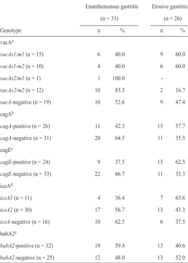
The distribution of the cagA, cagE, vacA, iceA and babA2
genes in relation to the endoscopic diagnoses is described in Table 1. A statistically signiicant association was found between the cagE gene and the diagnosis of erosive gastritis (p=0.029). However, between the cagA, vacA, iceA and babA2
genes and the clinical manifestations, no statistically signiicant association was observed, although a biological signiicance was suggested.
DISCUSSION
TABLE 1 - Distribution of the vacA, cagA, cagE, iceA and babA2 genes in gastric biopsy samples from patients with different gastric disorders
Enanthematous gastritis Erosive gastritis (n = 31) (n = 26)
Genotype n % n %
vacAa
vacAs1/m1 (n = 15) 6 40.0 9 60.0
vacAs1/m2 (n = 10) 4 40.0 6 60.0
vacAs2/m1 (n = 1) 1 100.0
-vacAs2/m2 (n = 12) 10 83.3 2 16.7
vacA-negative (n = 19) 10 52.6 9 47.4 cagAb
cagA-positive (n = 26) 11 42.3 15 57.7
cagA-negative (n = 31) 20 64.5 11 35.5 cagEc
cagE-positive (n = 24) 9 37.5 15 62.5
cagE-negative (n = 33) 22 66.7 11 33.3 iceAd
iceA1 (n = 11) 4 36.4 7 63.6
iceA2 (n = 30) 17 56.7 13 43.3
iceA-negative (n = 16) 10 62.5 6 37.5 babA2e
babA2-positive (n = 32) 19 59.4 13 40.6
babA2-negative (n = 25) 12 48.0 13 52.0
ap-value of the Chi-square test = 0.136; bp-value of the Chi-square test = 0.094; cp-value
of the Chi-square test = 0.029; dp-value of the Chi-square test = 0.381; ep-value of the
Chi-square test = 0.392.
TABLE 2 - Relationship between cagA and cagE genes in gastric biopsy samples
cagE – positive cagE – negative
Genotype (n = 24) (n = 33)
cagA – positive (n = 26) 92.3% (24/26) 7.7% (2/26)
cagA – negative (n = 31) 0% (0/31) 100% (31/31) p value of the Chi-square test < 0.001 .
TABLE 3 - Relationship between the cagA, cagE and vacA genes versus babA2 in gastric biopsy samples
babA2 – positive babA2 – negative
Genotype (n = 32) (n = 25)
cagAa
cagA – positive (n = 26) 80.8% (21/26) 19.2% (5/26)
cagA – negative (n = 31) 35.5% (11/31) 64.5% (20/31) cagEb
cagE – positive (n = 24) 79.2% (19/24) 20.8% (5/24)
cagE – negative (n = 33) 39.4% (13/33) 60.6% (20/33) vacAc
vacAs1/m1 (n = 15) 73.3% (11/15) 26.7% (4/15)
vacAs1/m2 (n = 10) 90.0% (9/10) 10.0% (1/10)
vacAs2/m1 (n = 1) 100.0% (1/1)
-vacAs2/m2 (n = 12) 83.3% (10/12) 16.7% (2/12)
vacA-negative (n = 19) 5.3% (1/19) 94.7% (18/19) ap-value of the Chi-square test < 0.001; bp-value of the Chi-square test = 0.003; cp-value of the Chi-square test < 0.001.
(Table 3). We evaluated the distribution of genes in all patients. The combination cagA/cagE was detected in 62.5% (15/24) and 37.5% (9/24) of patients with erosive and enanthematous gastritis, respectively. The biomarkers cagA/cagE/babA2/
vacAs1m1/iceA1combined was present in 15.4% (4/26) of patients with erosive gastritis. In patients with enanthematous gastritis, the combination babA2/vacAs2m2/iceA2 was detected in 22.6% (7/31). Among patients H. pylori-positive, 28.1% (16/57) did not show any of the biomarkers studied here.
Helicobacter pylori infection has been related to severe gastroduodenal diseases. There is an increasing evidence that the presence of H. pylori genes and their different genotypic combinations are related to development of gastric diseases11.
The cagA gene was detected in 57.7% (15/26) of gastric biopsy samples from patients with erosive gastritis. This gene has often been associated with the apoptosis of T helper type 1 (Th1) cells, increased IL-8 production, increased inlammation in the gastric mucosa and a higher risk for developing peptic ulcers or gastric cancers27.
On the other hand, the cagE gene was identiied in 62.5% (15/24) of gastric biopsy samples from patients with erosive gastritis. This study found a statistically signiicant association between cagE and erosive gastritis, a more severe mucosal injury. This may be due to the fact that this gene is directly connected with an increase in the production of IL-8 in the gastric cells and with the intensity of epithelial damage28.
When evaluating the effect of the combination of genes with the type of gastritis, the presence of the cagA/cagE
combination in patients with erosive gastritis was 62.5% (15/24). In patients with enanthematous gastritis, this combination was detected in 37.5% (9/24). The relation between the variables was statistically signiicant (p=0.047). These results permit to infer that cagE is an important marker of pathogenicity alone or combined with cagA.
Concerning the vacA gene, the combination s1/m1 was mostly detected in gastric biopsy samples derived from patients with an endoscopic diagnosis of erosive gastritis. However, the combination s2/m2 of the vacA gene was frequently observed in samples from patients with enanthematous gastritis. In general,
amounts of vacuolating toxin and induce a higher vacuolating activity in gastric epithelial cells than vacAs2/m2 strains12,31.
Regarding the iceA gene, the iceA1 allele was more commonly found in samples from patients with erosive gastritis (63.6%), whereas the iceA2 allele was more commonly identiied in specimens from patients with enanthematous gastritis (56.7%), but no statistically signiicant association was observed. A previous study demonstrated that iceA1 expression was signiicantly related to the host mucosal response, which led to the hypothesis that the levels of transcription within the host environment may contribute to disease development. In contrast,
iceA2 expression may be more inluenced by the gene structure, which has a repeated protein structure but is not homologous with known proteins7.
A statistically signiicant association was observed between
cagA, cagE, vacA genes and babA2 (p<0.05) (Table 3), but other authors did not ind any association between these pathogenicity genes in the samples they investigated23,32,33.Our
data, however, supports the relationship between the genes
cagA, cagE, vacA and babA2 that was described in previous reports34-37.
The association of biomarkers cagA/cagE/babA2/vacAs1m1/
iceA1 was detected in 15.4% (4/26) of patients with erosive gastritis, this is such an important evidence, considering that these genotypes are more pathogenic. Similar percentages were found by studies conducted in Colombia and in Brazil. In the latter, only the iceA gene was discordant38,39.In patients with
enanthematous gastritis, the combination of babA2/vacAs2m2/
iceA2 was detected in 22.6% (7/31). In a previous study in southern Brazil, the vacAs2m2 and iceA2 alleles were also related with enanthematous gastritis40.
According to the results, we concluded that the detection of
H. pylori is not in itself suficient to assess the development of gastric mucosal damage, but the presence of pathogenicity genes is able to give such information. Although the small number of samples can be a limitation in this study, these indings highlight the importance of the detection of biomarkers to evaluate the need of treatment for the microorganism eradication, since in some cases the elimination can lead to the development of other pathologies such as gastric esophageal relux, asthma and obesity41.The cagE gene can be used as a risk biomarker
for gastric lesions contributing to a better assessment of the pathogenic potential of H. pylori and for the infection prognosis of the gastric mucosa.
ACKNOWlEDGMENTS
FINANCIAl SUPPORT
REFERENCES
We thank Dr. Ernani Pinho de Moraes and Dra. Marcia Fernandes by collecting samples; Dr. Obirajara Rodrigues and Dr. Carlos Renan Varela Juliano for the histological analysis.
The authors declare that there is no conlict of interest.
CONFlICT OF INTEREST
This study was supported by the Fundação de Apoio ao Hospital de Ensino do Rio Grande (FAHERG), Coordenação de Aperfeiçoamento de Pessoal de Nível Superior (CAPES) and Conselho Nacional de Desenvolvimento Científico e Tecnológico (CNPq).
1. Godoy APO, Miranda MCB, Paulino LC, Mendonça S, Ribeiro ML, Pedrazzoli JRJ. Análise das impressões digitais de DNA e de fatores de virulência de linhagens de Helicobacter pylori. Arq Gastroenterol 2007; 44:107-112.
2. Miszputen SJ. Guias de Medicina Ambulatorial e Hospitalar UNIFESP. 2rd ed. Barueri(SP): Manoli; 2007.
3. Kusters JG, Van Vliet AHM, Kuipers EJ. Pathogenesis of Helicobacter pylori
Infection. Clin Microbiol Rev 2006;19:449-490.
4. Covacci A, Censini S, Bugnoli M, Petracca R, Burroni D, Macchia G, et al. Molecular characterization of the 128-kDa immunodominant antigen of
Helicobacter pylori associated with cytotoxicity and duodenal ulcer. Proc Natl Acad Sci USA 1993; 90:5791-5795.
5. Censini S, Lange C, Xiang Z, Crabtree JE, Ghiara P, Borodovsky M, et al.
cag, a pathogenicity island of Helicobacter pylori, encodes type I-speciic and disease-associated virulence factors. Proc Natl Acad Sci USA 1996; 93:14648-14653.
6. Cover TL. The vacuolating cytotoxin of Helicobacter pylori. Mol Microbiol 1996; 20:241-246.
7. Peek Jr RM, Thompson SA, Donahue JP, Tham KT, Atherton JC, Blaser MJ, et al. Adherence to gastric epithelial cells induces expression of a Helicobacter pylori gene, iceA, that is associated with clinical outcome. Proc Assoc Am Physicians 1998; 110:531-544.
8. Gerhard M, Lehn N, Neumayer N, Borén T, Rad R, Schepp W, et al. Clinical relevance of the Helicobacter pylori gene for blood-group antigen-binding adhesin. Proc Natl Acad Sci USA 1999; 96:12778-12783.
9. Bittencourt PFS, Rocha GA, Penna FJ, Queiroz DMM. Gastroduodenal peptic ulcer and Helicobacter pylori infection in children and adolescents. J Pediatr 2006; 82:325-334.
10. Blaser MJ, Berg DE. Helicobacter pylori genetic diversity and risk of human disease. J Clin Invest 2001; 107:767-773.
11. Lima VP, Rabenhorst SHB. Genes Associados à Virulência de Helicobacter pylori. Rev Bras Cancerologia 2009; 55:389-396.
12. Van Doorn L-J, Figueiredo C, Sanna R, Pena S, Midolo P, Ng EK, et al.
Expanding allelic diversity of Helicobacter pylori vacA. J Clin Microbiol 1998; 36:2597-2603.
13. Cavalcante MQ, Silva CIS, Braga-Neto MB, Fialho ABC, Fialho AN, Barbosa AMC, et al. Helicobacter pylori vacA and cagA genotypes in patients from northeastern Brazil with upper gastrointestinal diseases. Mem Inst Oswaldo Cruz 2012; 107:561-563.
14. Van Doorn L-J, Figuereido C, Sanna R, Plaisier A, Schneeberger P, De Boer W, et al. Clinical relevance of the cagA, vacA, and iceA status of Helicobacter pylori. Gastroenterology 1998; 115:58-66.
15. Sampaio A, Santos P. Factores genéticos do Helicobacter pylori e do hospedeiro na carcinogénese gástrica. Rev Port Ciências Biomédicas 2008; 3:72-78. 16. Oliveira AG, Santos A, Guerra JB, Rocha GA, Rocha AMC, Oliveira CA,
et al. babA2- and cagA-Positive Helicobacter pylori Strains Are Associated with Duodenal Ulcer and Gastric Carcinoma in Brazil. J Clin Microbiol 2003; 41:3964-3966.
17. Tytgat GNJ. The Sydney System: Endoscopic Division. Endoscopic appearances in gastritis/duodenitis. J Gastroenterol Hepatol 1991; 6:223-234.
19. Clayton CL, Kleanthous H, Coates PJ, Morgan DD, Tabaqchali S. Sensitive detection of Helicobacter pylori by using polymerase chain reaction. J Clin Microbiol 1992; 30:192-200.
20. Rota CA, Pereira-Lima JC, Blaya C, Nardi NB. Consensus and variable region PCR analysis of Helicobacter pylori 3 region of cagA gene in isolates from individuals with or without peptic ulcer. J Clin Microbiol 2001; 39:606-612. 21. Bukanov NO, Berg DE. Ordered cosmid library and high-resolution
physical-genetic map of Helicobacter pylori strain NCTC 11638. Mol Microbiol 1994; 11:509-523.
22. Sozzi M, Tomasini ML, Vindigni C, Zanussi S, Tedeschi R, Basaglia G, et al. Heterogeneity of cag genotypes and clinical outcome of Helicobacter pylori
infection. J Lab Clin Med 2005; 146:262-269.
23. Mattar R, Santos AF, Eisig JN, Rodrigues TN, Silva FM, Lupinacci RM, et al. No Correlation of babA2 with vacA and cagA Genotypes of Helicobacter pylori and Grading of Gastritis from Peptic Ulcer Disease Patients in Brazil. Helicobacter 2005; 10:601-608.
24. Yamaoka Y, Kodama T, Gutierrez O, Kim JG, Kashima K, Graham DY. Relationship between Helicobacter pylori iceA, cagA and vacA status and clinical outcome: Studies in four different countries. J Clin Microbiol 1999; 37:2274-2279. 25. Benenson S, Halle D, Rudensky B, Faber J, Schlesinger Y, Branski D,
et al. Helicobacter pylori Genotypes in Israeli Children: The Signiicance of Geography. J Pedriatr Gastroenterol Nutr 2002; 35:680-684.
26. Sheu BS, Sheu SM, Yang HB, Huang AH, Wu JJ. Host gastric Lewis expression determines the bacterial density of Helicobacter pylori in babA2 genopositive infection. Gut 2003; 52:927-932.
27. Boyanova L, Markovska R, Yordanov D, Marina M, Ivanova K, Panayotov S, et al. High prevalence of virulent Helicobacter pylori strains in symptomatic Bulgarian patients. Diagn Micr Infec Dis 2009; 64:374-380.
28. Ladeira MSP, Salvadori DMF, Rodrigues MAM. Biopatologia do Helicobacter pylori. J. Bras Patol Med Lab 2003; 39:335-342.
29. Nogueira C, Figueiredo C, Carneiro F, Gomes AT, Barreira R, Figueira P, et al. Helicobacter pylori genotypes may determine gastric histopathology. Am J Pathol 2001; 158:647-654.
30. Martins LC, Corvelo TCO, Demachki S, Araujo MTF, Assumpção MB, Vilar SCAJ, et al. Clinical and pathological importance of vacA allele heterogeneity and cagA status in peptic ulcer disease in patients from north Brazil.Mem Inst Oswaldo Cruz 2005; 100:875-881.
31. Letley DP, Atherton JC. Natural Diversity in the N Terminus of the Mature Vacuolating Cytotoxin of Helicobacter pylori Determines Cytotoxin Activity. J Bacteriol 2000; 182:3278-3280.
32. Gatti LL, Módena JLP, Payão SLM, Smith MAC, Fukuhara Y, Módena JLP,
et al. Prevalence of Helicobacter pylori cagA, iceA and babA2 alleles in Brazilian patients with upper gastrointestinal diseases. Acta Trop 2006; 100:232–240.
33. Paniagua GL, Monroy E, Rodríguez R, Arroniz S, Rodríguez C, Cortés JL, et al. Frequency of vacA, cagA and babA2 virulence markers in Helicobacter pylori strains isolated from Mexican patients with chronic gastritis. Ann Clin Microbiol Antimicrob 2009; 8:1-6.
34. Erzin Y, Koksal V, Altun S, Dobrucali A, Aslan M, Erdamar S, et al. Prevalence of Helicobacter pylori vacA, cagA, cagE, iceA, babA2 Genotypes and Correlation with Clinical Outcome in Turkish Patients with Dyspepsia. Helicobacter 2006; 11:574-580.
35. Chiarini A, Calà C, Bonura C, Gullo A, Giuliana G, Peralta S, et al. Prevalence of virulence-associated genotypes of Helicobacter pylori and correlation with severity of gastric pathology in patients from western Sicily, Italy. Eur J Clin Microbiol Infect Dis 2009; 28:437-446.
36. Torres LE, Melián K, Moreno A, Alonso J, Sabatier CA, Hernández M, et al. Prevalence of vacA, cagA and babA2 genes in Cuban Helicobacter pylori
isolates. World J Gastroenterol 2009; 15:204-210.
37. Boyanova L, Yordanov D, Gergova G, Markovska R, Mitov I. Association of
iceA and babA genotypes in Helicobacter pylori strains with patient and strain characteristics. Antonie van Leeuwenhoek 2010; 98:343-350.
38. Galvis AA, Trespalacios-Rangel AA, Otero W, Mercado-Reyes MM, Poutou-Piñales RA. Prevalence of cagA, vacA, babA2 and iceA Genes in H. pylori
Strains Isolated from Colombian Patients with Functional Dyspepsia. Polish J Microbiol 2012; 61:33-40.
39. Garcia GT, Aranda KR, Gonçalves ME, Cardoso SR, Iriya K, Silva NP, et al. High prevalence of clarithromycin resistance and cagA, vacA, iceA2 and babA2
genotypes of Helicobacter pylori in Brazilian children. J Clin Microbiol 2010; 48:4266-4268.
40. Ramis IB, Fonseca TL, Moraes EP, Fernandes MS, Mendoza-Sassi R, Rodrigues O, et al. Molecular Basis of pathogenicity in Helicobacter pylori clinical isolates. J Clin Microbiol 2010; 48:3776-3778.