Cop
yright
© ABE&M t
odos os dir
eit
os r
eser
vados
.
869
case report
Arq Bras Endocrinol Metab. 2014;58/8
Breast cancer metastasis
to the pituitary gland
Metástase de câncer de mama para a hipóise
Julia Fragoso Magalhães1, Renata Prota Bacchin1, Priscila Scatena Costa1,
Gisele Malavazi Alves1, Fadlo Fraige Filho1, Lenira Cristina Stella1
SUMMARY
Metastatic tumors to the pituitary gland are an unusual complication typically seen in elderly patients with diffuse malignant disease. Breast and lung are the commonest sites of the primary tumor. Prognosis of patients with breast cancer metastasis is poor and depends on the primary neoplastic extension. We report a 54 year-old woman with breast cancer metastasis to the pitui-tary stalk irst diagnosed because of visual disturbance with no other symptoms. Pituipitui-tary gland stalk metastasis is a very uncommon ind and this case report includes a literature review. Arq Bras Endocrinol Metab. 2014;58(8):869-72
SUMÁRIO
Os tumores hipoisários malignos são raros e geralmente se constituem de metástases de neo-plasias disseminadas. Câncer de mama e pulmão são os sítios primários mais frequentes e o prognóstico depende do grau de comprometimento da doença. Este é o relato do caso de uma mulher de 54 anos que apresentou uma lesão tumoral restrita à haste hipoisária, que se revelou como metástase do câncer de mama previamente conhecido. O acometimento da haste hipoi-sária é muito raro, motivo pelo qual descrevemos o caso com a revisão da literatura especíica.
Arq Bras Endocrinol Metab. 2014;58(8):869-72
1 Department of Endocrinology,
Hospital Beneicência Portuguesa de São Paulo, São Paulo, SP, Brazil
Correspondence to: Lenira Cristina Stella
Rua Maestro Cardim, 889 – Bela Vista 01323-001 – São Paulo, SP, Brazil [email protected]
Received on Aug/21/2013 Accepted on Sep/1/2014
DOI: 10.1590/0004-2730000002950
INTRODUCTION
P
ituitary gland adenomas correspond to 5-20% of cen-tral nervous system tumors with an incidence of 94 ca-ses per 100.000 in the adult population (1). Data from au-topsies suggests a prevalence of approximately 12.6-20%, suggesting they are underdiagnosticated (2,3). The majo-rity of pituitary tumors are benign and primary carcinoma are equivalent to 0.1-0.2% of cases (4). Of all the pituitary tumor resections, approximately 1% corresponds to metas-tatic tumors (5,6). The pituitary is not a common site of metastasis. In the autopsy series, pituitary metastases were found in 1-3.6% of patients with malignant tumors (6,7).In this report, we describe a rare case of a metastatic breast cancer to the pituitary, with an uncommon charac-teristic of stalk involvement.
CASE REPORT
A 54-year-old female was admitted in the oncology ser-vice of our hospital, presenting with blurred vision and diminished visual acuity in the last two weeks.
Her past personal history included invasive ductal breast carcinoma diagnosed 10 years before, treated with a left mastectomy, axillary dissection and adjuvant che-motherapy. In the previous year, lung and multiple bone metastases were diagnosed and she was treated with zole-dronic acid, anastrazole and palliative radiotherapy.
The ophthalmic exam displayed a discreet papille-dema in the left eye, which motivated a cranial MRI scan request. MRI revealed a mass with its epicenter in the pituitary stalk, with heterogeneous paramagnetic contrast enhancement, predominantly peripheral, de-limiting the central areas of cystic degeneration mea-suring 1.6 x 1.0 x 1.1 cm, intimately related to the op-tic chiasm (Figure 1).
Cop
yright
© ABE&M t
odos os dir
eit
os r
eser
vados
.
870 Arq Bras Endocrinol Metab. 2014;58/8
(serum sodium of 148 mg/dL). Synthetic vasopres-sin analogue (DDAVP – nasal solution 0.1 mg/mL) was given at a dose of 10 mcg 2x/day for only one day, with normalization of clinical and laboratory status.
Anatomopathological exam was consistent with in-iltrative carcinoma and immunohistochemistry revealed the primary site in the breast. Markers as AE1/AE3, CK7, estrogen receptor, GCDFP-15, C-erbB-2 (score 3+/3+) and Ki67 in 5-10% of cells (Figure 2) were positive.
The patient improved clinically and restore ophthal-mological disturbance without acuity examination after admission. Hospital discharge was accompanied by ra-diotherapy scheduling because the incomplete tumor resection due to technical dificulty and prednisone prescription. Outpatient follow-up has been maintai-ned in the last 5 months and the patient is clinically
Figure 1. (A-C) Cranial MRI in the preoperative axial, coronal and sagittal, respectively cuts. Expansive lesion epicenter in the pituitary stalk, showing heterogeneous and predominantly peripheral uptake of the paramagnetic contrast medium, delimiting the central areas of cystic degeneration measuring 1.6 x 1.0 x 1.1 cm, intimately related to the optic chiasm. (D-F) Cranial MRI after 1 week of transphenoidal surgery in the axial, coronal and sagittal, respectively cuts.
A
D
B
E
C
F
Pituitary metastasis of breast cancer
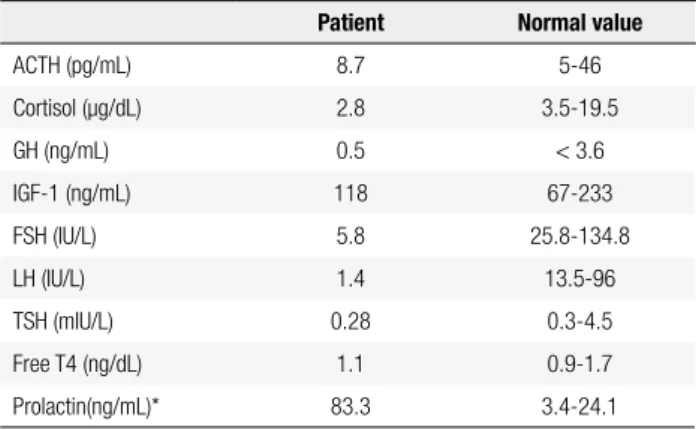
Table 1. Pituitary function evaluation before surgery
Patient Normal value
ACTH (pg/mL) 8.7 5-46
Cortisol (μg/dL) 2.8 3.5-19.5
GH (ng/mL) 0.5 < 3.6
IGF-1 (ng/mL) 118 67-233
FSH (IU/L) 5.8 25.8-134.8
LH (IU/L) 1.4 13.5-96
TSH (mIU/L) 0.28 0.3-4.5
Free T4 (ng/dL) 1.1 0.9-1.7
Prolactin(ng/mL)* 83.3 3.4-24.1
* Absence of macroprolactin.
Cop
yright
© ABE&M t
odos os dir
eit
os r
eser
vados
.
871 Arq Bras Endocrinol Metab. 2014;58/8
DISCUSSION
Pituitary metastases are most prevalent in the sixth and seventh decades of life (8). Breast tumors (20-30%) and lung tumors (30-50%) (9) are the most common pri-mary sites, but others are also described, such as the gastrointestinal tract, prostate, kidney, thyroid, pancre-as, lymphompancre-as, leukemias and plasmacytomas.
Generally, pituitary metastases are a part of wides-pread metastatic disease, associated with 5 or more sites of metastases, especially bone (6). Occasionally, breast cancer metastasizes to the pituitary, with reported ra-tes ranging between 6 and 8% of cases (5), majority asymptomatic. Hypophysectomy of patients with breast cancer found 5.5% to 25% of metastases not previously diagnosed (7).
The present patient had multiple metastases from the breast carcinoma besides the pituitary stalk one. Immunohistochemical proile of pituitary stalk mass disclosed the presence of the estrogen receptor, which is consistent with the primary site in the breast. The presence of estrogen receptors becomes able to hormo-nal therapy (10). The positivity of c-erbB-2 and Ki67 are correlated with a worse prognosis. The c-erbB-2 proto-oncogene occurs in about 20% of all breast can-cers and Ki67 is a nuclear non-histone protein that re-fers to a poorly differentiated tumor (10).
About metastasis location, the posterior lobe of the pituitary gland is most common site (69-79%) (11) due to its larger area of contact with the adjacent dura-ma-ter and the lack of blood supply to the andura-ma-terior lobe (5,7,9,12,13).
Mc Cormick and cols., reviewing the location of
pi-tuitary metastases in 201 cases, found the involvement of the posterior lobe alone or in combination with ante-rior lobe in 84.6%, and the involvement of the anteante-rior lobe alone in 15.4% (13). However, especially in breast cancer, metastases occur preferentially to the anterior pituitary gland (6,11). Hematogenous dissemination is the most important mechanism of development of these metastases, but also occur by contiguity of adja-cent bone metastasis or meningeal spread through the suprasellar cistern (9).
Pituitary metastases are symptomatic in only 2.5 to 18.2% of cases (5-7) because most occur in patients too debilitated by cancer with poor survival rates to beco-me clinically evident. Still then, the systemic compli-cations of malignancy, including nonspeciic symptoms of weakness, vomiting and weight loss as well as the involvement of the CNS, may mask the dysfunction of the anterior pituitary. So the majority of cases have been discovered accidentally during autopsies (6). Ho-wever, in some patients, symptoms related to pituitary metastasis may be the irst manifestation of an occult malignancy (5).
The most noticeable symptom is diabetes insipi-dus (DI), reinforcing the predominance of metasta-ses to the posterior lobe. DI occurred in 45.2% of cases among 190 patients with pituitary metastases (6) reaching 61% to 70% in other series (13,14). Ac-cording to Houck and cols. 20% of patients
diagno-sed with DI may present pituitary metastases (15). However, the clinical presentation is variable (16), and may be associated with ophthalmoplegia, heada-che, visual ield abnormalities and anterior pituitary dysfunction (5), and could also present with seizures, paralysis of III and IV cranial pairs and hyperprolac-tinemia.
Among the visual alterations, bilateral hemianopia is the most common (6). Given the invasiveness of tu-mors, visual loss may result from suprasellar extension and can result in painful ophthalmoplegia due to inil-tration of the cavernous sinus (5).
Treatment is essentially palliative and depends on the extent of disease and symptoms. Radiotherapy and/ or chemotherapy are recommended as initial treatment, especially in patients with disseminated metastases in combination with replacement of pituitary hormones for relief of symptoms (7,13,15). Surgical exploration and decompression alone or in combination with radio-therapy is essential if the pathological diagnosis would
Pituitary metastasis of breast cancer
Cop
yright
© ABE&M t
odos os dir
eit
os r
eser
vados
.
872 Arq Bras Endocrinol Metab. 2014;58/8
guide the therapy plan or if suprasellar extension is cau-sing pain or progressive visual deterioration.
There is no consensus on improved rates of survi-val with surgical resection, but there is improvement of symptoms by local decompression of the tumor al-though it does require the combination of different therapeutic modalities (17).
Prognosis is poor, not because of the location it-self, but due to the aggressiveness of the primary tumor (18). Median survival rate was 6-7 months in clinical series (7,14,15). Among 72 patients in literature re-view, Ntyonga Pono and cols. (19) found only 10% of survivors one year forward diagnosis, with a maximum survival of 3 years. When the pituitary lesion is a single metastasis, survival can improve (7,14). Patients with more than 65 years of age at presentation, small cell lung cancer as the primary site and a short interval (less than 1 year) between the initial diagnosis of cancer and invasion of pituitary are related to worse prognosis (14). Pituitary stalk invasion presentation has a wor-se prognosis as 2-4 months of survival, approximately (5,20).
Disclosure: no potential conlict of interest relevant to this article was reported.
REFERENCES
1. Daly AF, Rixhon M, Adam C, Dempegioti A, Tichomirowa MA, Beckers A. High prevalence of pituitary adenomas: a cross-sec-tional study in the province of Liege, Belgium. J Clin Endocrinol Metab. 2006;91(12):4769-75.
2. Aghakhani K, Kadivar M, Kazemi-Esfeh S, Zamani N, Moradi M, Sanaei-Zadeh H. Prevalence of pituitary incidentaloma in the Ira-nian cadavers. Indian J Pathol Microbiol. 2011;54(4):692-4. 3. Ironside JW. Best Practice No 172: pituitary gland pathology. J
Clin Pathol. 2003;56(8):561-8.
4. Pichard C, Gerber S, Laloi M, Kujas M, Clemenceau S, Ponvert D, et al. Pituitary carcinoma: report of an exceptional case and review of the literature. J Endocrinol Invest. 2002;25(1):65-72. 5. Fassett DR, Couldwell WT. Metastases to the pituitary gland.
Neu-rosurg Focus. 2004;16(4):E8.
6. Komninos J, Vlassopoulou V, Protopapa D, Korias S, Konto-georgos G, Sakas DE, et al. Tumors metastatic to the pituitary gland: case report and literature review. J Clin Endocrinol Metab. 2004;89(2):574-80.
7. Sioutos P, Yen V, Arbit E. Pituitary gland metastases. Ann Surg On-col. 1996;3(1):94-9.
8. Max MB, Deck MD, Rottenberg DA. Pituitary metastasis: inci-dence in cancer patients and clinical differentiation from pituitary adenoma. Neurology. 1981;31(8):998-1002.
9. Spinelli GP, Lo Russo G, Miele E, Prinzi N, Tomao F, Antonelli M, et al. Breast cancer metastatic to the pituitary gland: a case report. World J Surg Oncol. 2012;10:137.
10. Eisenberg ALA, Koifan S. Câncer de mama: marcadores tumorais e revisão da literatura. Rev Bras Cancerol. 2001;47(4):377-88. 11. Kim YH, Lee BJ, Lee KJ, Cho JH. A case of pituitary metastasis
from breast cancer that presented as left visual disturbance. J Korean Neurosurg Soc. 2012;51(2):94-7.
12. Kramer CK, Ferreira N, Silveiro SP, Gross JL, Dora JM, Azevedo MJ. Pituitary gland metastasis from renal cell carcinoma present-ed as a non-functioning macroadenoma. Arq Bras Endocrinol Metabol. 2010;54(5):498-501.
13. McCormick PC, Post KD, Kandji AD, Hays AP. Metastatic carcino-ma to the pituitary gland. Br J Neurosurg. 1989;3(1):71-9. 14. Morita A, Meyer FB, Laws ER Jr. Symptomatic pituitary
metasta-ses. J Neurosurg. 1998;89(1):69-73.
15. Houck WA, Olson KB, Horton J. Clinical features of tumor metas-tasis to the pituitary. Cancer. 1970;26(3):656-9.
16. Marsh JC, Garg S, Wendt JA, Gielda BT, Turian JV, Herskovic AM. Intracranial metastatic disease rarely involves the pituitary: retro-spective analysis of 935 metastases in 155 patients and review of the literature. Pituitary. 2010;13(3):260-5.
17. Branch CL Jr, Laws ER Jr. Metastatic tumors of the sella turcica masquerading as primary pituitary tumors. J Clin Endocrinol Metab. 1987;65(3):469-74.
18. Delattre JY, Castelain C, Davila L, Schadeck B, Poisson M. [Metas-tasis to the pituitary stalk in a case of breast cancer]. Rev Neurol (Paris). 1990;146(6-7):455-6.
19. Ntyonga-Pono MP, Thomopoulos P, Luton JP. [Pituitary metasta-ses. 3 cases]. Presse Med. 1999;28(29):1567-71.
20. Kurkjian C, Armor JF, Kamble R, Ozer H, Kharfan-Dabaja MA. Symptomatic metastases to the pituitary infundibulum resulting from primary breast cancer. Int J Clin Oncol. 2005;10(3):191-4.