Hydroxylase Genes Caused by a XylR/DmpR-Type
Regulator MphR in
Acinetobacter calcoaceticus
Haiying Yu1,2., Zixin Peng2,3., Yuhua Zhan2
, Jin Wang2, Yongliang Yan2,4, Ming Chen2, Wei Lu2, Shuzhen Ping2, Wei Zhang2,4, Zhonglin Zhao2, Shuying Li2, Masahiro Takeo3*, Min Lin1,2*
1College of Biological Sciences, China Agricultural University, Beijing, China,2Key Laboratory of Crop Biotechnology, Biotechnology Research Institute, Chinese Academy of Agricultural Sciences, Ministry of Agriculture, Beijing, China,3Department of Materials Science and Chemistry, Graduate School of Engineering, University of Hyogo, Himeji, Hyogo, Japan,4National Centre for Plant Gene Research, Beijing, China
Abstract
Acinetobacter calcoaceticusPHEA-2 utilizes phenol as its sole carbon and energy source and has a multi-component phenol hydroxylase-encoding gene operon (mphKLMNOP) for phenol degradation. Two additional genes, mphR and mphX, were found upstream and downstream of mphKLMNOP, respectively. The mphR gene encodes a XylR/DmpR-type regulator-like protein and is transcribed in the opposite direction to mphKLMNOP. The mphX gene is transcribed in the same direction as mphKLMNOP and encodes a protein with 293 amino acid residues showing weak identity with some unknown proteins encoded in the meta-cleavage pathway gene clusters for aromatic compound degradation. Disruption of mphR by homologous recombination resulted in the loss of phenol degradation while disruption of mphX caused significantly faster phenol degradation than in the wild type strain. Transcriptional assays for mphK, mphR, and mphX revealed that mphR activated mphKLMNOP transcription in the presence of phenol, but mphX partially repressed this activation. Gel mobility-shift assay demonstrated a direct interaction of MphR with the mphK promoter region. These results indicate the involvement of a novel repressor protein MphX in transcriptional regulation of phenol hydroxylase genes caused by a XylR/ DmpR-type regulator MphR.
Citation:Yu H, Peng Z, Zhan Y, Wang J, Yan Y, et al. (2011) Novel Regulator MphX Represses Activation of Phenol Hydroxylase Genes Caused by a XylR/DmpR-Type Regulator MphR inAcinetobacter calcoaceticus. PLoS ONE 6(3): e17350. doi:10.1371/journal.pone.0017350
Editor:Roland Roberts, King’s College London, United Kingdom
ReceivedSeptember 27, 2010;AcceptedJanuary 31, 2011;PublishedMarch 24, 2011
Copyright:ß2011 Yu et al. This is an open-access article distributed under the terms of the Creative Commons Attribution License, which permits unrestricted use, distribution, and reproduction in any medium, provided the original author and source are credited.
Funding:This work was supported by grants from the National Natural Science Foundation of China (No. 30925002 and 30800022), the National Basic Research (973) Program of China (No. 2007CB707805 and 2010CB126504) and the National High-Tech (863) Program of China (No. 2007AA021304 and 2010AA10A203). The funders had no role in study design, data collection and analysis, decision to publish, or preparation of the manuscript.
Competing Interests:The authors have declared that no competing interests exist.
* E-mail: [email protected] (MT); [email protected] (ML)
.These authors contributed equally to this work.
Introduction
Besides being important industrially for the synthesis of chemical products such as resins, medicines, and dyes, phenol is also a major toxic contaminant in industrial wastewaters from oil refineries, petrochemical plants, and phenolic resin plants. Thus, efforts to improve the efficiency of wastewater treatment via phenol degradation have gained special attention. Such efforts have led to a deeper understanding of phenol biodegradation mechanisms [1,2].
A large variety of soil bacteria are able to degrade aromatic compounds by employing different strategies for regulatory control at the level of gene expression. In aerobic phenol degradation by microorganisms, phenol is normally converted by phenol hydrox-ylase into catechol, which flows into anortho-cleavage pathway or a meta-cleavage pathway after the cleavage of an aromatic-ring by catechol 1,2-dioxygenase or catechol 2,3-dioxygenase [2]. Phenol hydroxylase, catalyzing the initial reaction in phenol degradation, is a key rate-limiting enzyme. Many phenol hydroxylases from soil microorganisms can be classified into three groups: single- [3,4], two- [5,6], and multi- [7,8,9] component types. Microorganisms
activator binding tos54-dependent promoter regions of respective target operons [14,24,25]. In addition to XylR/DmpR-type mediated regulation, co-regulation of mPH gene expression by different types of regulators has also been observed inComamonas testosteroni strains TA441 and R5 [7,23,28,29]. Furthermore, carbon catabolite repression and some host-dependent global regulations have been reported to affect the primary regulation by XylR/DmpR-type proteins [14,30,31].
In most cases, the catabolic pathway-specific regulators act as transcriptional activators of gene expression with few exceptions of GntR-type proteins, which are exclusively described as repressors. The use of repressors in the regulation of phenol degradation is not yet fully understood. We previously isolated phenol and benzoate degradation gene clusters spanning a 15.5-kb region from Acinetobacter calcoaceticus PHEA-2 [32,33]. This region contains at least four putative transcriptional units, mphKLMNOP, benM, benABCDE, and benKP, in this order. The complete genome of A. calcoaceticus PHEA-2 was sequenced recently and has been deposited in GenBank under accession number CP002177. Establishment of the complete genome sequence enabled mapping of the entire catabolic gene cluster in the PHEA-2 chromosome. In this study, we compared organization of the phenol catabolic gene cluster in A. calcoaceticus PHEA-2 (mph) and with the other phenol degrading genes cluster. In addition, the result of the growth test on phenol and measurements of phenol-oxygenat-ing activities suggested that phenol degradation in PHEA-2 was inducible and that mphR and mphX encoded an activator and a repressor, respectively, affecting expression of the mPH-encoding genes.
Results
Involvement of themphgenes in phenol degradation
As described in the introduction, two gene candidates,mphR and mphX, were found upstream and downstream of themphoperon, respectively. From sequence analysis,mphKLMNOP was found to encode mPH proteins, catalyzing the initial reaction in phenol degradation (Fig. 1A). This multi-component enzymes exhibits 38–72% and 58–93% identity with proteins encoded by the cognate genes dmpKLMNOP of Pseudomonas sp. CF600 [9] and
mopKLMNOP of A. calcoaceticus NCIB8250 [8], respectively. A mphN-deletion mutant lacking the center portion of the mPH-encoding operon (Fig. 1B) was constructed by homologous recombination and named A2N. It lost its phenol degradation activity (Fig. 2), but was still able to assimilate catechol (data not shown), showing thatmphKLMNOPis essential for the initial step of phenol degradation in PHEA-2.
The MphR protein showed significant identity with XylR/ DmpR-type regulators MopR (70% aa sequence identity), PhlR (52% identity), DmpR (51% identity), PhhR (51% identity), TouR (51% identity), CapR (51% identity) and XylR (48% identity), and expected to encode a XylR/DmpR-type regulatory protein homolog (Fig. 3A). To clarify the function of mphR in phenol degradation, a mphR-deletion mutant was constructed from PHEA-2 by homologous recombination and named A2R. The deletion was confirmed by PCR (the expected deleted segment is shown in Fig. 1B). As shown in Fig. 2, A2R was unable to grow on or degrade phenol at all. Introduction of the wild-type mphR into A2R using a complementation plasmid resulted in recovery of phenol degradation activity (data not shown). These results indicate thatmphRencodes an activator for the expression ofmphKLMNOPrequired for phenol degradation in PHEA-2.
In addition, we compared organization of the phenol catabolic gene cluster inA. calcoaceticusPHEA-2 (mph) and with the other phenol degrading genes cluster in A. calcoaceticus NCIB8250 (mop),C. testosteroniTA441 (aph) andC. testosteroni R5 (phc) (Fig. 1B). We found that an 882-bp open reading frame namedmphXwas detected betweenmphKLMNOP andbenM and its gene product showed no significant similarity with any known functional proteins. To investigate the involvement of mphX in phenol degradation, an mphX-deletion mutant was constructed from PHEA-2 by homologous recombination and named A2X. Interestingly, mphX-deletion mutant A2X showed faster growth on phenol and faster phenol degradation than the wild-type PHEA-2 strain (Fig. 2). The mphX-complemented strain recovered the normal ability to grow on phenol with the growth rate similar to that of wild type (data not shown). In addition, in the phenol-oxygenating activity assay using cell suspensions, phenol-induced A2X cells showed higher activity (1.9760.14mmol/min/g-dry cell) compared to phenol-induced
Figure 1. Phenol degradation inA. calcoaceticusPHEA-2.(A) Catabolic payhway for degradation of phenol to catechol. (B) Organization of the phenol catabolic gene cluster inA. calcoaceticusPHEA-2 (mph) and comparison with the other phenol degrading genes cluster inA. calcoaceticus NCIB8250 (mop),C. testosteroniTA441 (aph) andC. testosteroniR5 (phc). Genes are denoted by open arrows. The region of a deleted segment in mphR,mphNormphX, respectively, is indicated by gray-shaded box.
PHEA-2 cells (1.3860.11mmol/min/g-dry cell). However,
lactate-grown cells of both strains showed similar basal level of activities (,0.05mmol/min/g-dry cell). Therefore, it is
possible that mphX encodes a repressor protein for the expression of the mPH genes. The MphX protein is comprised of 293 aa residues and is expected to have a molecular mass of 32.2 kDa. It shows weak identity with proteins encoded by orfX (27.8% aa sequence identity) in the phenol degradation gene cluster of Comamonas testosteroni TA441 [34] and orfU (26.3% identity) in the aniline degradation gene cluster of Delftia tsuruhatensis AD9 [35] whose functions are unknown. Searches found several homologous bacterial proteins with more than 30% identity, but the findings are annotated as putative or hypothetical proteins (Fig. 3B). MphX shows little identity (15% or less) with members of known regulator families (data not shown). Therefore, if MphX is a repressor, it can be classified as a new type of regulatory protein.
Sequence analysis of the intergenic region between mphRandmphKand determination of the transcriptional start site formphK
As shown in Fig. 4A, in the intergenic region betweenmphRand mphK, three incomplete inverted repeat sequences (IR1, IR2, and IR3) [36], a long A/T-rich sequence (spanning from2134 to229 relative to the putative transcriptional start site formphK), and a putative s54-dependent 212/224 promoter sequence (59 -TTGGCATA-N4-TTGTA-39, N = any base) for mphK were
found. The putative promoter sequence is very similar to the consensus sequence of the 212/224 promoters (59 -YTGG-CACG-N4-TTGCW-39, Y = C or T, W = A or T) described by
Wigneshwerarajet al.[37]. To identify the transcriptional start site for mphK, primer extension analysis was performed using total RNA isolated from phenol-induced PHEA-2 cells. A single band corresponding to a G residue located 39 bp upstream from the translation initiation codon of mphK was detected (Fig. 4B). Considering the location of the212/224 promoter formphK, this position seems suitable for the transcriptional start site formphK.
In contrast, a putative s70
-dependent 210/235 promoter sequence for mphR (59-ATGAAG-N23-ATTTAT-39) was also
detected (Fig. 4A). This sequence is similar to thes70
-dependent promoter (59-GTGAAG-N19-TAATAT-39) of the A. calcoaceticus
NCIB8250mopR gene [22], suggesting that mphR is transcribed from the s70-dependent promoter. The arrangement of mphR-KLMNOPand the above promoter/operator elements are nearly identical to those of several other phenol degradation gene clusters with a XylR/DmpR-type regulatory gene, such asdmp,mop,phc, aph,phl(strain H) andphh[7,19,20,21,22,28].
Regulation of the transcription ofmphKLMNOP,mphR, and mphX
To evaluate the transcriptional activities ofPmphK,PmphRandPmphX under various conditions,PmphK-,PmphR-, andPmphX-lacZfusions were constructed in pGD926 and the resulting plasmids, pGDP, pGDRP and pGDPX, were independently introduced into the wild-type strain PHEA-2, the mphR-deletion mutant A2R and the mphX -deletion mutant A2X. After 6-h incubation with or without phenol, cell suspensions were prepared and b-galactosidase activity was measured as transcriptional activity using the cell suspensions. The result is summarized in Table 1. From the result, transcriptional regulation of these keymphgenes can be explained as follows:
(i) Transcription ofmphK(the mphKLMNOP operon): Phenol-induced PHEA-2 cells showed 10 times higherPmphKactivity than that of uninduced PHEA-2 cells, indicating that the transcription of mphK was phenol-inducible in PHEA-2. Irrespective of induction conditions, however,mphRdeletion resulted in complete loss ofPmphK activity with or without phenol. This result demonstrated thatmphRis essential for the transcription ofmphKand encoded an activator protein. In contrast, thePmphKactivity of phenol-induced A2X cells was 2.4 times higher than that of the phenol-induced PHEA-2 cells, suggesting thatmphX partially represses the mphR -dependent activation in PHEA-2 cells.
(ii) Transcription ofmphR: Phenol-induced PHEA-2 cells showed 1.8 times higherPmphRactivity than that of uninduced PHEA-2 cells. This indicates that the transcription ofmphRwas weakly inducible in the presence of phenol. However, since a considerable level of thePmphR activity was detected even in uninduced cells, a small amount of MphR is probably always produced even in the absence of effectors in PHEA-2 and may serve as a sensor for effectors coming from outside of the cells. Figure 2. Growth and phenol degradation of A. calcoaceticus
Low PmphR activity observed in both phenol-induced and uninduced A2R cells suggests that MphR is also an activator for its own gene expression. Furthermore, the absence of mphX resulted in a 40% increase inPmphR activity (calculated from comparison of thePmphRactivity in phenol-induced A2X cells with that in phenol-induced PHEA-2 cells), suggesting that mphXalso represses the transcription ofmphRin PHEA-2 cells. This result is in good agreement with the result of the measurement of the phenol-oxygenating activities shown above. (iii) Transcription of mphX: Phenol-induced of PHEA-2 cells showed a high level of thePmphXactivity compared to that of uninduced PHEA-2 cells. However, in the absence ofmphR ormphX(in A2R or in A2X),PmphXactivities were at a basal level even under induction by phenol. These results indicate that the transcription of mphX was phenol-inducible and absolutely dependent on the presence of mphR and mphX itself. Very low PmphX activity in A2X cells even in the presence ofmphRand phenol demonstrates thatmphXmight be in a transcriptional unit separate frommphKLMONP.
Binding of MphR to themphR-mphKintergenic region
As shown in Fig. 5A, a region spanning IR2 and IR3 appears to contain the upstream activating sequences (UASs) ofPmphKbecause
it is similar to those found in the promoter regions of the mopKLMNOP,dmpKLMNOP, andxylupper pathway gene clusters [22]. In order to investigate the direct interaction of MphR with the intergenic region betweenmphR and mphK, we designed a DNA fragment P380 completely covering themphKpromoter region from positions2326 to+49 relative to the transcription start site (Fig. 4A). The fragment was mixed with the purified His-MphR (Fig. 5C). Then, the mixtures of DNA/MphR complex and free DNA were resolved by native PAGE. The purified His-MphR protein with P380 caused a band shift of P380. As the amount of protein increased, shifted band was detected, indicating that the MphR protein specifically retards DNA fragments containing themphR -mphKpromoter region (Fig. 5B). This observation provides evidence for a direct interaction of MphR with the mphR-mphK promoter region, suggesting an activation mechanism similar to that proposed for a well-studied XylR/DmpR-type regulator MopR in the phenol-degrading bacteriumA. calcoaceticusNCIB8250 [22].
Discussion
regulators consist of four functional domains: the N-terminal signal reception domain (A-domain), the central activation domain with ATPase activity (C-domain), the C-terminal DNA-binding domain with a helix-turn-helix (HTH) motif (D-domain), and a short flexible linker domain between the A-domain and the C-domain (B-domain) [27,38]. Multiple alignment of MphR with XylR/
DmpR-type regulators showed that MphR has an NTP-binding motif (GxxGxGKExxAxxxHxxS, where x is any amino acid, aa 272–aa 289) [15,39] and an AAA+ protein family signature (GAYTGA, aa 319–aa 324) [40] with one amino acid difference (the complete motif is GAFTGA) in the C-domain. The former is involved in the ATP binding and hydrolysis on the C-domain, Figure 4. Analysis of the promoter region and transcriptional start site for mphK.(A) Intergenic region betweenmphRandmphK. The first nucleotide in the transcript is shown in bold. Arrows and dashed arrows indicate incomplete inverted repeats (named IR1, IR2, and IR3) and the translational start codons formphRandmphK, respectively. Double-underlines mean the putatives54-dependent212/224 promoter consensus sequence and thes70-dependent210/235 promoter consensus sequences. An arrowhead with+1 indicates the transcriptional start site formphK. A grey-shaded box indicates the putative ribosome-binding site for themphKtranscription. (B) Mapping of the transcriptional start site formphKby primer extension. A sequence ladder was generated with the same primer (lanes T, A, C, and G).
while the latter is expected to interact withs54-RNAP. Recently, the molecular mechanism of ATP hydrolysis-mediated transcrip-tional activation was discussed based on the structures of bacterial enhancer-binding proteins PspF and NtrC by Rappaset al.[41]. These proteins have several common amino acid residues important in ATP binding and hydrolysis (K42, E43, N64, D107, E108, and R227 in PspF) and they are all conserved in MphR as K278, E279, N300, D343, E344, and R462, respectively. In the B-domain of MphR, a heptad-repeats signature-like sequence with regularly spaced hydrophobic amino acids is present (MxxxLxxLxxx-LxxLxxxIxxxSxxY, aa 213–aa 239), which is involved in dimer formation and prevention of ATP hydrolysis without effector binding [42]. Moreover, the HMMPFAM motif search program (http://hmmpfam.ddbj.nig.ac.jp/top-e.html) predicted the pres-ence of a HTH DNA binding motif in the D-domain (aa 514–aa 555) of MphR. Conservation of these functionally important motifs demonstrates that MphR has many structural and mechanistic features common in NtrC family regulators including XylR/DmpR subclass members and the mechanism for the transcriptional activation by MphR is essentially identical to the above-mentioned mechanism by XylR/DmpR-type regulators.
Although some lines of experimental evidence confirming the repressor function of MphX have been obtained in this study, MphX shows little similarity with the members of known bacterial Table 1.b-Galactosidase activities ofmphKpromoter-,mphR
promoter-, ormphXpromoter-lacZtranscriptional fusion inA. calcoaceticusPHEA-2 andmphR- andmphX-deletion mutants (A2R and A2X).
b-Galactosidase activity (Miller unit)a
+Phenol 2Phenol
PHEA-2(PmphK-lacZ) 336633 3462
A2R(PmphK-lacZ) * *
A2X(PmphK-lacZ) 807658 3062
PHEA-2(PmphR-lacZ) 160620 92610
A2R(PmphR-lacZ) 2065 2163
A2X(PmphR-lacZ) 225633 176615
PHEA-2(PmphX-lacZ) 332650 *
A2R(PmphX-lacZ) * *
A2X(PmphX-lacZ) * *
aCells were grown in the presence (
+) or absence (2) of 2.0 mM phenol and assayed forb-galactosidase activities as described in Materials and Methods. Values shown are means6standard deviation of triplicate experiments. *No detectable activity.
doi:10.1371/journal.pone.0017350.t001
Figure 5. Electrophoretic mobility shift assay.(A) Comparison of the upstream region of themphKpromoter with the corresponding regions of themopgene cluster ofA. calcoaceticusNCIB8250, thexylupper gene cluster ofPseudomonas putidamt-2 (TOL), and thedmpgene cluster of Pseudomonassp. CF600. Grey-shaded boxes show the putative upstream activating sequences (UASs) in themph,mop,xyl, anddmpgene clusters to which the corresponding XylR/DmpR-type regulators bind. (B) gel retardation analysis of His-MphR binding to themphR-mphKintergenic region. Gel-mobility shift assay was performed as described in the text using a 380-bp fragment (P380) covering themphR-mphKintergenic region. Lane 1 is a control lane containing 2.5 nM of labeled P380 fragment only. Lane 2–7 contains 2.5 nM of the labeled P380 fragment incubated with 5, 10, 20, 30, 40 and 50 nM His-MphR protein. Lane 8 is containing 2.5 nM of labeled DNA fragment amplified using two primers, IRDye-labeled M13-F and M13-R, and plasmid pGEM T-vector as template, named P150. Lane 9–10 contains 2.5 nM of the labeled P150 fragment incubated with 30 and 50 nM His-MphR protein, respectively. The positions of the free DNA and the His-MphR-DNA complex are indicated. (C) SDS-PAGE of purified His-His-MphR protein from E. coliBL21 (pEMR). Lane 1: Molecular markers with their masses indicated; Lane 2: Uninduced of BL21 (pEMR); Lane 3: BL21 (pEMR) induced by 0.1 mM IPTG; Lane 4: BL21 (pEMR) induced by 0.5 mM IPTG; Lane 5: MphR purified by NTA resin and eluted by NTA-60; Lane 6: MphR purified by NTA resin and eluted by NTA-80.
regulator families. BLAST (http://blast.ddbj.nig.ac.jp/top-e.html) and CDART (http://www.ncbi.nlm.nih.gov/Structure/lexington/ lexington.cgi) searches suggest that MphX belongs to the COG4313 family in the COG (Clusters of Orthologous Group) database [43]. Although most members of the COG4313 family are annotated as hypothetical or putative proteins, several proteins are designated as ‘‘involved in meta-pathway of phenol degradation’’, including those encoded byorfX(identity with the encoded protein: 27.8%) in the phenol degradation gene cluster ofC. testosteroni TA441 [34],orf1 (27.0%) in the aniline degradation gene cluster ofP. putidaUCC22 [44], andorfU(26.3%) in the aniline degradation gene cluster ofD. tsuruhatensis AD9 [35]. In the phenol degradation gene cluster of TA441 (aphSRKLMNOPQBorfXYaphTCEFGHJI), orfX is located downstream of a catechol 2,3-dioxygenase gene (aphB) [34]. Arai et al.[34] pointed out that theorfXgene product (OrfX) has a typical amino-terminal signal sequence for membrane translocation, suggesting that OrfX is a protein in the periplasm. Sequence analysis by SignalIP 3.0 (http://www.cbs.dtu.dk/services/SignalP/ ) predicted that MphX also has a signal peptide sequence at its amino-terminus (aa 1–aa 23, signal peptide probability = 1.000) and its most likely cleavage site (A21F22A23-T24E25, maximum cleavage site probability = 0.999). Thus, MphX has at least one feature specific to periplasmic proteins, but at present the actual location of the MphX protein in PHEA-2 is not known. The same authors found that anorfXYmutant of TA441 grew poorly on phenol and accumulated 2-hydroxymuconic semialdehyde [34]. To our knowledge, this is the only description of an MphX homolog-coded gene function. The mutant phenotype might be caused by a mutation inorfX, but there are also other possibilities, i.e.a mutation inorfYor a polar effect for the expression of the downstream meta-cleavage pathway genes. In the aniline degra-dation gene clusters ofD. tsuruhatensisAD9 andP. putidaUCC22, MphX homolog-coded genes, orfU and orf1, are also located between two sets of genes encoding a ferredoxin-like protein and a catechol 2,3-dioxygenase (tdnD1C-tdnD2C2 and tadD1C1 -tadD2C2) [35,44]. However, the functions of these genes remain unknown. Several MphX homolog-coded genes have been found near alcohol dehydrogenase genes, such as orf1 near the 4-nitrobenzoate degradation gene cluster of Pseudomonas sp. TW3 [45],qbdBin the short-chain alcohol degradation gene cluster of P. putida HK5 [46], and chnX in the cyclohexanol degradation gene cluster of Acinetobacter sp. SE19 [47]. These gene products showed 21–22% aa sequence identity with MphX. However, the functions of these genes have not been elucidated yet. Accordingly, MphX is the only protein in this COG4313 family whose function has been demonstrated as a repressor. We looked for DNA binding motifs in MphX using several motif search programs (e.g., http://motif.genome.jp/), but we were unable to detect any HTH motif or other motifs specific to regulatory proteins in MphX.
Comamonas testosteroni strains TA441 and R5 have their respective mPH-encoding operon (aphKLMNOP and phcKLMNOP) and the counterpart XylR/DmpR-type regulatory gene (aphR andphcR) is in almost the same gene arrangement as that of PHEA-2 [29,34]. However, strain TA441 has an additional regulatory gene, aphS, just downstream of aphR, whereas strain R5 has two additional regulatory genes,phcSand phcT, downstream ofphcR. MphX shows little sequence similarity with these regulatory proteins. In strain TA441,aphScompletely represses the transcriptional activation ofaphKLMNOPcaused by aphR in the presence of phenol. Consequently, strain TA441 is unable to grow on phenol, but itsaphS-deficient mutant is able to grow on phenol due to the de-repression [7]. Interestingly, it was found that the transcription of aphKLMNOPis also caused by a
gratuitous inducer, acetate, and that aphS represses this gratuitous activation, suggesting that aphS prevents wasteful expression ofaphKLMNOPinduced by such a ubiquitous carbon source [7]. In strain R5, phcS also represses the transcriptional activation of phcKLMNOP caused by phcR in the presence of phenol and similar gratuitous expression by acetate [29]. However, thephcgene cluster contains another regulatory gene, phcT, and the presence of phcT enabled strain R5 to overcome the repression caused byphcSand to grow on phenol [28]. The aa sequence analysis of these regulatory gene products indicated that AphS and PhcS belong to the GntR regulator family [7,29] while PhcT belongs to the AraC/XylS regulator family [28]. One of the AphS binding sites (consensus motif, TATCGTAT) determined by gel-mobility shift assay is located around the putative IHF (Integration Host Factor)-binding site, but not within the putative UASs [7]. Similar binding motifs were also found in the putative IHF-binding site of the phcR-phcK intergenic region [28]. Hence, it is suggested that these proteins could bind to the putative IHF binding sites and hamper the binding of IHF [15,28,48]. However, the IHF-binding consensus motif is absent in the A+T-rich sequence of themph operon of strain PHEA-2. In particular, we were unable to detect any HTH motifs in the MphX protein using several motif search programs (e.g., http://motif.genome.jp/). Moreover, all attempts to perform electrophoretic mobility shift analysis using MphX have failed. So far we have not succeeded in the purification of MphX due to its easily-aggregating property. We presume that the mechanism by which MphX partially repressesmphKLMNOP transcription differs from those used by AphS and PhcS; however, regulatory mechanism of themphXgene as a repressor requires further investigation.
Materials and Methods
Bacterial strains, plasmids, primers, and growth conditions used
The bacterial strains and plasmids used in this study are listed in Table 2 and the sequences of all the primers used are listed in Table 3.Acinetobacterstrains were grown at 30uC in mineral salts (MS) medium (NaNO30.5 g, K2HPO40.65 g, KH2PO40.17 g,
MgSO4 0.10 g per liter) supplemented with phenol or sodium
lactate, or in Luria-Bertani (LB) medium. Escherichia coli strains were grown at 37uC in LB medium. When necessary, ampicillin (20mg/ml), kanamycin (50mg/ml), and tetracycline (50mg/ml) were added to the medium.
Construction ofmphR-,mphN-andmphX-deletion mutants
In order to disrupt key genes in phenol degradation, the upstream and downstream regions ofmphR,mphN, andmphXwere generated by PCR using the total DNA of PHEA-2 as the template and several primer sets shown in Table 3. The amplified PCR products were purified and ligated into the pMD18-T vector (Takara). The resulting plasmids were double-digested with EcoRI and SalI (for plasmids carrying the upstream region) or SalI and BamHI (for those carrying the downstream region) and purified. The upstream and downstream fragments for each gene were cloned together into the EcoRI/BamHI sites of pK18mobsacB [51]. The resulting plasmids were introduced into E. coli JM109 and then transferred into PHEA-2 by triparental mating [52]. Integration of the introduced plasmid into the chromosome by the first crossover was selected on LB plates containing kanamycin (25mg/ml) and ampicillin (20mg/ml). The second crossover cells were selected by culture on LB plates
containing 10% (w/v) sucrose and ampicillin (20mg/ml). The
selected cells were analyzed by PCR to confirm that gene replacement had occurred (data not shown). The resultingmphR, mphNandmphXdeletion mutants were designated as A2R, A2N and A2X, respectively.
Phenol degradation test
Acinetobacterstrains were grown overnight in LB medium and the cultures were harvested by centrifugation (8,0006g, 4uC, 5 min).
The cells were washed twice with MS medium and suspended at an OD600of 0.15 with MS medium containing 2 mM phenol. Cell
suspensions were further incubated with shaking at 200 rpm on a rotary shaker. Aliquots of the cultures were sampled at specific intervals and centrifuged (13,0006g, 4uC, 1 min) to remove the
cells. Following cell removal, phenol concentrations in the supernatants were determined using the 4-aminoantipyrine colorimetric method. This analysis was performed according to the procedures described in Folsomet al.[1].
Assay for phenol-oxygenating activity
Acinetobacter strains were grown in LB medium to the late-log phase and the cultures were harvested by centrifugation (8,0006g,
4uC, 5 min). The cells were washed twice with MS medium, suspended at an OD600of 0.7 with MS medium containing 2 mM
phenol or 5 mM sodium lactate and further incubated at 30uC for 6 h for induction. Then, the cells were again collected by centrifugation, washed with MS medium, and resuspended at an OD600 of 1.5 in 40 ml potassium phosphate buffer (100 mM,
pH 7.5). After measurement of endogenous oxygen uptake for 5 min, reactions were started by adding phenol to the cultures to a final concentration of 0.1 mM. Oxygen concentrations in the cultures were monitored at 25uC for 3 min using a YSI 5000
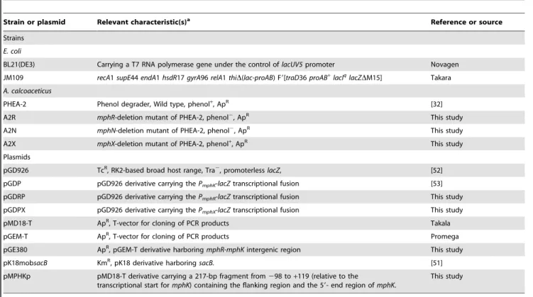
Table 2.Bacterial strains and plasmids used in this study.
Strain or plasmid Relevant characteristic(s)a Reference or source
Strains
E. coli
BL21(DE3) Carrying a T7 RNA polymerase gene under the control oflacUV5promoter Novagen
JM109 recA1supE44endA1hsdR17gyrA96relA1thiD(lac-proAB) F9[traD36proAB+
lacIq
lacZDM15] Takara
A. calcoaceticus
PHEA-2 Phenol degrader, Wild type, phenol+, ApR [32]
A2R mphR-deletion mutant of PHEA-2, phenol2, ApR This study
A2N mphN-deletion mutant of PHEA-2, phenol2, ApR This study
A2X mphX-deletion mutant of PHEA-2, phenol+, ApR This study
Plasmids
pGD926 TcR, RK2-based broad host range, Tra2, promoterlesslacZ, [52]
pGDP pGD926 derivative carrying thePmphK-lacZtranscriptional fusion [53]
pGDRP pGD926 derivative carrying thePmphR-lacZtranscriptional fusion This study
pGDPX pGD926 derivative carrying thePmphX-lacZtranscriptional fusion This study
pMD18-T ApR, T-vector for cloning of PCR products Takala
pGEM-T ApR, T-vector for cloning of PCR products Promega
pGE380 ApR, pGEM-T derivative harboringmphR-mphKintergenic region This study
pK18mobsacB KmR, pK18 derivative harboring
sacB. [51]
pMPHKp pMD18-T derivative carrying a 217-bp fragment from298 to+119 (relative to the
transcriptional start formphK) containing the flanking region and the 59- end region ofmphK.
This study
aphenol+
, growth on phenol; phenol2, no growth on phenol; ApR, ampicillin resistance; TcR, tetracycline resistance; KmR, kanamycin resistance.
Clark-type oxygen electrode (Yellow Springs Instruments, Yellow Springs, USA) and oxygen uptake rates were corrected for endogenous respiration. Phenol-oxygenating activity was ex-pressed as mmoles of O2 uptake per min per gram of dry cell.
The final data were obtained from three independent experiments and shown as mean6standard error.
Construction of plasmids containingmphR,mphN and mphXpromoter-lacZ transcriptional fusion
The construction of mphR or mphX promoter-lacZ transcrip-tional fusion was similar to that ofPmphK-lacZfusion [33,53]. The flanking region and the 59-end region of the mphR and mphX genes were generated by PCR using the total DNA of PHEA-2 as the template and primer sets shown in Table 3. PCR products were purified and ligated into the pGEM-T vector (Promega, Madison, USA). The resulting plasmids were double-digested with BamHI and HindIII. The digested fragments were ligated into the BamHI/HindIII site of a RK2-based broad host range vector, pGD926, with a promoterlesslacZ gene [52] in order to develop promoter-lacZ fusions and to introduce them into Acinetobacter strains. The resulting plasmids with the PmphR- or PmphX-lacZ fusion were designated as pGDRP and pGDPX, respectively.
b-Galactosidase assay
For the measurement ofb-galactosidase activity inAcinetobacter strains, overnight cultures were washed twice with MS medium and suspended at an OD600of 0.2 with MS medium containing
5 mM sodium lactate with and without 2 mM phenol and further incubated for 6 h with shaking at 200 rpm on a rotary shaker for induction. b-Galactosidase activity of cell suspensions prepared from three independently grown cultures was measured as
described by Miller [54] and the values obtained were averaged and expressed in Miller units (U).
Primer extension analysis
In order to map the transcriptional start site formphK, the total RNA of PHEA-2 was isolated from mid-log phase cells grown in LB medium containing 1 mM phenol using a RNAspin Mini kit (GE Healthcare Bio-Sciences, Tokyo, Japan) according to the supplier’s instructions. For primer extension, an oligonucleotide labeled with FAM at its 59 end (named Pk-FAM, 59 -CTCTGCATTTAAGTCGCCAGTG-39) was custom-synthe-sized by and purchased from Gene Design Inc. (Osaka, Japan). Annealing and primer extension were performed using a Quanti Tect Reverse Transcription Kit (Qiagen, Tokyo, Japan) according to the supplier’s instructions. Detection and sequencing of the DNA fragments extended from the FAM-labeled primer were carried out using a DSQ-2000L autosequencer (Shimadzu, Kyoto, Japan) and a 8% (v/v) polyacrylamide (Long Ranger, Takara) gel. The sequencing reaction was carried out using a Thermo Sequenase Labeled Primer Cycle Sequencing Kit with 7-deaza-dGTP (GE Healthcare Bio-Sciences) according to the manufac-ture’s instructions. The pMPHKp plasmid (Table 2), which has a 217-bp insert fragment spanning the flanking region and the 59 -end region ofmphK(from298 to+119), was used as the template for sequencing.
Purification of histidine-tagged MphR
The full-lengthmphR gene was ligated into the pET-28 vector [53] and transformed intoE. coliBL21 (DE3) to express the His-tagged MphR. The transformants were cultured in LB medium containing kanamycin and induced with 0.5 mM isopropyl-b -D-thiogalactopyranoside (IPTG) for 16 h at 10uC. The cells were Table 3.Primers used in this study.
Name Sequence (59-39)a Purpose
PR-F AAGGATCCTTTAACCCAGCTTGATAACC Amplification ofmphRpromoter region
PR-R CGAATTCGGGATCCTCTGCATTTAAGTC
PX-F GTAAGCTTGGAGATAGTCGCACG Amplification ofmphXpromoter region
PX-R ACGGATCCACACCACATATGATTG
R1-F GCGAATTCAAGTCTTCAACATTAAC Construction ofmphR–deletion mutant
R1-R AAGTCGACTTTGAGCAATTGTC
R2-F TGCGTATCGTCCCGTTGTA
R2-R TCGGCTGTGTCCTGTAAAC
N1-F ATGAATTCGCCACGTTGAACAAAC Construction ofmphN–deletion mutant
N1-R TTGTCGACGATTGATCATCCTTAAATC
N2-F ATGTCGACAAGCATGGCTACCTG
N2-R TTGGATCCAGCTTGGAATTCGATTG
X1-F GCAGCGACTACTTATGTGC Construction ofmphX–deletion mutant
X1-R ATTTGGAAGGCAGAACTCCT
X2-F ATGTCGACTATAGCTTGGGTTATC
X2-R TTGGATCCGTGAATTGATGAAGATG
MphR188-F TCGAGCCATGAGTGATCTGTT Amplification of the 380-bpmphR-mphKintergenic region
MphK207-R GTACTCCATCAAGACATGGTC
M13-fwd CACGACGTTGTAAAACGAC Amplification of the 380-bpmphR-mphKintergenic region
M13-rev GGATAACAATTTCACACAGG
aRestriction sites for BamHI (59-GGATCC-39), HindIII (59-AAGCTT-39), EcoRI (59-GAATTC-39), and SalI (59-GTCGAC-39) are shown in bold and italic.
harvested by centrifugation and disrupted by sonication (160W, 2 s bursts with a 3 s interval between every two bursts) for 10 min on ice. The lysate was centrifuged (20,0006g, 4uC, 20 min) and
the supernatant was immediately used for protein purification by the nickel-nitrilotriacetic acid (Ni-NTA) columns (Qiagen) accord-ing to the manufacture’s recommendations. Protein concentrations were measured by the Bradford method [55].
Gel mobility-shift assay
For the gel mobility-shift assay, a fragment (380 bp) containing the intact mphR-mphK intergenic region was prepared (Fig. 4A). First, the intergenic region was amplified by PCR using two primers, MphR188-F and MphK207-R (Table 3), and PHEA-2 total DNA as the template. Then, the amplified fragment was purified and ligated into the pGEM-T vector (Promega). The resulting construct was designated as pGE380 (Table 2). The sequence of the insert fragment was confirmed by sequencing. Secondly, the intergenic region was amplified again using a different primer set (M13-fwd and M13-rev) labeled with IRD800 infrared dye at the 59end (Table 3) (Li-cor) and pGE380 as the template. At the same time, the control DNA fragment was amplified using the same primer set and the pGEM-T vector as
the template. The amplified fragment was electrophoretically purified from a 2% (w/v) low temperature-melting agarose gel (Cambrex, East Rutherford, USA) according to the protocol and named P380. Binding reaction mixtures contained 20 mM Tris-HCl (pH 7.5), 50 mM KCl, 6 mM DTT, 0.5% (v/v) Tween-20, 0.05mg/ml poly (dI-dC) (Roche, Mannheim, Germany), 2.5 nM DNA fragment and the purified histidine-tagged (His-) MphR with the increasing concentrations in a final volume of 10ml. After incubation for 20 min at 30uC in darkness, 1ml of 106Orange
loading dye (0.02 g orange G, 6 ml 50% [v/v] glycerol, 1.2 ml 0.5 M EDTA [pH 8.0], 2.87 ml sterile water) was added to the mixtures, mixed briefly, and then loaded onto a 8% polyacryl-amide gel. Electrophoresis was carried out at 10 V/cm for about 3 h in 0.56TBE buffer (45 mM Tris borate, 1 mM EDTA) in darkness. Scanning of the gel was conducted using an Odyssey Imager (LI-COR) under the supplier’s recommended condition.
Author Contributions
Conceived and designed the experiments: ML. Performed the experiments: HY ZP YZ ZZ SL JW YY MC. Analyzed the data: JW YY MC WL SP WZ. Wrote the paper: HY ZP MT ML.
References
1. Folsom BR, Chapman PJ, Pritchard PH (1990) Phenol and trichloroethylene degradation by Pseudomonas cepacia G4: kinetics and interactions between substrates. Appl Environ Microbiol 56: 1279–1285.
2. Heinaru E, Truu J, Stottmeister U, Heinaru A (2000) Three types of phenol and p-cresol catabolism in phenol- and p-cresol-degrading bacteria isolated from river water continuously polluted with phenolic compounds. FEMS Microbiol Ecol 31: 195–205.
3. Kalin M, Neujahr HY, Weissmahr RN, Sejlitz T, Johl R, et al. (1992) Phenol hydroxylase from Trichosporon cutaneum: gene cloning, sequence analysis, and functional expression inEscherichia coli. J Bacteriol 174: 7112–7120.
4. Kukor JJ, Olsen RH (1992) Complete nucleotide sequence of tbuD, the gene encoding phenol/cresol hydroxylase from Pseudomonas pickettii PKO1, and functional analysis of the encoded enzyme. J Bacteriol 174: 6518–6526. 5. Duffner FM, Kirchner U, Bauer MP, Muller R (2000) Phenol/cresol
degradation by the thermophilic Bacillus thermoglucosidasius A7: cloning and sequence analysis of five genes involved in the pathway. Gene 256: 215–221. 6. Omokoko B, Jantges UK, Zimmermann M, Reiss M, Hartmeier W (2008)
Isolation of the phe-operon fromG. stearothermophiluscomprising the phenol degradative meta-pathway genes and a novel transcriptional regulator. BMC Microbiol 8: 197.
7. Arai H, Akahira S, Ohishi T, Kudo T (1999) Adaptation ofComamonas testosteroni TA441 to utilization of phenol by spontaneous mutation of the gene for a trans-acting factor. Mol Microbiol 33: 1132–1140.
8. Ehrt S, Schirmer F, Hillen W (1995) Genetic organization, nucleotide sequence and regulation of expression of genes encoding phenol hydroxylase and catechol 1,2-dioxygenase inAcinetobacter calcoaceticusNCIB8250. Mol Microbiol 18: 13–20. 9. Nordlund I, Powlowski J, Shingler V (1990) Complete nucleotide sequence and polypeptide analysis of multicomponent phenol hydroxylase fromPseudomonassp. strain CF600. J Bacteriol 172: 6826–6833.
10. Sandhu A, Halverson LJ, Beattie GA (2009) Identification and genetic characterization of phenol-degrading bacteria from leaf microbial communities. Microb Ecol 57: 276–285.
11. Li D, Yan Y, Ping S, Chen M, Zhang W, et al. (2010) Genome-wide investigation and functional characterization of the beta-ketoadipate pathway in the nitrogen-fixing and root-associated bacteriumPseudomonas stutzeriA1501. BMC Microbiol 10: 36.
12. O’Neill E, Wikstrom P, Shingler V (2001) An active role for a structured B-linker in effector control of the sigma54-dependent regulator DmpR. EMBO J 20: 819–827.
13. Park SM, Park HH, Lim WK, Shin HJ (2003) A new variant activator involved in the degradation of phenolic compounds from a strain ofPseudomonas putida. J Biotechnol 103: 227–236.
14. Shingler V (2003) Integrated regulation in response to aromatic compounds: from signal sensing to attractive behaviour. Environ Microbiol 5: 1226–1241. 15. Sze CC, Laurie AD, Shingler V (2001) In vivo and in vitro effects of integration
host factor at the DmpR-regulated sigma(54)-dependent Po promoter. J Bacteriol 183: 2842–2851.
16. Butler JE, He Q, Nevin KP, He Z, Zhou J, et al. (2007) Genomic and microarray analysis of aromatics degradation in Geobacter metallireducens and comparison to a Geobacter isolate from a contaminated field site. BMC Genomics 8: 180.
17. Sarand I, Skarfstad E, Forsman M, Romantschuk M, Shingler V (2001) Role of the DmpR-mediated regulatory circuit in bacterial biodegradation properties in methylphenol-amended soils. Appl Environ Microbiol 67: 162–171. 18. Powlowski J, Shingler V (1994) Genetics and biochemistry of phenol degradation
byPseudomonassp. CF600. Biodegradation 5: 219–236.
19. Shingler V, Bartilson M, Moore T (1993) Cloning and nucleotide sequence of the gene encoding the positive regulator (DmpR) of the phenol catabolic pathway encoded by pVI150 and identification of DmpR as a member of the NtrC family of transcriptional activators. J Bacteriol 175: 1596–1604. 20. Muller C, Petruschka L, Cuypers H, Burchhardt G, Herrmann H (1996) Carbon
catabolite repression of phenol degradation inPseudomonas putidais mediated by the inhibition of the activator protein PhlR. J Bacteriol 178: 2030–2036. 21. Ng LC, Poh CL, Shingler V (1995) Aromatic effector activation of the NtrC-like
transcriptional regulator PhhR limits the catabolic potential of the (methyl)phe-nol degradative pathway it controls. J Bacteriol 177: 1485–1490.
22. Schirmer F, Ehrt S, Hillen W (1997) Expression, inducer spectrum, domain structure, and function of MopR, the regulator of phenol degradation in Acinetobacter calcoaceticusNCIB8250. J Bacteriol 179: 1329–1336.
23. Teramoto M, Futamata H, Harayama S, Watanabe K (1999) Characterization of a high-affinity phenol hydroxylase from Comamonas testosteroni R5 by gene cloning, and expression inPseudomonas aeruginosaPAO1c. Mol Gen Genet 262: 552–558.
24. Cases I, de Lorenzo V (2005) Promoters in the environment: transcriptional regulation in its natural context. Nat Rev Microbiol 3: 105–118.
25. Ramos JL, Marques S, Timmis KN (1997) Transcriptional control of the PseudomonasTOL plasmid catabolic operons is achieved through an interplay of host factors and plasmid-encoded regulators. Annu Rev Microbiol 51: 341–373. 26. Diaz E, Prieto MA (2000) Bacterial promoters triggering biodegradation of
aromatic pollutants. Curr Opin Biotechnol 11: 467–475.
27. Tropel D, van der Meer JR (2004) Bacterial transcriptional regulators for degradation pathways of aromatic compounds. Microbiol Mol Biol Rev 68: 474–500.
28. Teramoto M, Ohnishi K, Harayama S, Watanabe K (2002) An AraC/ XylS family member at a high level in a hierarchy of regulators for phenol-metabolizing enzymes in Comamonas testosteroni R5. J Bacteriol 184: 3941–3946.
29. Teramoto M, Harayama S, Watanabe K (2001) PhcS represses gratuitous expression of phenol-metabolizing enzymes in Comamonas testosteroni R5. J Bacteriol 183: 4227–4234.
30. Laurie AD, Bernardo LM, Sze CC, Skarfstad E, Szalewska-Palasz A, et al. (2003) The role of the alarmone (p)ppGpp in sigma N competition for core RNA polymerase. J Biol Chem 278: 1494–1503.
31. Petruschka L, Burchhardt G, Muller C, Weihe C, Herrmann H (2001) The cyo operon ofPseudomonas putidais involved in carbon catabolite repression of phenol degradation. Mol Genet Genomics 266: 199–206.
32. Xu Y, Chen M, Zhang W, Lin M (2003) Genetic organization of genes encoding phenol hydroxylase, benzoate 1,2-dioxygenase alpha subunit and its regulatory proteins inAcinetobacter calcoaceticusPHEA-2. Curr Microbiol 46: 235–240. 33. Zhan Y, Yu H, Yan Y, Chen M, Lu W, et al. (2008) Genes involved in the
34. Arai H, Ohishi T, Chang MY, Kudo T (2000) Arrangement and regulation of the genes for meta-pathway enzymes required for degradation of phenol in Comamonas testosteroniTA441. Microbiology 146(Pt 7): 1707–1715.
35. Liang Q, Takeo M, Chen M, Zhang W, Xu Y, et al. (2005) Chromosome-encoded gene cluster for the metabolic pathway that converts aniline to TCA-cycle intermediates inDelftia tsuruhatensisAD9. Microbiology 151: 3435–3446. 36. Peng Z, Yan Y, Xu Y, Takeo M, Yu H, et al. (2010) Improvement of anE. coli
bioreporter for monitoring trace amounts of phenol by deletion of the inducible sigma54-dependent promoter. Biotechnol Lett 32: 1265–1270.
37. Wigneshweraraj SR, Burrows PC, Bordes P, Schumacher J, Rappas M, et al. (2005) The second paradigm for activation of transcription. Prog Nucleic Acid Res Mol Biol 79: 339–369.
38. Ng LC, O’Neill E, Shingler V (1996) Genetic evidence for interdomain regulation of the phenol-responsive final sigma54-dependent activator DmpR. J Biol Chem 271: 17281–17286.
39. Morett E, Segovia L (1993) The sigma 54 bacterial enhancer-binding protein family: mechanism of action and phylogenetic relationship of their functional domains. J Bacteriol 175: 6067–6074.
40. Dago AE, Wigneshweraraj SR, Buck M, Morett E (2007) A role for the conserved GAFTGA motif of AAA+transcription activators in sensing promoter DNA conformation. J Biol Chem 282: 1087–1097.
41. Rappas M, Bose D, Zhang X (2007) Bacterial enhancer-binding proteins: unlocking sigma54-dependent gene transcription. Curr Opin Struct Biol 17: 110–116.
42. Garmendia J, de Lorenzo V (2000) The role of the interdomain B linker in the activation of the XylR protein ofPseudomonas putida. Mol Microbiol 38: 401–410. 43. Tatusov RL, Galperin MY, Natale DA, Koonin EV (2000) The COG database: a tool for genome-scale analysis of protein functions and evolution. Nucleic Acids Res 28: 33–36.
44. Fukumori F, Saint CP (2001) Complete nucleotide sequence of the catechol metabolic region of plasmid pTDN1. J Gen Appl Microbiol 47: 329–333. 45. James KD, Hughes MA, Williams PA (2000) Cloning and expression of ntnD,
encoding a novel NAD(P)(+)-independent 4-nitrobenzyl alcohol dehydrogenase fromPseudomonassp. Strain TW3. J Bacteriol 182: 3136–3141.
46. Toyama H, Fujii T, Aoki N, Matsushita K, Adachi O (2003) Molecular cloning of quinohemoprotein alcohol dehydrogenase, ADH IIB, fromPseudomonas putida HK5. Biosci Biotechnol Biochem 67: 1397–1400.
47. Cheng Q, Thomas SM, Kostichka K, Valentine JR, Nagarajan V (2000) Genetic analysis of a gene cluster for cyclohexanol oxidation inAcinetobactersp. Strain SE19 by in vitro transposition. J Bacteriol 182: 4744–4751.
48. Hales LM, Gumport RI, Gardner JF (1994) Determining the DNA sequence elements required for binding integration host factor to two different target sites. J Bacteriol 176: 2999–3006.
49. Takahashi Y, Shintani M, Li L, Yamane H, Nojiri H (2009) Carbazole-degradative IncP-7 plasmid pCAR1.2 is structurally unstable in Pseudomonas fluorescensPf0-1, which accumulates catechol, the intermediate of the carbazole degradation pathway. Appl Environ Microbiol 75: 3920–3929.
50. Schirmer F, Hillen W (1998) TheAcinetobacter calcoaceticusNCIB8250 mop operon mRNA is differentially degraded, resulting in a higher level of the 39 CatA-encoding segment than of the 59phenolhydroxylase-encoding portion. Mol Gen Genet 257: 330–337.
51. Schafer A, Tauch A, Jager W, Kalinowski J, Thierbach G, et al. (1994) Small mobilizable multi-purpose cloning vectors derived from the Escherichia coli plasmids pK18 and pK19: selection of defined deletions in the chromosome of Corynebacterium glutamicum. Gene 145: 69–73.
52. Ditta G, Schmidhauser T, Yakobson E, Lu P, Liang XW, et al. (1985) Plasmids related to the broad host range vector, pRK290, useful for gene cloning and for monitoring gene expression. Plasmid 13: 149–153.
53. Zhan Y, Yu H, Yan Y, Ping S, Lu W, et al. (2009) Benzoate catabolite repression of the phenol degradation inAcinetobacter calcoaceticusPHEA-2. Curr Microbiol 59: 368–373.
54. Miller JH (1972) Experiments in molecular genetics. N.Y.: Cold Spring Harbor Laboratoty, Cold Spring Harbor.