Development in Mice
Brad M. Reinholt, Xiaomei Ge, Xiaofei Cong, David E. Gerrard, Honglin Jiang*
Department of Animal and Poultry Sciences, Virginia Polytechnic Institute and State University, Blacksburg, Virginia, United States of America
Abstract
The goal of this study was to identify novel factors that mediate skeletal muscle development or function. We began the study by searching the gene expression databases for genes that have no known functions but are preferentially expressed in skeletal muscle. This search led to the identification of the Src homology three (SH3) domain and cysteine rich (C1) domain 3 (Stac3)gene. We experimentally confirmed thatStac3mRNA was predominantly expressed in skeletal muscle. We determined ifStac3plays a role in skeletal muscle development or function by generatingStac3knockout mice. AllStac3 homozygous mutant mice were found dead at birth, were never seen move, and had a curved body and dropping forelimbs. These mice had marked abnormalities in skeletal muscles throughout the body, including central location of myonuclei, decreased number but increased cross-sectional area of myofibers, decreased number and size of myofibrils, disarrayed myofibrils, and streaming Z-lines. These phenotypes demonstrate that theStac3gene plays a critical role in skeletal muscle development and function in mice.
Citation:Reinholt BM, Ge X, Cong X, Gerrard DE, Jiang H (2013) Stac3 Is a Novel Regulator of Skeletal Muscle Development in Mice. PLoS ONE 8(4): e62760. doi:10.1371/journal.pone.0062760
Editor:Petras Dzeja, Mayo Clinic, United States of America
ReceivedJanuary 31, 2013;AcceptedMarch 25, 2013;PublishedApril 23, 2013
Copyright:ß2013 Reinholt et al. This is an open-access article distributed under the terms of the Creative Commons Attribution License, which permits unrestricted use, distribution, and reproduction in any medium, provided the original author and source are credited.
Funding:This work was supported in part by Agriculture and Food Research Initiative Competitive Grant no. 2012-67015-19452 from the USDA National Institute of Food and Agriculture. The funders had no role in study design, data collection and analysis, decision to publish, or preparation of the manuscript. No additional external funding was received for this study.
Competing Interests:The authors have declared that no competing interests exist. * E-mail: [email protected]
Introduction
Skeletal muscle is essential to human health and well-being and is at the core of the animal production industry. It is responsible for approximately 40 percent of body mass in mammals and provides power for respiration, locomotion, and structural support essential for survival. The basic unit of skeletal muscle is the myofiber, a multinucleated cell created by the fusion of single-nucleated muscle progenitor cells, myoblasts. Formation of myofibers from myoblasts (i.e., myogenesis) is chiefly regulated by the myogenic regulatory factors (MRFs), including MyoD, Myf5, myogenin, and MRF4 [1–5]. MyoD and Myf5 are required for myogenic lineage determination [5,6], whereas myogenin and MRF4 regulate differentiation of myoblasts into functional myofibers [5,7–9].
Myogenesis is a complex process. Despite the identification of the aforementioned MRFs, this process is likely controlled by additional factors. To identify novel factors that regulate myogenesis, we searched the gene expression databases for genes that are preferentially expressed in skeletal muscle using the GeneNote program [10]. This search led to the identification of the Src homology three (SH3) domain and cysteine rich (C1) domain 3 (Stac3) gene. The Stac3gene is the third member of the
Stacgene family that also includesStacandStac2[11]. UnlikeStac3, StacandStac2are expressed specifically in neurons [12]. Prior to the present study, no function had been associated with any member of theStacfamily.
Because it is expressed predominantly in skeletal muscle and because it is predicted to encode two important functional domains, we hypothesized that Stac3 might play an important
role in skeletal muscle development and function. We tested this hypothesis by disrupting Stac3 gene transcription in mice. Our data show that Stac3 is essential for development of functional skeletal muscle and viable mice, and thatStac3mutant mouse is a novel mammalian model for myopathies.
Materials and Methods
RT-PCR and real-time PCR
Total RNA from mouse tissue samples was isolated with an RNeasy Mini Kit from QIAGEN (Valencia, CA), following the manufacturer’s instructions. cDNA was transcribed from total RNA using random primers and the ImProm-IITM reverse transcriptase (Promega, Madison, WI), according to the manu-facturer’s instructions. PCR and real-time PCR were performed using the PCR Master Mix (Promega) and the SyberGreen PCR Master Mix (Applied Biosystems Inc., Foster City, CA), re-spectively. In these PCRs, Gapdh mRNA or 18s rRNA was amplified as an internal control. The Stac3, Gapdh, and 18s
cDNAs were amplified using the following PCR primers: 59 -TACAGCGACCAACAGTACGC-39and reverse 59 -TCTGCATTGTTTCCATCCTG-39; 5-‘ACCCAGAA-GACTGTGGATGG-39 and 59 -GGATGCAGGGAT-GATGTTCT-39; and 59-TTAAGAGGGACGGCCGGGGG-39
Generation ofStac3mutant mice
Stac3 mutant mice were generated by the Knockout Mouse Project (KOMP; University of California at Davis) using the knockout-first strategy [13]. Two Stac3-targeted (Stac3 tm1a(-KOMP)Wtsi
) embryonic stem (ES) clones (EPD0101_1_A09, EPD0101_1_A10) on a C57BL/6N background were injected into blastocysts from C57BL/6 Albino mice. In both clones, the
Stac3 gene is inserted with the ‘‘SA-bgeo-pA’’ trapping cassette between exons 1 and 2, and this insertion is expected to disrupt the generation of Stac3 mRNA and Stac3 protein. Seven male offspring from injecting the A09 ES cells and one male from injecting the A10 cells were found to be chimeric (30–95%). Two males with 95% and 85% chimerism were selected to breed with C57BL/6 Albino females. The 95% male chimera successfully transmitted the targetedStac3locus to its offspring as evidenced by the coat color (black) and by genotyping (see below). Pairs of mice heterozygous for the targetedStac3allele (Stac3+/2) were mated to generate Stac3 homozygous mutant (Stac32/2) mice. Mice were housed at designated facility on a timed 12 h light/dark schedule with free access to standard rodent diet and water. All procedures involving animals were approved by the Virginia Tech In-stitutional Animal Care and Use Committee.
Genotyping
Genomic DNA was isolated from ear notches or tail clips using a DNeasy Blood & Tissue Kit from QIAGEN. Mouse genotypes were determined by PCR using two pairs of primers. One pair of primers (forward 59-CTCCATAGCTCTACCGCAGTC-39 and reverse 59-CTCTGCCTTGTGAGTGTGGA-39) was designed to flank the third loxP site in the trapping cassette. This pair of primers was expected to generate a 344 bp PCR product from the targetedStac3allele and a 317 bp product from the wild-typeStac3
allele. The second pair of primers (forward 59 -TGTTGGGCTTCTTCGTCTCT-39 and reverse 59 -TGGTACCTTGTGTGGTGGTG-39) was designed to amplify a 468 bp intron region of the wild-typeStac3allele. These PCR reactions were set up using the PCR Master Mix from Promega.
Sequences of all PCR products were confirmed by DNA sequencing.
Histology
Whole mouse fetuses or dissected hind limbs were fixed in 10% neutral buffered formalin for 48 h, embedded in paraffin, and sectioned on a microtome according to standard procedures. Serial 6mm sagittal sections were cut from the whole body. Serial 6mm longitudinal and cross sections were cut from the limbs. For limbs, sections were collected until representative sections from the widest girth of the limb were captured. Sections were mounted on Superfrost Plus micro slides (VWR International, Radnor, PA) and were deparaffinized using standard paraffin clearing and re-hydration methods. Slides were stained with hematoxylin and eosin (H&E) following standard procedures. Stained sections were photographed using a Nikon eclipse E600 microscope connected to a QColor3 OLYMPUS digital camera.
Electron microscopy
Transmission Electron Microscopy (TEM) was used to evaluate the ultrastructure of skeletal muscle. Lower leg samples from newborn mice were fixed in 3% glutaraldehyde dissolved in phosphate buffered saline (PBS). The remaining steps were performed by the Morphology Service Laboratory of the Virginia-Maryland Regional College of Veterinary Medicine (Blacksburg, VA), following standard protocols. Both thin (60– 90 nm) and thick (5mm) sections were cut. The thick sections were stained with H&E to verify the location of the extensor digitorum longus (EDL) muscle, and the thin sections were stained with uranyl acetate and lead citrate for electron microscopy. Electron micrographs were captured using a Ziess 10 CA electron microscope at a voltage of 60 KV (Ziess Electron Microscopy, Thornwood, NY) on a AMT 4.10 & PCDIG side mount camera using the AMT V601 Image Capture Engine software (Advanced Microscope Technologies, Woburn, MA).
Whole-mountb-galactosidase staining
This was done as described previously [14]. Briefly, mouse embryos were fixed in 4% glutaraldehyde dissolved in PBS supplemented with 5 mM EGTA and 2 mM MgCl2for 30 min.
Fixed embryos were washed in PBS supplemented with 5 mM EGTA, 2 mM MgCl2, 0.01% sodium desoxycholate, and 0.02%
Nonidet P-40. To stain for b-galactosidase, embryos were incubated in 0.1% X-gal in PBS supplemented with 2 mM MgCl2, 5 mM EGTA, 5 mM K3Fe(CN)6, 5 mM K4Fe(CN)6, and
0.01% Nonidet P-40. All steps were performed at room temperature. Pictures of stained embryos were taken with a S2-CTV OLYMPUS anatomical microscope or a Nikon Eclipse 80i microscope connected to a QColor3 OLYMPUS digital camera.
Immunohistochemistry
Antigen retrieval was performed by boiling slides in 10 mM citrate buffer adjusted to pH 6 at 95uC for 30 min and then cooling them at room temperature for 20 min. Nonspecific binding was blocked by incubating slides with 5% goat serum (Sigma-Aldrich, St. Louis, MO) diluted in PBS for 1 h at room temperature. Myofibers were detected by incubating slides with 1:200 diluted sarcomeric myosin heavy chain antibody MF20 (Developmental Studies Hybridoma Bank, University of Iowa, Iowa City, IA) and then with 1:200 diluted DyLight 594-conjugated goat anti-mouse secondary antibody (Fisher Scientific, Pittsburgh, PA). Cell membranes were stained with 1:400 diluted fluorescein-conjugated wheat germ agglutinin (Invitrogen, Grand
Figure 1. Skeletal muscle-predominant expression of Stac3
mRNA in mice. Tissues from three adult C57BL/6 male mice were analyzed by real-time RT-PCR. In this analysis,18srRNA was used as an internal control. Data are expressed as mean6SEM (n = 3). Bars not sharing the same letter labels are different (P,0.05). The y-axis is displayed on a log10 scale. M: molecular ladder; SOL: soleus; EDL:
extensor digitorum longus; TA: tibialis anterior; GAS: gastrocnemius. doi:10.1371/journal.pone.0062760.g001
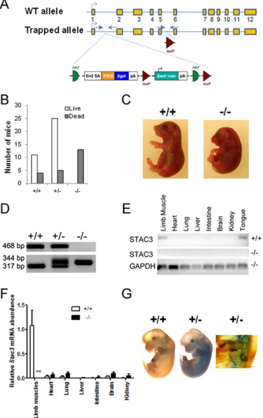
Figure 2. Generation and analyses ofStac3mutant mice. A, Schematic representation of the targetedStac3allele. The wild-type (WT)Stac3
allele has 12 exons, with exon 3 containing the translation start codon. The targeted allele is inserted with a trapping cassette between exons 1 and 2, and this insertion is expected to disrupt the generation of normalStac3 mRNA. Arrows indicate locations of PCR primers for genotyping.B, Genotypes and life of newborn offspring from intercrossing mice heterozygous for the targetedStac3allele. ‘‘+/+’’, ‘‘+/2’’, and ‘‘2/2’’ indicate wild-type mice, and mice heterozygous and homozygous for the targetedStac3allele, respectively. A total of 10 litters were included in this analysis. C, Images of aStac3homozygous mutant mouse and a wild-type littermate. Note the curved body shape and dropping forelimbs in the mutant.D,
Genotyping by PCR. See panel A for the locations of two pairs of primers.E, Validation of diminished expression ofStac3mRNA inStac32/2mice by standard RT-PCR. Shown is a representative image.F, Real-time RT-PCR validation of diminished expression ofStac3mRNA inStac32/2mice. Data are expressed as mean6SEM (n = 4). **P,0.01vs.‘‘+/+’’.G, LacZ staining of E13 wild-type andStac3+/2embryos. Shown are representative images. The
image on the right highlights the stained somites in an E13Stac3+/2embryo.
Island, NY). Nuclei were stained with DAPI (Invitrogen). Aqueous Prolong Gold antifade reagent mounting medium was applied over the sections for preservation (Invitrogen). Fluorescent images were captured with a Nikon eclipse TS100 connected to a Cool-SNAP HQ2 monochrome camera (Photometrics, Tucson, AZ) using a NIS-Elements AR Ver3.1 software program (Nikon Imaging Inc., Melville, NY).
Quantitative analyses of myofibers
The EDL muscle was used to determine muscle fiber characteristics due to its uniformity and easy recognition. Total number and average cross sectional area of myofibers in an EDL muscle were determined by counting and averaging all myosin heavy chain (MyHC) positive fibers on three serial cross sections at the widest girth of the EDL muscle using the NIS-Elements AR Ver3.1 software.
Statistical analyses
Data were analyzed using ANOVA followed by Tukey HSD multiple comparison of means in the JMP software (SAS, Cary, NC). A difference was considered statistically significant when
P,0.05. Data are presented as means 6 standard error of the mean (SEM).
Results
Stac3mRNA was predominantly expressed in skeletal muscle
Searching the gene expression database at GeneNote with ‘‘skeletal muscle’’ [10], the program generated a list of human genes that were predicted to be specifically expressed in skeletal muscle. This list containedStac3. We became more interested in this gene than the other genes on the list because it had no known function but was predicted to encode a protein containing a SH3 domain and a C1 domain, two classical functional domains [15,16]. We examined the expression profile ofStac3mRNA in adult mice by quantitative RT-PCR. As shown in Fig. 1, Stac3 mRNA was abundantly expressed in skeletal muscle of four different locations, but was not expressed or expressed at very low levels in any of non-skeletal muscle tissues or organs examined, including brain, heart, and the smooth muscle-containing stomach.
Stac3mutant mice died at birth
Mice heterozygous for the targetedStac3allele (Stac3+/2) were generated by injecting blastocysts with theStac3-targted ES cells (Fig. 2A). Both male and female Stac3+/2mice were viable and fertile and grew normally compared to their wild-type littermates. Breeding between Stac3+/2 mice resulted in normal-sized litters with a near Mendelian representation of genotypes, but all of the mice homozygous for the targeted Stac3 allele (Stac32/2) were found dead at birth (Fig. 2B). AllStac32/2mice had a curved body and dropping forelimbs (Fig. 2C).Stac32/2fetuses were never seen move and did not respond to touch, but they had heart beating when dissected out of the uterus. Genotypes ofStac32/2orStac3+/ 2 mice were confirmed by PCR (Fig. 2D). At E18.5, Stac32/2
fetuses weighed approximately 11% and14% less than their heterozygous and wild-type littermates, respectively (P,0.05).
Stac3mRNA expression was markedly diminished in
Stac3mutant mice
Insertion of the trapping cassette between exon 1 and exon 2 of theStac3allele was expected to disrupt the generation of normal
Stac3transcript (Fig. 2A). This was confirmed by the fact thatStac3
mRNA was barely detectable in any tissues examined, including skeletal muscle, in E18.5Stac32/2fetuses (Fig. 2E and 2F). As in adult wild-type mice (Fig. 1), Stac3 mRNA was preferentially expressed in skeletal muscle or skeletal muscle-containing organs such as the tongue in E18.5 wild-type fetuses (Fig. 2E and 2F). The trapping cassette contained ab-galactosidase gene, and this gene was expected to be expressed under the control of the original
Stac3promoter (Fig. 2A). LacZ staining of E13 embryos confirmed
Figure 3. Histological analyses of skeletal muscles in newborn
Stac3 homozygous mutant mice (2/2) and wild-type (+/+) littermates. A–H, Hematoxylin and eosin staining of diaphragm, tongue, tibialis anterior (TA), and extensor digitorum longus (EDL) muscles. Note the difference in the location of myonuclei between the two genotypes.IandJ, Immunohistochemical staining of EDL muscle for myosin heavy chain protein (red) and nuclei (blue). Shown are representative images taken from matched areas. Scale bars = 50mm for micrographs A, B, I, and J. Scale bars = 25mm for micrographs C-H. doi:10.1371/journal.pone.0062760.g003
the expected activity of b-galactosidase in somites and limb muscles inStac3+/2but not in wild-type embryos (Fig. 2G).
Skeletal muscle ofStac3 mutant mice had abnormal morphology
Based on gross dissection and H&E staining,Stac32/2mice had no obvious structural aberrations in viable organs such as heart, liver, kidney, and the gastrointestinal tract, and had no edema or necrosis, disorders that are typically associated within uterodeath. The diaphragms of newbornStac32/2mice were slightly thinner than those of their wild-type littermates (Fig. 3A and 3B). In newborn wild-type mice, most myofibers had peripherally located nuclei (Fig. 3A, 3C, 3E, 3G, and 3I) and were polygonally shaped on cross sections (Fig. 3A, 3C, and 3I). However, in theirStac32/2
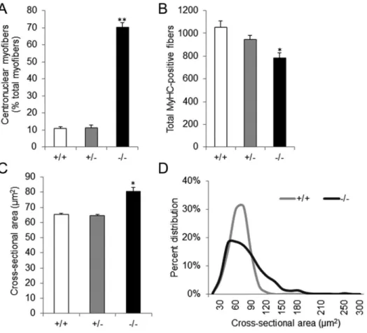
littermates, the majority of myofibers had centrally located nuclei (Fig. 3B, 3D, 3F, 3H, and 3J) and appeared round on cross sections (Fig. 3B, 3D, and 3J). Quantitative analyses of the EDL muscle revealed that more than 70% of myosin heavy chain-positive cells in newborn Stac32/2 mice had centrally located nuclei, whereas that number was only 11% in wild-type or heterozygous littermates (P,0.01) (Fig. 4A).
Skeletal muscle ofStac3 mutant mice had fewer but larger myofibers
The total number of myofibers in the EDL muscle of newborn
Stac32/2 mice was 26% or 18% less than that of wild-type or heterozygous littermates, respectively (P,0.05, Fig. 4B). However,
the average cross-sectional area (CSA) of myofibers in the EDL muscle ofStac32/2mice was 20% greater than that of wild-type or
Stac3+/2 mice (P,0.01, Fig. 4C). The myofibers in the EDL muscle of newborn wild-type or Stac3+/2 mice had a normal distribution of CSA (Fig. 4D). Those inStac32/2mice, however, had a skewed-to-the right distribution (Fig. 4D). Despite these differences, the average size of the EDL muscle was not different between Stac32/2 mice and their wild-type or heterozygous littermates (P.0.1).
Myofibers ofStac3mutant mice had fewer, smaller, and disorganized myofibrils
We examined the ultrastructure of skeletal muscle with trans-mission electron microscopy (TEM). Myofibers in the EDL muscle of newborn wild-type mice had abundant, well-aligned, and near-uniform myofibrils and sarcomeres (Fig. 5A and 5C). In contrast, those of newborn Stac32/2 littermates had fewer, smaller, disarrayed, and sometimes fragmented myofibrils (Fig. 5B and 5D). Compared to sarcomeres in the EDL muscle of wild-type mice (Fig. 5C), those of Stac32/2 mice had streaming Z-lines (Fig. 5D). The TEM also confirmed the central location of myonuclei in newbornStac32/2mice (Fig. 5B) and the peripheral location of myonuclei in newborn wild-type mice (Fig. 5A).
Discussion
In this study, we showed that the functionally unknownStac3
gene was specifically expressed in skeletal muscle, and we investigated the potential role of this gene in skeletal muscle development and function by disrupting its transcription in mice. AllStac32/2 mice were found dead at birth. This demonstrates thatStac3is essential to the development of viable mice. Whether at birth or dissected from the uterus, theStac32/2 mice had an abnormal body curvature and dropping forelimbs, and did not respond to prodding. These phenotypes are similar to those caused by mutation of the myogenin gene [7,8], the ryanodine receptor gene [17], and the dihydropyridine receptor gene [18], genes that are known to play a critical role in either the development of functional skeletal muscle or contraction of skeletal muscle. When a mouse is born, it becomes dependent on respiration for oxygen intake, which requires contraction of diaphragm and intercostal muscles [19,20]. Therefore, it is likely thatStac3mutant mice died at birth because their dysfunctional diaphragm and intercostal muscles could not support breathing. Because Stac3 is not
expressed in the nervous system, it is unlikely thatStac3mutant mice die from dysfunctional neural control.
Histological analyses revealed that the skeletal muscle of newbornStac32/2 mice had several abnormalities compared to that of wild-type mice, including: 1) most of their nuclei remained in the center of myofibers; 2) they had fewer yet larger myofibers; 3) they had a disproportional number of myofibers with large cross-sectional areas; 4) their myofibers had fewer, smaller, and less organized myofibrils. With these abnormalities, a skeletal muscle is unlikely able to function. Among these abnormalities, the most striking was that most of the myonuclei were located in the center of myofibers at a developmental stage (i.e. birth) when they should have been relocated to the periphery of myofibers [21]. While having centrally located nuclei is typically indicative of regeneration in adult skeletal muscle [5,22], it is unlikely that skeletal muscle in fetalStac32/2 mice was undergoing regenera-tion because every skeletal muscle examined had centrally located nuclei. Translocation of myonuclei from the center to the periphery is the hallmark of myofiber maturation during late embryonic development [21]. Therefore, it is possible thatStac3
Figure 5. Representative electron micrographs of extensor digitorum longus (EDL) muscle of newbornStac32/2mice andStac3+/+
littermates.Note the differences in the location of myonuclei and the size and arrangement of myofibrils betweenStac3homozygous mutant and wild-type muscles. Arrows point to myonuclei (micrographs A and B), and arrowheads indicate Z-lines (micrographs C and D). Scale bars = 10mm for
micrographs A and B; Scale bars = 500 nm for micrographs C and D. doi:10.1371/journal.pone.0062760.g005
gene mutation disrupted myofiber maturation. This possibility is also supported by the observation that Stac32/2 myofibers had fewer and smaller myofibrils than wild-type myofibers at the same developmental stage. It is also possible that Stac3 disruption disrupted the ill-defined mechanism that controls the translocation of myonuclei during skeletal muscle development.
Myofibers of Stac32/2 mice had greater cross-sectional areas than those of wild-type orStac3+/2littermates. This difference was probably not due to myofiber hypertrophy in Stac32/2 mice because myofibers in those mice indeed had fewer and smaller myofibrils than in wild-type orStac3+/2. The difference was more likely caused by increased fusion of myoblasts into myotubes in
Stac3 mutant mouse embryos. The relative numbers of pro-liferating myoblasts versus differentiating myoblasts during muscle development can determine the total number of myofibers formed in a mature muscle [23,24]. If myoblasts withdraw from the cell cycle and begin differentiation before an adequate number of founder myoblasts have developed, this would decrease the overall number of myofibers formed. Therefore, an increase in myoblast fusion could also explain why aStac32/2skeletal muscle had fewer myofibers than a wild-type or a Stac3+/2 skeletal muscle. Myogenesis is a highly orchestrated program, and premature myoblast fusion could lead to the formation of dysfunctional muscle [25,26]. Therefore, increased fusion of myoblasts may also explain why skeletal muscle in Stac3mutant mice was unable to contract.
Unlike the master regulators of skeletal muscle development, the MRFs (MyoD, myogenin, MRF4, Myf5), Stac3 is not a transcrip-tion factor and does not appear to have direct DNA-binding ability. The Stac proteins are believed to be confined to the cytosolic domain of the cell with no evidence for enzymatic activity [15,27]. However, Stac3 does possess two domains (SH3 and C1) that are frequent components of signaling proteins [16,28].
Interestingly, two known regulators of myoblast fusion, Mbc and DCrk, also contain the SH3 domain [29,30]. Therefore, Stac3 might be involved in the process of myoblast fusion through the SH3 domain. However, both the SH3 domain and the C1 domain can interact with multiple partners in a cell [29,30]. The multiple skeletal muscle phenotypes caused byStac3gene disruption may be mediated by different Stac3-protein interactions.
In summary, we have demonstrated that the Stac3 gene is exclusively expressed in skeletal muscle and that it is essential for development of functional skeletal muscle and mice viable at birth. The domain structures of Stac3 protein suggest that it is a signaling protein, and further investigation into the molecular and cellular cause of abnormalities inStac3mutant skeletal muscle may reveal novel signaling mechanisms that regulate skeletal muscle de-velopment or function. The centrally located myonuclei and lack of muscle contraction phenotypes of Stac3 mutant mice are hallmarks of centronuclear/myotubular myopathies in humans [31,32]. Therefore, theStac3mutant mouse may represent a useful animal model for understanding the pathophysiology of this muscle disease. While this manuscript was being prepared, a study published in JBC indicates that Stac3 is essential for myotube formation in zebrafish [33]. Our study, however, shows thatStac3
gene inactivation does not prevent myotube formation in mice (Fig. 3, Fig. 5). The reason for this difference is not clear, but it may be related to the use of different model systems between the two studies.
Author Contributions
Conceived and designed the experiments: HJ. Performed the experiments: BMR XG XC. Analyzed the data: BMR XG XC DEG HJ. Wrote the paper: BMR XG DEG HJ.
References
1. Rudnicki MA, Braun T, Hinuma S, Jaenisch R (1992) Inactivation of MyoD in Mice Leads to up-Regulation of the Myogenic HLH Gene Myf-5 and Results in Apparently Normal Muscle Development. Cell 71: 383–390.
2. Braun T, Buschhausen-Denker G, Bober E, Tannich E, Arnold HH (1989) A novel human muscle factor related to but distinct from MyoD1 induces myogenic conversion in 10T1/2 fibroblasts. EMBO J 8: 701–709.
3. Wright WE, Sassoon DA, Lin VK (1989) Myogenin, a factor regulating myogenesis, has a domain homologous to MyoD. Cell 56: 607–617. 4. Rhodes SJ, Konieczny SF (1989) Identification of MRF4: a new member of the
muscle regulatory factor gene family. Genes Dev 3: 2050–2061.
5. Charge SB, Rudnicki MA (2004) Cellular and molecular regulation of muscle regeneration. Physiological Reviews 84: 209–238.
6. Rudnicki MA, Schnegelsberg PNJ, Stead RH, Braun T, Arnold HH, et al. (1993) MyoD or Myf-5 Is Required for the Formation of Skeletal-Muscle. Cell 75: 1351–1359.
7. Nabeshima Y, Hanaoka K, Hayasaka M, Esumi E, Li S, et al. (1993) Myogenin gene disruption results in perinatal lethality because of severe muscle defect. Nature 364: 532–535.
8. Hasty P, Bradley A, Morris JH, Edmondson DG, Venuti JM, et al. (1993) Muscle deficiency and neonatal death in mice with a targeted mutation in the myogenin gene. Nature 364: 501–506.
9. Yoon JK, Olson EN, Arnold HH, Wold BJ (1997) Different MRF4 knockout alleles differentially disrupt Myf-5 expression: cis-regulatory interactions at the MRF4/Myf-5 locus. Dev Biol 188: 349–362.
10. Yanai I, Benjamin H, Shmoish M, Chalifa-Caspi V, Shklar M, et al. (2005) Genome-wide midrange transcription profiles reveal expression level relation-ships in human tissue specification. Bioinformatics 21: 650–659.
11. Kawai J, Suzuki H, Hara A, Hirose K, Watanabe S (1998) Human and mouse chromosomal mapping of Stac, a neuron-specific protein with an SH3 domain. Genomics 47: 140–142.
12. Legha W, Gaillard S, Gascon E, Malapert P, Hocine M, et al. (2010) stac1 and stac2 genes define discrete and distinct subsets of dorsal root ganglia neurons. Gene Expr Patterns 10: 368–375.
13. Skarnes WC, Rosen B, West AP, Koutsourakis M, Bushell W, et al. (2011) A conditional knockout resource for the genome-wide study of mouse gene function. Nature 474: 337–342.
14. Buchberger A, Nomokonova N, Arnold HH (2003) Myf5 expression in somites and limb buds of mouse embryos is controlled by two distinct distal enhancer activities. Development 130: 3297–3307.
15. Abrams CS, Zhao W (1995) SH3 domains specifically regulate kinase activity of expressed Src family proteins. Journal of Biological Chemistry 270: 333–339. 16. Colon-Gonzalez F, Kazanietz MG (2006) C1 domains exposed: from
diacylglycerol binding to protein-protein interactions. Biochim Biophys Acta 1761: 827–837.
17. Takeshima H, Iino M, Takekura H, Nishi M, Kuno J, et al. (1994) Excitation-contraction uncoupling and muscular degeneration in mice lacking functional skeletal muscle ryanodine-receptor gene. Nature 369: 556–559.
18. Pai AC (1965) Developmental Genetics of a Lethal Mutation, Muscular Dysgenesis (Mdg), in the Mouse. I. Genetic Analysis and Gross Morphology. Developmental Biology 11: 82–92.
19. Copp AJ (1995) Death before birth: clues from gene knockouts and mutations. Trends in Genetics 11: 87–93.
20. Turgeon B, Meloche S (2009) Interpreting neonatal lethal phenotypes in mouse mutants: insights into gene function and human diseases. Physiological Reviews 89: 1–26.
21. Ontell M, Kozeka K (1984) The organogenesis of murine striated muscle: a cytoarchitectural study. American journal of anatomy 171: 133–148. 22. Mitchell CA, McGeachie JK, Grounds MD (1992) Cellular differences in the
regeneration of murine skeletal muscle: a quantitative histological study in SJL/J and BALB/c mice. Cell and Tissue Research 269: 159–166.
23. Dlugosz AA, Tapscott SJ, Holtzer H (1983) Effects of phorbol 12-myristate 13-acetate on the differentiation program of embryonic chick skeletal myoblasts. Cancer Research 43: 2780–2789.
24. Atreya KB, Fernandes JJ (2008) Founder cells regulate fiber number but not fiber formation during adult myogenesis in Drosophila. Developmental Biology 321: 123–140.
25. Bentzinger CF, Wang YX, Rudnicki MA (2012) Building muscle: molecular regulation of myogenesis. Cold Spring Harb Perspect Biol 4.
27. Suzuki H, Kawai J, Taga C, Yaoi T, Hara A, et al. (1996) Stac, a novel neuron-specific protein with cysteine-rich and SH3 domains. Biochem Biophys Res Commun 229: 902–909.
28. Mayer BJ (2001) SH3 domains: complexity in moderation. Journal of Cell Science 114: 1253–1263.
29. Dworak HA, Sink H (2002) Myoblast fusion in Drosophila. Bioessays 24: 591– 601.
30. Chen EH, Olson EN (2004) Towards a molecular pathway for myoblast fusion in Drosophila. Trends Cell Biol 14: 452–460.
31. Jungbluth H, Wallgren-Pettersson C, Laporte J (2008) Centronuclear (myotub-ular) myopathy. Orphanet J Rare Dis 3: 26.
32. Romero NB (2010) Centronuclear myopathies: a widening concept. Neuromus-cular Disorders 20: 223–228.
33. Bower NI, de la Serrana DG, Cole NJ, Hollway GE, Lee HT, et al. (2012) Stac3 is required for myotube formation and myogenic differentiation in vertebrate skeletal muscle. Journal of Biological Chemistry 287: 43936–43949.