Quantitative LC-MS/MS Analysis of Proteins
Involved in Metastasis of Breast Cancer
Rieko Goto1,2*, Yasushi Nakamura1, Tomonori Takami2, Tokio Sanke1, Zenzaburo Tozuka3
1Department of Clinical Laboratory Medicine, Wakayama Medical University,Wakayama, Japan,2JCL Bioassay Corporation, Nishiwaki, Hyogo, Japan,3Graduate School of Pharmaceutical Science Osaka University, Suita, Osaka, Japan
Abstract
The purpose of this study was to develop quantitative liquid chromatography-tandem mass spectrometry (LC-MS/MS) methods for the analysis of proteins involved in metastasis of breast cancer for diagnosis and determining disease prognosis, as well as to further our understand of metastatic mechanisms. We have previously demonstrated that the protein type XIV collagen may be specifically expressed in metastatic tissues by two dimensional LC-MS/MS. In this study, we developed quantitative LC-MS/MS methods for type XIV colla-gen. Type XIV collagen was quantified by analyzing 2 peptides generated by digesting type XIV collagen using stable isotope-labeled peptides. The individual concentrations were equivalent between 2 different peptides of type XIV collagen by evaluation of imprecise tran-sitions and using the best transition for the peptide concentration. The results indicated that type XIV collagen is highly expressed in metastatic tissues of patients with massive lymph node involvement compared to non-metastatic tissues. These findings were validated by quantitative real-time RT-PCR. Further studies on type XIV collagen are desired to verify its role as a prognostic factor and diagnosis marker for metastasis.
Introduction
The 5-year breast cancer survival rate is generally greater than 90% but it is roughly 55% in women with numerous lymph node metastases [1–3]. The prognosis of breast cancer associ-ated with nodal metastasis, age, tumor size (pT), histological grade of tumor, estrogen and pro-gesterone receptors status, human epidermal growth factor receptors (HERs) status, and breast cancer (BRCA) gene mutations. Nodal status (including number and location of nodes) corre-lates with disease-free and overall survival better than any other prognostic factor. Also, long-term prognosis depends on tumor stage. Treatment of breast cancer relies on surgical excision and various forms of systemic therapy. However, as metastasized and recurrent breast cancers (especially breast cancer with massive nodal metastasis) are difficult to cure, the development of new therapeutic agents including molecular targeted drugs is awaited. We have searched for the role of lymph node- and metastasis-related biomarkers [4]. Lymph nodes are the first site
OPEN ACCESS
Citation:Goto R, Nakamura Y, Takami T, Sanke T, Tozuka Z (2015) Quantitative LC-MS/MS Analysis of Proteins Involved in Metastasis of Breast Cancer. PLoS ONE 10(7): e0130760. doi:10.1371/journal. pone.0130760
Editor:William B. Coleman, University of North Carolina School of Medicine, UNITED STATES
Received:March 2, 2015
Accepted:May 22, 2015
Published:July 15, 2015
Copyright:© 2015 Goto et al. This is an open access article distributed under the terms of the Creative Commons Attribution License, which permits unrestricted use, distribution, and reproduction in any medium, provided the original author and source are credited.
Data Availability Statement:All relevant data are within the paper.
Funding:JCL Bioassay Corporation, Nishiwaki, Hyogo, Japan provided support in the form of salaries for authors RG & TT, but did not have any additional role in the study design, data collection and analysis, decision to publish, or preparation of the manuscript.
of metastasis for several carcinomas, and the extent of lymph node metastasis is a major crite-rion for evaluating patient prognosis.
In previous studies in which we screened for metastasis-specific proteins, breast cancer tis-sues from a non-metastasis group and a massive lymph node metastasis group were compared based on comprehensive analysis using 2-dimensional liquid chromatography-tandem mass spectrometry (2D LC-MS/MS); 34 proteins were screened as specific candidate proteins involved in metastasis by 2D LC-MS/MS analysis. Type XIV collagen in particular was found to be selectively expressed in specimens from the massive lymph node metastasis group; there-fore the protein may be a potential biomarker of metastasis and useful for evaluating patient prognosis. The difference in expression was confirmed by immunohistochemistry [5–8]. How-ever, in the 2D LC-MS/MS analysis, the 34 proteins were screened by protein score based on XCorr, which is measured from mass and MS/MS peaks and correlates with peptide sequence [9,10]; the proteins have not yet been demonstrated by quantitative analysis. The discovery of candidate biomarker proteins is often hampered by false-positive results caused by impreci-sion, and quantification of targeted proteins is needed for screening of candidate protein bio-markers [9–15]. Therefore, development of quantitative of methods for directly measuring expression of these proteins, including type XIV collagen, could be useful for diagnosis, prog-nosis, and understanding of the mechanisms of metastasis. Multiple reaction monitoring (MRM)-based and selected reaction monitoring (SRM)-based quantification using LC-MS/MS have been used for quantitative simultaneous analysis of multiple proteins without antibodies [16–20]. In MRM and SRM analysis, ion suppression of small molecules–i.e., reduction of sen-sitivity due to ionization of endogenous substances in the biological fluid matrix–is a major issue that must be considered. To avoid the influence of ion suppression on quantification, sta-ble isotope-labeled peptide, which co-elutes with the endogenous peptide, is used [21–23]. The evaluation of imprecise transitions is important in protein quantification because interference in biological fluids due to impurities affects quantification [24,25].
Our purpose in the present study was to develop quantitative LC-MS/MS methods for the analysis of proteins involved in metastasis of breast cancer, so as to facilitate diagnosis and determination of disease prognosis, as well as to shed light on the mechanisms involved in metastasis. In this preliminary study, specific candidate proteins were selected, and as model of quantification of the proteins, type XIV collagen was quantified with stable isotope-labeled peptides by LC-MS/MS and the expression compared between massive lymph node metastasis tissues and non-metastasis tissues.
Materials and Methods
1. Ethics statement
Institutional review board of Wakayama Medical University approved this study, and all patients had given written informed consent.
2. Subjects and samples
Fresh tumor tissue specimens of primary lesions were obtained after surgical resection from 220 patients with primary breast cancer. Each tumor specimen was stored at−80°C prior to
invasive cancer but was free of lymph node metastasis; no lymph node metastases were detected on pathological examination, and the samples were confirmed to be negative for cyto-keratin 19 by the one-step nucleic acid amplification (OSNA) method. The other group con-sisted of patients with massive lymph node involvement with more than 10 lymph node metastases detected on pathological examination. This type of cancer is highly aggressive and generally has an extremely low survival rate. In this study, the non-metastasis group was com-prised of six cases (subjects 1–6), and the massive lymph node metastasis group included 13 cases (subjects 7–19).
3. Quantitative LC-MS/MS analysis
3.1. Protein purification. Each tissue specimen (a slice600μm thick) was dissociated
into single cells by addition of 150μL collagenase solution [500μg/mL collagenase, 137 mM NaCl, 10 mM HEPES (pH 7.5) 5 mM KCl, 5 mM CaCl2, 4 mM NaHCO3, 0.8 mM Na2
H-PO4.2H2O, 0.5 mM NaH2PO4.H2O, and 2μL protease inhibitor cocktail], followed by incuba-tion at 37°C for 10 min. The cells were washed with 450μL minimum essential medium (MEM) and separated by centrifugation (10 × g, for 2 min at 4°C). The supernatant was removed, and the residue containing the cells was washed twice in 600μL PBS buffer. The cells were recovered by centrifugation (160 × g, for 5 min at 4°C), resuspended in 600μL buffer A [10 mM HEPES-NaOH (pH 7.9), 10 mM KCl, 1.5 mM MgCl2, 0.5 mM DTT, 0.5 mM PMSF,
2μL protease inhibitor cocktail], and recovered again by centrifugation (1000 × g, for 10 min at 4°C). The residue containing the cells was placed in 340μL of new buffer A and homoge-nized slowly by rotating 20 times on ice at 70 rpm. The homogenates were centrifuged at 1000 × g for 10 min at 4°C. The supernatant containing the cytoplasm was stored at−80°C
until analysis. The total protein content in purified samples was determined by a protein assay based on the Bradford method (Bio-Rad Laboratories, Hercules, CA, USA), using bovine serum albumin as a standard.
3.2. Protein digestion. Purified samples were diluted with 50 mM NH4HCO3to 40μL
total volume and reduced with 10μL of 100 mM DTT in 50 mM NH4HCO3for 60 min at
50°C. Cysteine residues were alkylated with 20μL of 100 mM iodoacetamide in 50 mM NH4HCO3in the dark at room temperature for 30 min. Each mixture was proteolyzed to
pep-tides with 20μL of 5μg/mL trypsin (Promega, Madison, WI, USA) in 50 mM NH4HCO3for
16 h at 37°C. Proteolysis was stopped by addition of 10μL of 10% formic acid.
3.3. Protein and peptide selection for quantification. Proteins with higher proteotypic
peptide response were screened as specific candidate proteins by in silico digestion using Pin-point software (version 1.0 Thermo Fisher ScientiFIc). MS/MS spectra of 4 to 27 amino acid peptides were composited with in silico digestion. MS/MS spectra of 2D LC-MS/MS were com-pared to the composite MS/MS spectra with Xcorr values above 2.0, 2.0, and 3.3 for 1+, 2+, and 3+ charge states of the peptide. Five MS/MS spectra with high intensity in each peptide of the candidate protein were selected from m/z 600–1250 for protein quantification.
3.4. Peptide selection by MRM analysis without internal standard. To confirm
target peptide peaks originating from the protein [20]. Based on the difference in peptide abun-dance between the 2 groups, candidate proteins whose retention times were the same among multi-transition chromatograms and transitions with peak height greater than 1000 cps were selected. Three transitions for each target peptide of the candidate proteins were selected based on the formation of higher intensity product ions.
3.5. Quantification with stable isotope-labeled peptides. For the development of
quanti-tative analytical methods, 2 stable isotope-labeled peptides AQUA peptides corresponding to type XIV collagen (purchased from Thermo Fisher Scientific) were used as internal standards.
Purified samples in triplicate (6μg total protein per replicate) from each subject were
digested in the presence of stable isotope-labeled peptides (2 pmol), and the digestion samples (3μg total protein) were quantitatively analyzed by MRM using stable isotope-labeled peptides.
Equipment used was the same as that for MRM analysis without internal standard. LC meth-ods, masses, collision energy (CE), declustering potential (DP), and collision cell exit potential (CXP) were optimized for quantitative analysis using each AQUA peptide. Elution was per-formed at a flow rate of 0.2 mL/min with a linear gradient of 3% to 65% acetonitrile containing 0.1% formic acid for 29 minutes. Initially, we set the following 2 criteria to determine the con-centration of a protein: 1) the retention times of peaks in multichromatograms of transitions should be the same between endogenous peptide, thereby confirming that the peptide origi-nated from the target protein [20]; and 2) the transition should have an average peak height for endogenous peptide above 1000 cps in the massive lymph node metastasis group.
In addition to these criteria, we subsequently added a third to reduce the influence of matrix-derived interference, which became apparent later. The third criterion was exclusion of a transition from the peptide concentration calculation if the evaluation of imprecise transi-tions was influenced by interference. For example, when there is a transition for a subject whose coefficient of variation (CV) of intra-assay reproducibility (n = 3) is above 50, the transi-tion should be excluded from peptide concentratransi-tion calculatransi-tions. Finally, the best transitransi-tion with high specificity and high sensitivity was selected from transitions which met the 3 criteria, and concentrations of the best transition were used as the peptide concentrations. The peptide concentrations were interpreted as protein concentrations.
4. Validation by Quantitative real time Reverse Transcription
Polymerase Chain Reaction (real-time RT-PCR)
COL14A1 or GAPDH was purchased from Applied Biosystems, and PCR was carried out according to the manufacturer’s protocol.
Results
1. Quantitative LC-MS/MS analysis
1.1 Protein and peptide selection for quantification. By comprehensive analysis using
2D LC-MS/MS, 34 proteins were screened as specific candidate proteins expressed highly in massive lymph node metastasis breast cancer [5]. From these 34 proteins, those whose transi-tions were confirmed by Pinpoint software were selected for MRM analysis; i.e., 104 MRM transitions for 22 peptides of 7 proteins (4–5 transitions per peptide).
1.2. Peptide selection by MRM analysis without internal standard. In order to confirm
differential abundance of a specific candidate protein, the 104 MRM transitions for 22 peptides of 7 proteins from each subject were analyzed using a QTRAP5500 system. From the results of the 104 MRM transitions, 58 MRM transitions for 13 peptides of 7 proteins (3–5 transitions per peptide) were selected as candidates, since the fragment ions had higher intensities. From the results of the 58 MRM transitions, 28 MRM transitions for 9 peptides of 7 proteins (3 tran-sitions per peptide) were selected as the best trantran-sitions for each peptide, since the fragment ions had higher intensities. In the results of the 28 MRM transitions for 9 peptides of 7 pro-teins, the retention time of the peaks differed among multiple transition of 1 peptide. There-fore, the protein for which the retention time between peptides differed was excluded, and the remaining 6 proteins were selected as specific candidate proteins. The proteins were type XIV collagen, hexokinase I, MSTP161, angiomotin, alpha-2 type I collagen, and alpha-1 type I collagen.
1.3. Quantification with stable isotope-labeled peptides. Type XIV collagen was
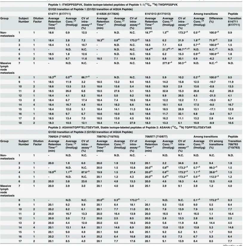
mea-sured by quantitative LC-MS/MS with stable isotope-labeled peptides via analyzing 12 MRM transitions for 2 peptides generated by digesting type XIV collagen (3 transitions per peptide) within 1 injection. The quantitative results of purified samples in triplicate from each subject (subjects 1–6, non metastasis; subjects 7–18, massive lymph node metastasis) are listed in
Table 1. Seven samples were diluted as necessary, and the sample for subject 19 was not ana-lyzed due to insufficient quantity. All transitions had the same retention time between the endogenous peptide and the stable isotope-labeled peptides, and the average peak heights of endogenous peptide in the massive lymph node metastasis group were greater than 1000 cps, which satisfied the criteria.
Fig 1shows the average intra-assay concentrations (n = 3) in all transitions (614/711, 614/826 and 614/1013) for peptide ITWDPPSSPVK, 2 transitions (614/711 and 614/826) for peptide ITWDPPSSPVK, and all transitions (708/624, 708/763 and 708/877) for peptide ASA-HAITGPPTELITSEVTAR of type XIV collagen. Although, theoretically, the concentration of peptides that originated from the same protein should be the same, the difference in the aver-age intra-assay concentration between peptides ITWDPPSSPVK and ASAHAITGPPTELIT-SEVTAR of type XIV collagen was remarkable in subjects 4 and 7 (Fig 1A and 1B, and
Table 1). This discrepancy arose from an extremely high concentration of transition 614/1013 of peptide ITWDPPSSPVK.
Table 1. Concentrations of type XIV collagen determined by quantitative LC-MS/MS analysis with stable isotope-labeled peptides.
Peptide 1: ITWDPPSSPVK, Stable isotope-labeled peptides of Peptide 1: I (13C
6,15N) TWDPPSSPVK
Q1/Q3 transition of Peptide 1 (Q1/Q3 transition of AQUA Peptide)
614/711 (617/711) 614/826 (617/826) 614/1013 (617/1013) Among transitions Peptide Group Subject Number Dilution Factor Average Retention Time*1
(min)
Average Conc.*2
(fmol/μg) CV of intra-assay*3
(%)
Average Retention Time*1
(min)
Average Conc.*2
(fmol/μg) CV of intra-assay*3
(%)
Average Retention Time*1
(min)
Average Conc.*2
(fmol/μg) CV of intra-assay*3
(%)
Average Conc.*6
(fmol/μg)
Difference (%) Transition 614/711 (617/711) Non-metastasis
1 1 18.6 0.9 12.5 - N.D. N.C. 18.7#1 1.5#1 173.2 *5 0.5
*5 100.0
*5 0.9
2 1 18.4 2.8 7.2 18.3#1 0.8#1 173.2
*5 18.5 6.2 31.9 1.8
*5 71.9
*5 2.8
3 1 18.4 1.5 10.7 - N.D. N.C. 18.5 7.1 6.6 0.7*5 100.0
*5 1.5
4 1 - N.D. N.C. - N.D. N.C. 18.4#2 21.2#2,
*4 96.7 *4,
*5 N.D. N.C.
*5 N.D.
5 1 18.5 1.8 12.6 - N.D. N.C. 18.6 6.2 15.3 0.9*5 100.0
*5 1.8
6 2 18.5 6.7 11.6 18.5 7.1 18.8 18.5 8.8 30.1 6.9 -6.2 6.7 Massive
lymph node metastasis
7 1 - N.D. N.C. - N.D. N.C. 18.6 277.8*4 50.3
*4 N.D. N.C.
*5 N.D.
8 1 18.3#2 0.5#2 86.7
*5 - N.D. N.C. 18.5 5.9 10.2 0.2
*5 100.0
*5 0.5
9 1 18.5 11.9 3.2 18.5 13.2 9.4 18.5 14.2 15.8 12.5 -10.7 11.9 10 2 18.6 13.5 2.5 18.6 13.6 5.4 18.6 18.9 2.9 13.6 -0.8 13.5 11 2 18.5 26.0 6.6 18.5 27.6 5.1 18.5 32.8 15.3 26.8 -6.2 26.0 12 1 18.4 4.1 8.2 18.4 5.0 9.2 18.5 9.9 39.5 4.5 -21.5 4.1 13 2 18.4 6.7 17.4 18.4 7.4 10.5 18.4 12.2 12.2 7.1 -10.3 6.7 14 4 18.4 16.7 4.8 18.4 18.2 6.6 18.4 19.1 6.6 17.5 -9.0 16.7 15 1 18.4 12.0 5.1 18.4 14.1 11.3 18.4 18.5 28.9 13.1 -17.1 12.0 16 1 18.6 9.7 6.7 18.6 10.0 0.5 18.6 11.7 32.1 9.8 -3.4 9.7 17 2 18.5 13.4 7.0 18.5 13.0 4.5 18.5 16.2 11.1 13.2 2.8 13.4 18 2 18.3 10.5 12.1 18.4 11.4 27.9 18.3 13.3 6.1 10.9 -8.9 10.5
Peptide 2: ASAHAITGPPTELITSEVTAR, Stable isotope-labeled peptides of Peptide 2: ASAHAI (13C
6,15N) TGPPTELITSEVTAR
Q1/Q3 transition of Peptide 2 (Q1/Q3 transition of AQUA Peptide)
708/624 (710/627) 708/763 (710/763) 708/877 (710/877) Among transitions Peptide Group Subject Number Dilution Factor Average Retention Time*1
(min)
Average Conc.*2
(fmol/μg) CV of intra-assay*3
(%)
Average Retention Time*1
(min)
Average Conc.*2
(fmol/μg) CV of intra-assay*3
(%)
Average Retention Time*1
(min)
Average Conc.*2
(fmol/μg) CV of intra-assay*3
(%)
Average Conc.*6
(fmol/μg)
CV among transitions (%) Transition 708/763 (710/763) Non-metastasis
1 1 - N.D. N.C. - N.D. N.C. - N.D. N.C. N.D. N.C. N.D. 2 1 20.0 1.9 6.0 20.0 1.9 13.2 20.1 2.2 34.6 2.0 9.4 1.9 3 1 - N.D. N.C. 20.0 1.5 15.8 20.0#1 0.8#1 173.2
*5 0.7
*5 99.3
*5 1.5
4 1 19.9#2 1.1#2 87.6
*5 19.9 1.5 27.4 20.0#1 0.6#1 173.2 *5 1.1
*5 39.8
*5 1.5
5 1 - N.D. N.C. 20.1 1.2 4.3 20.0#1 0.4#1 173.2 *5 0.5
*5 112.5
*5 1.2
6 2 20.0 3.8 23.6 20.1 4.8 13.3 20.0 4.6 18.3 4.4 12.5 4.8 Massive
lymph node metastasis
7 1 20.0 3.9 3.0 20.1 4.0 3.8 20.1 3.9 9.1 3.9 1.2 4.0
8 1 - N.D. N.C. 20.0#1 0.3#1 173.2
*5 - N.D. N.C. 0.1
*5 173.2
*5 0.3
9 1 20.1 9.2 9.9 20.1 8.4 18.1 20.1 9.3 13.6 9.0 5.5 8.4 10 2 20.2 8.2 6.1 20.1 7.7 11.0 20.1 7.8 13.9 7.9 3.5 7.7 11 2 20.0 16.7 13.3 20.0 16.4 13.9 20.0 16.5 9.1 16.5 1.1 16.4 12 1 20.0 3.0 7.2 20.0 2.5 8.5 20.0 2.8 13.3 2.8 8.6 2.5 13 2 20.0 4.4 12.0 20.0 4.5 18.3 20.0 5.6 11.9 4.8 14.4 4.5 14 4 20.1 13.1 9.4 20.1 14.6 6.9 20.0 13.8 12.0 13.8 5.3 14.6 15 1 20.1 9.0 4.9 20.1 9.0 6.6 20.1 9.2 6.2 9.1 1.7 9.0 16 1 20.2 7.1 5.4 20.2 6.4 10.8 20.2 6.5 7.3 6.7 5.3 6.4 17 2 20.1 8.5 4.0 20.1 7.7 17.6 20.1 9.1 13.9 8.4 8.5 7.7
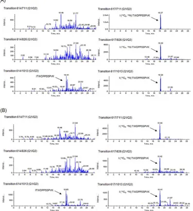
Fig 3A and 3Bshow representative MRM chromatograms for peptide ITWDPPSSPVK and the internal standard for subjects 4 and 7, respectively. As shown inFig 3, only the endogenous peptide area of transition 614/1013 in subjects 4 and 7 was detected, while other transitions (614/711 or 614/826) showed no peaks.
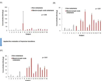
Therefore, interfering substances with fragment ions of 614/1013 seemed to be eluted in peaks overlapping with peptide ITWDPPSSPVK, resulting in miscalculated erroneously high concentrations of the target peptide in subjects 4 and 7 due to the influence of interfering substances. Furthermore, the CVs of intra-assay reproducibility (n = 3) for transition 614/ 1013 in subjects 4 and 7 were 96.7% and 50.3%, which were extremely high (Table 1). These results indicate that transition 614/1013 has low specificity. By excluding the transition 614/ 1013, the concentrations of peptide ITWDPPSSPVK corresponded to below the detection limit in all measurements for subject 4 (Table 1). Furthermore the T-testpvalue for peptide ITWDPPSSPVK was changed fromp<0.05 top<0.01, which correlated with peptide
ASA-HAITGPPTELITSEVTAR, as shown inFig 1A, 1A' and 1B.
With respect to the intra-assay reproducibility (n = 3) of each transition, there were some subjects that were occasionally detected and occasionally not detected based on difference of the transition, because the detected values were near the detection limit. By the existence of samples not detected occasionally the average concentration of transitions was shifted to the lower, the difference between 2 transitions for peptide ITWDPPSSPVK was high (71.9% and 100.0%), and the CVs among transitions for peptide ASAHAITGPPTELITSEVTAR were high (39.8–173.2%), as shown inTable 1.
By excluding the low-specificity transition 614/1013 and samples that were not detected in some measurements, the CVs of intra-assay reproducibility (n = 3) improved from 0.5–173.2 to 0.5–27.9 for peptide ITWDPPSSPVK, and from 2.7–173.2 to 2.7–34.6 for peptide
ASA-HAITGPPTELITSEVTAR (Table 1). Therefore, concentrations of transition 614/711 were used as the concentrations of peptide ITWDPPSSPVK and concentrations of transition 708/ 763 were used as the concentrations of peptide ASAHAITGPPTELITSEVTAR, since the tran-sitions have high specificity and high sensitivity. That is, the concentrations of the best transi-tion with high specificity and high sensitivity were used as the peptide concentratransi-tions, avoiding calculation of average from all transitions.
Table 1. (Continued)
18 2 20.0 8.3 20.8 20.0 8.0 5.5 20.0 7.9 2.7 8.1 2.8 8.0
*1: Average retention time; Average retention time of triplicate samples (#1: Retention time of one sample,#2: Average retention time of duplicate
samples.-: All samples were below detection limit.).
*2: Average Conc.; Average intra-assay concentration (n = 3) (#1: Concentration of one sample,#2: Average concentration of duplicate samples, N.D.: Not
detected because concentrations of all triplicate samples were not detected, and the concentration was calculated as 0.0.).
*3: CV of intra-assay; Coefficient of variation of intra-assay reproducibility (n = 3) (N.C.: Not calculated because concentrations of all tripricate samples were not detected.)
*4: Value including interferences
*5: Value including detected samples and non-detected samples
*6: Average conc.; Average concentration from 2 transitions (614/711and 614/826) for peptide 1 and 3 transitions (708/624, 708/763, 708/877) for peptide
2.
*7: Difference of peptide 1; Difference (%) = (Conc. of 614/711-Conc. of 614/826)×100 /Conc. of 614/711, (N.C.: Not calculated because concentration of 614/711 was 0.)
#1: Value of 1 sample because other duplicate samples were not detected. #2: Average of duplicate samples because another sample was not detected.
Fig 4shows the individual concentrations of peptide ITWDPPSSPVK and peptide ASA-HAITGPPTELITSEVTAR after evaluation of imprecise transitions and using the best transi-tion for the peptide concentratransi-tion. The T test p-value for peptide ITWDPPSSPVK was
changed fromp<0.05 top<0.01, which is equivalent to that for peptide
ASAHAITGPPTELIT-SEVTAR.Fig 5shows that the individual concentrations calculated from the best transitions were correlated between peptide ITWDPPSSPVK and peptide ASAHAITGPPTELITSEVTAR (p<0.001;r= 0.9558). The results show that both peptides ASAHAITGPPTELITSEVTAR and
ITWDPPSSPVK of type XIV collagen are specific to the massive lymph node metastasis group.
2. Validation by quantitative RT-PCR
The m-RNA expression of type XIV collagen was analyzed by quantitative real-time RT-PCR.
Fig 6shows the correlation between type XIV collagen m-RNA expression and protein
Fig 1. Average intra-assay concentration of peptide ITWDPPSSPVK and peptide ASAHAITG PPTELITSEVTAR of type XIV collagen.Peptides from each subject (subjects 1–6: non metastasis, subjects 7–18: massive lymph node metastasis) were quantified (n = 3) by LC-MS/MS. The average intra-assay concentration and SD of peptide concentrations from individual proteins are calculated from all transitions (614/711, 614/826 and 614/1013) for peptide ITWDPPSSPVK of Protein 12 (A), 2 transitions (614/711 and 614/826) for peptide ITWDPPSSPVK (A'), and all transitions (708/624, 708/763 and 708/877) for peptide ASAHAITGPPTELITSEVTAR (B) of type XIV collagen. By excluding the low specificity transition 614/1013, the concentration of peptide ITWDPPSSPVK was changed (A) to (A'), which correlated with the concentrations of (B). Significant difference between non metastasis and massive lymph node metastasis in (A') and (B).
doi:10.1371/journal.pone.0130760.g001
Fig 2. Average peak area of internal standards of peptide ITWDPPSSPVK and peptide ASAHAITGPPTELITSEVTAR.The internal standards of peptide ITWDPPSSPVK and peptide ASAHAITGPPTELITSEVTAR were peptide I (13C
6,15N) TWDPPSSPVK and peptide ASAHAI (13C6,15N)
TGPPTELITSEVTAR, respectively. The peak areas of peptide I(13C
6,15N)TWDPPSSPVK were 8% in
subject 4 and 1% in subject 7, which were less than 10% of the values in other subjects (A). The peak area of peptide ASAHAI(13C6,15N)TGPPTELITSEVTAR was equivalent in all subjects.
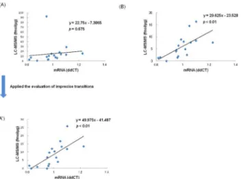
expression. One sample (Subject 8) was not analyzed by quantitative real-time RT-PCR due to insufficient quantity. Although protein expression calculated from the average concentration of all transitions including imprecise transition was not correlated with m-RNA expression (p= 0.675;r= 0.1099,Fig 6A), m-RNA expression was correlated with protein expression cal-culated from the best transition (614/711) for peptide ITWDPPSSPVK (p<0.01;r= 0.7190,Fig
6A') and with protein expression calculated from the best transition (708/763) for peptide
Fig 3. MRM chromatograms for peptide ITWDPPSSPVK of subject 4and subject 7.Transitions 614/711, 614/826 and 614/1013 were for endogenous peptide ITWDPPSSPVK, and transitions 617/711, 617/826, and 617/1013 were for stable isotope-labeled peptide, peptide I (13C
6,15N) TWDPPSSPVK. (A) subject 4 (B)
subject 7. Only the endogenous peptide area of transition 614/1013 in subjects 4 and 7 was as high as that in other subjects, while other transitions (614/711 or 614/826) showed no peaks.
doi:10.1371/journal.pone.0130760.g003
Fig 4. Type XIV collagen concentrations calculated from the best transitions of peptide
ITWDPPSSPVK and peptide ASAHAITGPPTELITSEVTAR.After evaluation of imprecise transitions and using the best transition for the peptide concentration, concentrations of transition 614/711 were used as the concentrations of peptide ITWDPPSSPVK, and concentrations of transition 708/763 were used as the concentrations of peptide ASAHAITGPPTELITSEVTAR.
ASAHAITGPPTELITSEVTAR (p<0.01;r= 0.6538,Fig 6B). The results indicate that type XIV
collagen protein and m-RNA expression are correlated, and specific expression of type XIV collagen in massive lymph node metastasis was validated by quantitative real-time RT-PCR.
Fig 5. Correlation of type XIV collagen concentrations calculated from the best transitions of peptide ITWDPPSSPVK and peptide ASAHAITGPPTELITSEVTAR.The x-axis represents protein concentration of type XIV collagen calculated from the best transition (708/763) of peptide ASAHAITGPPTELITSEVTAR as determined by LC-MS/MS. The y-axis represents protein concentration of type XIV collagen calculated from the best transition (614/711) of peptide ITWDPPSSPVK as determined by LC-MS/MS. In the scatter plot, each data point represents protein concentration.
doi:10.1371/journal.pone.0130760.g005
Fig 6. Correlation of concentrations between LC-MS/MS and quantitative real-time RT-PCR for validation of type XIV collagen.The x-axis represents m-RNA expression of type XIV collagen as determined by quantitative real-time RT-PCR. The y-axis represents protein concentration of type XIV collagen as determined by LC-MS/MS. In the scatter plot, each data point represents protein concentration and m-RNA expression. (A) The protein concentrations are average concentrations of all peptide transitions (614/711, 614/826, and 614/1013) of peptide ITWDPPSSPVK. (A') The protein concentrations are calculated from the best transition (614/711) for peptide ITWDPPSSPVK. (B) The protein concentrations are calculated from the best transition (708/763) for peptide ASAHAITGPPTELITSEVTAR.
3. Identification of specific proteins
To examine the peptide-sequence specificity of type XIV collagen, a homology search was car-ried out for peptides ITWDPPSSPVK and ASAHAITGPPTELITSEVTAR by NCBI BLAST search against the human database. Only undulin and type XIV collagen share 100% amino acid sequence homology. Undulin is an alternative name for type XIV collagen, and the centration of peptides ITWDPPSSPVK and ASAHAITGPPTELITSEVTAR represent the con-centration of type XIV collagen.
Discussion
In this study, type XIV collagen was identified as a protein specific for massive lymph node metastasis of breast cancer tissue as determined by quantitative LC-MS/MS and quantitative RT-PCR.
Type XIV collagen plays an adhesive role by integrating collagen bundles. It has been sug-gested that the large globular domain of Type XIV collagen protrudes from the bundles into the extracellular matrix where it interacts with cancer cells [26–28]. Although type XIV colla-gen may not be an appropriate target of antibody drugs because it is present in normal extracel-lular matrix, it may be a potential biomarker of metastasis and useful in evaluation of patient prognosis. Type XIV collagen has been previously reported to be expressed in odontogenic, brain, and pancreatic tumors [29], but not in breast cancer, and the significance of this expres-sion remains unclear. Our findings suggest that type XIV collagen is a novel protein that is specific for massive lymph node metastasis breast cancer, in which it may play a role as an adhesion factor. Further studies on type XIV collagen are desired to verify its role as a prognos-tic factor and diagnosprognos-tic marker for metastasis.
The 5 proteins aside from type XIV collagen that were expressed in massive lymph node metastasis of breast cancer as determined by MRM analysis without an internal standard included 2 types of type I collagen, MSTP161, angiomotin and hexokinase type I. Like type XIV collagen, type I collagen and MSTP161 are extracellular matrix proteins. The adhesive role of type XIV collagen is probably associated with the surface of collagen fibrils via type I colla-gen, and it might interact with other matrix molecules or cell surface receptors [30]. MSTP161 is also known as proline/arginine-rich end leucine-rich repeat protein (PRELP) or prolargin. PRELP binds collagen type I and type II through its leucine-rich repeat domain, and its func-tion as a molecule anchoring basement membranes to the underlying connective tissue has been proposed [31,32]. Angiomotin, localized on the cell surface, maintains tight junctions of protein complexes and regulates endothelial cell migration and tube formation [33,34]. There-fore, the adhesive role of type XIV collagen may interact with type I collagen, PRELP and angiomotin. Hexokinase I is related to glycolysis [35]. Hexokinase II, which belong to the same hexokinase family, relates to invasion and metastasis because it facilitates and promotes the high-glycolytic tumor phenotype [36]. Further studies using LC-MS/MS quantification with internal standard and immunohistochemistry are desired to determine the specificity of these proteins in massive lymph node metastasis.
The results of peptide ITWDPPSSPVK analysis by LC-MS/MS in Subjects 4 and 7 showed that the influence of proteomic interference is a much greater challenge than that of small mol-ecule quantification in samples that can be purified by deproteinization. Interference occurred at random in individual clinical samples and occasionally caused the miscalculation of errone-ously high concentrations. When the presence of interference in peptides and subjects was not predicted in protein quantification, monitoring multiple transitions, evaluating imprecise tran-sition using CV, and using the best trantran-sition for the peptide concentration were effective for high reliability quantification. Although 3 transitions per peptide were used in this study, it is feasible to use 5 transitions per peptide given the performance of the QTRAP5500, and this method can be used when a transition has reliable specificity. Our results indicate that when the transition has not been evaluated and its specificity is unknown, evaluation of imprecise transitions is required. Transition evaluation and peptide concentration calculation are com-mon important points for protein quantification by LC-MS/MS in biological fluid.
Conclusions
We developed quantitative methods for the analysis of type XIV collagen, and demonstrated that type XIV collagen is specific to massive lymph node metastatic breast cancer. In a future study, we hope to quantify type XIV collagen and specific candidate proteins in plasma and lymph fluid of both non-metastasis and massive lymph node metastasis tissues, with the ulti-mate goal being to utilize this technique to facilitate diagnosis and early detection, and to fur-ther our understanding of the mechanisms involved in metastasis. The correlation between the expression of specific proteins and their corresponding genes may contribute to the develop-ment of new drugs for the treatdevelop-ment of massive lymph node metastatic breast cancer. The pro-tein was quantified with high reproducibility by evaluation of imprecise transitions and by using the best transition for peptide concentration. Analysis of imprecise transitions may also be useful for determining the pharmacokinetics of protein pharmaceuticals and the identifica-tion of protein biomarkers, as well as for the identificaidentifica-tion of specific proteins expressed in massive lymph node metastatic breast cancer.
Acknowledgments
We thank the patient tissue donors.
Author Contributions
Conceived and designed the experiments: RG YN TS ZT. Performed the experiments: RG YN. Analyzed the data: RG YN. Contributed reagents/materials/analysis tools: RG YN ZT. Wrote the paper: RG YN TT TS ZT.
References
1. Nemoto T, Vana J, Bedwani RN, Baker HW, McGregor FH, Murphy GP. Management and survival of female breast cancer: results of a national survey by the American College of Surgeons. Cancer 1980; 45(12): 2917–2924. PMID:7388735
2. SEER Cancer Statistics Review (CSR) 1975–2010. Available:http://seer.cancer.gov/csr/1975_2010/ browse_csr.php?sectionSEL=4&pageSEL=sect_04_table.14.html
3. Dings PJ, Elferink MA, Strobbe LJ, de Wilt JH. The prognostic value of lymph node ratio in node-posi-tive breast cancer: a Dutch nationwide population-based study. Ann Surg Oncol. 2013; 20(8):2607– 2614. doi:10.1245/s10434-013-2932-7PMID:23536053
5. Goto R, Nakamura Y, Shioyama S, Sanke T, Tozuka Z. Screening of Specific Proteins Involved in Breast Cancer Metastasis by 2D LC-ESI-MS/MS. J. Wakayama Med. Soc. 2014; 65(3)84–89.
6. Goto R, Kakudo K, Momiyama K, Nakamura Y, Takami T, Tozuka Z, et al. Detecting agent and thera-peutic agent for highly malignant breast cancer. PCT application PCT/JP2006/325008.
7. Nakamura Y, Yasuoka H, Tsujimoto M, Goto R, Tozuka Z, Momiyama K, et al. Detecting agent and therapeutic agent for highly malignant breast cancer. EP patent publication 1962094 (A1).
8. Nakamura Y, Yasuoka H, Tsujimoto M, Goto R, Tozuka Z, Momiyama K, et al. Detecting agent and therapeutic agent for highly malignant breast cancer. US patent publication 8030014 (B2).
9. Anderson DC, Li W, Payan DG, Noble WS. A new algorithm for the evaluation of shotgun peptide sequencing in proteomics: support vector machine classification of peptide MS/MS spectra and SEQUEST scores. J Proteome Res. 2003; 2(2):137–146. PMID:12716127
10. Elias JE, Gibbons FD, King OD, Roth FP, Gygi SP. Intensity-based protein identification by machine learning from a library of tandem mass spectra. Nat Biotechnol. 2004; 22(2):214–219. PMID: 14730315
11. López-Ferrer D, Martínez-Bartolomé S, Villar M, Campillos M, Martín-Maroto F, Vázquez J. Statistical model for large-scale peptide identification in databases from tandem mass spectra using SEQUEST. Anal Chem. 2004; 76(23):6853–6860. PMID:15571333
12. Razumovskaya J, Olman V, Xu D, Uberbacher EC, VerBerkmoes NC, Hettich RL, et al. A computa-tional method for assessing peptide-identification reliability in tandem mass spectrometry analysis with SEQUEST. Proteomics 2004; 4(4):961–969. PMID:15048978
13. Savitski MM, Nielsen ML, Zubarev RA. New data base-independent, sequence tag-based scoring of peptide MS/MS data validates Mowse scores, recovers below threshold data, singles out modified pep-tides, and assesses the quality of MS/MS techniques. Mol Cell Proteomics 2005; 4(8):1180–1188. PMID:15911534
14. Elias JE, Gygi SP. Target-decoy search strategy for increased confidence in large-scale protein identifi-cations by mass spectrometry. Nat Methods. 2007; 4(3):207–214. PMID:17327847
15. Rifai N, Gillette MA, Carr SA. Protein biomarker discovery and validation: the long and uncertain path to clinical utility. Nat Biotechnol. 2006; 24(8):971–983. PMID:16900146
16. Addona TA, Abbatiello SE, Schilling B, Skates SJ, Mani DR, Bunk DM, et al. Multi-site assessment of the precision and reproducibility of multiple reaction monitoring-based measurements of proteins in plasma. Nat Biotechnol. 2009; 27(7):633–641. doi:10.1038/nbt.1546PMID:19561596
17. Keshishian H, Addona T, Burgess M, Kuhn E, Carr SA. Quantitative, multiplexed assays for low abun-dance proteins in plasma by targeted mass spectrometry and stable isotope dilution. Mol Cell Proteo-mics 2007; 6(12):2212–2229. PMID:17939991
18. Kamiie J, Ohtsuki S, Iwase R, Ohmine K, Katsukura Y, Yanai K, et al. Quantitative atlas of membrane transporter proteins: development and application of a highly sensitive simultaneous LC/MS/MS method combined with novel in-silico peptide selection criteria. Pharm Res. 2008; 25(6):1469–1483. doi:10.1007/s11095-008-9532-4PMID:18219561
19. Anderson NL, Anderson NG, Haines LR, Hardie DB, Olafson RW, Pearson TW. Mass spectrometric quantitation of peptides and proteins using Stable Isotope Standards and Capture by Peptide Anti-bodies (SISCAPA). J Proteome Res. 2004; 3(2):235–244. PMID:15113099
20. Kiyonami R, Schoen A, Prakash A, Peterman S, Zabrouskov V, Picotti P, et al. Increased selectivity, analytical precision, and throughput in targeted proteomics. Mol Cell Proteomics 2011; 10(2): M110.002931. doi:10.1074/mcp.M110.002931PMID:20664071
21. Remane D, Meyer MR, Wissenbach DK, Maurer HH. Ion suppression and enhancement effects of co-eluting analytes in multi-analyte approaches: systematic investigation using ultra-high-performance liq-uid chromatography/mass spectrometry with atmospheric-pressure chemical ionization or electrospray ionization. Rapid Commun Mass Spectrom. 2010; 24(21):3103–3108. doi:10.1002/rcm.4736PMID: 20941756
22. Prakash C, Shaffer CL, Nedderman A. Analytical strategies for identifying drug metabolites. Mass Spectrom Rev. 2007; 26(3):340–369. PMID:17405144
23. Matuszewski BK, Constanzer ML, Chavez-Eng CM. Strategies for the assessment of matrix effect in quantitative bioanalytical methods based on HPLC-MS/MS. Anal Chem. 2003; 75(13):3019–3030. PMID:12964746
25. Reiter L, Rinner O, Picotti P, Hüttenhain R, Beck M, Brusniak MY, et al. mProphet: automated data pro-cessing and statistical validation for large-scale SRM experiments. Nat Methods 2011; 8(5):430–435. doi:10.1038/nmeth.1584PMID:21423193
26. Trueb J, Trueb B. Type XIV collagen is a variant of undulin. Eur J Biochem. 1992; 207(2):549–557. PMID:1339349
27. Gerecke DR, Foley JW, Castagnola P, Gennari M, Dublet B, Cancedda R, et al. Type XIV collagen is encoded by alternative transcripts with distinct 5' regions and is a multidomain protein with homologies to von Willebrand's factor, fibronectin, and other matrix proteins. J Biol Chem. 1993; 268(16):12177– 12184. PMID:8505337
28. Ehnis T, Dieterich W, Bauer M, Lampe B, Schuppan D. A chondroitin/dermatan sulfate form of CD44 is a receptor for collagen XIV (undulin). Exp Cell Res. 1996; 229(2):388–397. PMID:8986622
29. Löhr M, Trautmann B, Göttler M, Peters S, Zauner I, Maillet B, et al. Human ductal adenocarcinomas of the pancreas express extracellular matrix proteins. Br J Cancer. 1994; 69(1):144–151. PMID:8286197
30. Schuppan D, Cantaluppi MC, Becker J, Veit A, Bunte T, Troyer D, et al. Undulin, an extracellular matrix glycoprotein associated with collagen fibrils. J Biol Chem. 1990; 265(15):8823–8832. PMID:2187872
31. Bengtsson E, Mörgelin M, Sasaki T, Timpl R, Heinegard D, Aspberg A. The leucine-rich repeat protein PRELP binds perlecan and collagens and may function as a basement membrane anchor. J Biol Chem. 2002; 277(17):15061–15068. PMID:11847210
32. Amelinckx A, Castello M, Arrieta-Quintero E, Lee T, Salas N, Hernandez E, et al. Laser trabeculoplasty induces changes in the trabecular meshwork glycoproteome: a pilot study. J Proteome Res. 2009; 8 (7):3727–3736. doi:10.1021/pr900294gPMID:19432485
33. Troyanovsky B, Levchenko T, Mansson G, Matvijenko O, Holmgren L. Angiomotin: an angiostatin bind-ing protein that regulates endothelial cell migration and tube formation. J Cell Biol. 2001; 152(6):1247– 1254. PMID:11257124
34. Wells CD, Fawcett JP, Traweger A, Yamanaka Y, Goudreault M, Elder K, et al. A Rich1/Amot complex regulates the Cdc42 GTPase and apical-polarity proteins in epithelial cells. Cell. 2006; 125(3):535– 548. PMID:16678097
35. Rosano C, Sabini E, Rizzi M, Deriu D, Murshudov G, Bianchi M, et al. Binding of non-catalytic ATP to human hexokinase I highlights the structural components for enzyme-membrane association control. Structure. 1999; 7(11):1427–1437. PMID:10574795