Quim. Nova, Vol. 37, No. 3, 483-486, 2014
Artigo
http://dx.doi.org/10.5935/0100-4042.20140069
*e-mail: [email protected]
#Programa de Pós-graduação em Ciências Farmacêuticas
LC/ESI-MS METHOD APPLIED TO CHARACTERIZATION OF FLAVONOIDS GLYCOSIDES IN B. forficata subsp. pruinosa
Lidiane da Silveira Farias# and Andreas S. L. Mendez*,#
Curso de Farmácia, Universidade Federal do Pampa, BR 472 Km 585, 97500-970 Uruguaiana – RS, Brasil
Recebido em 08/08/2013; aceito em 28/10/2013; publicado na web em 26/02/2014
Bauhinia forficata is used in folk medicine for its hypoglycemiant effect. In the south of Brazil, the subspecies pruinosa is found in a region with the characteristic flora, pampa biome. This species has been consumed by the local population as a tea for diabetes treatment. We studied the chemical composition of hydroethanolic extracts using LC/ESI-MS. The leaf extracts were prepared by percolation with 50% (v/v) ethanol. The chromatographic analyses were performed using a reverse-phase system, gradient elution with acetonitrile:phosphoric acid 0.05%, and ESI-MS in the positive ion mode. The chemical profile of the flavonoids was suggested to involve four quercetin and kaempferol glycosides.
Keywords: Bauhinia forficata subsp. pruinosa; flavonoids; LC-ESI-MS.
INTRODUCTION
Bauhinia forficata is a medicinal plant largely consumed by the Brazilian population. It is popularly known as “pata de vaca” (cow’s hoof) because of the large and bilobed aspect of the leaves, a characteristic distinctive to this genus. Traditionally, the aerial parts are used in folk medicine for diabetes treatment, and it is available as a tea in local herbal stores.1-4 Two subspecies are also of interest, subsp. forficata and subsp. pruinosa. B. forficata subsp. forficata is well characterized with respect to its pharmacological aspects and phytochemical analysis.5-9 Their chemical composition is based on kaempferol and quercetin O-glycoside derivatives such as kaempferol 3,7-dirhamnoside (kaempferitrin) and quercetin 3,7-di-O-α-L-rhamnopyranoside.4,9,10 Several reports have discussed the hypoglycemiant potential.2,7,11 They mention different expe-rimental models, i.e. using alloxan-induced diabetic rats and the streptozotocin-induced diabetes model.2,7 Based on tests involving the crude extracts and their respective fractions, the observed effects demonstrated the biological potential for glucose and triglycerides level reduction, which are associated with improvement in the carbohydrate metabolism.2
B. forficata subsp. pruinosa is found in a characteristic region of South America (pampa biome) comprised of Argentina, Brazil, and Uruguay. In the south of Brazil, this plant is consumed by the local population as a tea for its known hypoglycemiant and diuretic effects. In recent work by Arigony,12 the hydroethanolic extracts obtained from this species were studied by LC-UV. Flavonoids were detected, and the compound, kaempferol-3-robinoside-7--rhamnoside, was preliminarily suggested as likely comprising the majority of the extract.
In the present work, an analytical investigation of hydroethanolic extracts obtained from B. forficata subsp. pruinosa was performed using LC/ESI-MS. The hyphenated system was applied to the cha-racterization of the chemical composition in flavonoids, observing the presence in a majority of compounds.
EXPERIMENTAL Chemicals
Acetonitrile was purchased from Tedia (Fairfield, OH, USA). Phosphoric acid, ethanol, and methanol were purchased from Merck (Darmstadt, Germany). Purified water was obtained using the Milli-Q Plus® system from Millipore (Milford, MA, USA). All other reagents used in this study were of analytical or HPLC grade.
Plant material
Leaves of Bauhinia forficata subsp. pruinosa (Vogel) Fortunato & Wunderlin were collected at Uruguaiana city (Rio Grande do Sul, Brazil) in October of 2011 (spring). This region is located near Argentina along the western border and is predominantly pam-pa biome flora. The plant was identified and voucher specimens (ICN 167491) were deposited at the ICN Herbarium (Instituto de Biociências, Universidade Federal do Rio Grande do Sul, Brazil).
Extracts
Air dried (at 35 °C) leaves of B. forficata subsp. pruinosa were reduced to a powder and submitted to extraction by percolation (1:1, v/v) using an ethanol-water mixture (1:2).8,13 The hydroethanolic extracts were then filtered and stored in a refrigerator until analysis. For chromatographic assays, the extracts were diluted in a 50% methanol solution at a ratio of 1:10 (v/v). All solutions were filtered through a 0.45 µm membrane filter from Millipore (Milford, MA, USA) before injection.
LC/ESI-MS
Farias and Mendez
484 Quim. Nova
The detection was done on a DAD detector set at 340 nm. The mobile phase was prepared daily, filtered through a 0.45 µm membrane filter (Millipore), and sonicated before use.
The MS analyses were performed on an Esquire plus 3000 (Bruker Daltonics, Billerica, MA, USA) ion trap mass spectrometer with an electrospray interface (ESI). The data acquisition software employed was Esquire CONTROL 5.2. The LC/ESI-MS was conducted in posi-tive-ion mode and operated according to defined conditions: nitrogen gas temperature - 320 °C; drying gas flow rate - 7 L min-1; capillary voltage - 4000 V; nebulizing pressure - 27 psi. Mass spectra were recorded using the full scan mode in the range of 200–800 Daltons.
RESULTS AND DISCUSSION
The phytoconstituents described for the Bauhinia species pre-dominantly include flavonoid O-glycosides, the majority of which are derivatives of kaempferol and quercetin.3,4,10B. forficata Link is known because of the presence of kaempferitrin, a flavonoid cited as a chemical marker for phytopreparations containing this species, and even mentioned because of its involvement with the observed
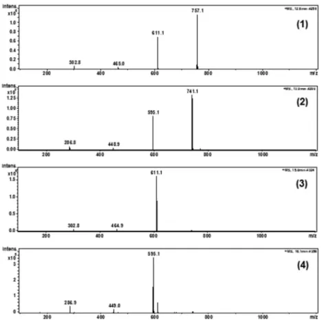
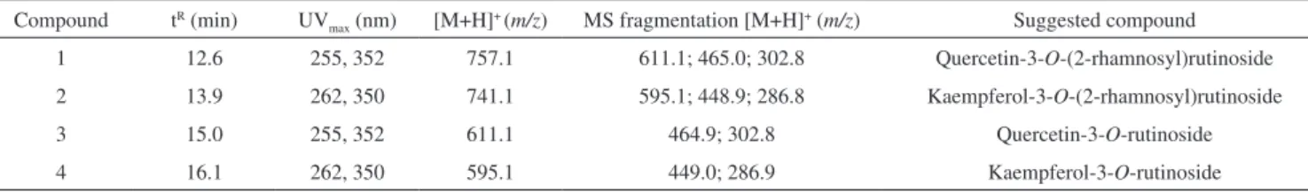
hypoglicemiant effect.2,9,11 The rich flavonoid content has also been reported and is associated with biological effects such as glycemic response, cholesterol metabolism, and antioxidant activity.14,15 Recently, our group studied the aqueous and hydroethanolic extracts from B. forficata subsp. pruinosa, evaluating the phenolic content and antioxi-dant potential using an in vitro experimental model based on a DPPH (2,2-diphenyl-1-picrylhydrazyl) assay and lipid peroxidation prevention by measuring TBARS (thiobarbituric acid reactive substances).13 The potential of this species to reduce lipid peroxidation in an experimental model of hyperglycemia in human erythrocytes in vitro was verified.16 Considering the reports on the effectiveness of this plant for the treatment of diabetes, we proposed an investigation on the chemical composition of flavonoids. The previous chromatographic analysis by LC-UV was performed using defined conditions that provided good separation and rapid elution.13 The experimental conditions were applied to LC-MS analysis, which provided a good separation profile and detected intense peaks at retention times of 12.6 (compound 1), 13.9 (compound 2), 15.0 (compound 3), and 16.1 min (compound 4) (Figure 1). Performing a detailed evaluation of the mass spectra (Figure 2), led to the suggestion of four phenolic compounds, all
Figure 1. Representative LC/ESI-MS chromatograms obtained from the analysis of Bauhinia forficata subsp. pruinosa. Peak numbers correspond to the sug-gested chemical compounds. (1) quercetin-3-O-(2-rhamnosyl)rutinoside; (2) kaempferol-3-O-(2-rhamnosyl)rutinoside; (3) quercetin-3-O-rutinoside; (4) kaempferol-3-O-rutinoside
LC/ESI-MS method applied to characterization of flavonoids glycosides in B. forficata subsp. pruinosa 485 Vol. 37, No. 3
containing a basic nucleus of quercetin or kaempferol. The identi-ties, UVmax, and m/z of the ions are described in Table 1. Initially, in previous experiments using chemical standards, the flavonoid, kaempferitrin, had a retention time similar to the compound retained at 16.1 min. This first analysis indicated their presence, which had not yet been confirmed by MS analysis.
The proposed MS fragmentation and chemical structures of fla-vonoids 1–4 are illustrated in Figure 3. The chemical fragmentation involves cleavage of interglycosidic bonds, resulting in an aglycone ion at a characteristic m/z. In our case, O-glycosides are suggested be-cause of the rearrangement and concomitant cleavage that occur at the interglycosidic bond during fragmentation, leading to the elimination of monossacharide residues and generation of an OH terminal group.17 Compounds 1 and 3 are identified as quercetin-3-O-(2-rhamnosyl) rutinoside and quercetin-3-O-rutinoside. Compound 1 displayed a pseudo-molecular ion [M+H]+ at m/z 757.1 and fragments at m/z 611.1 and 302.8, indicating the loss of monossacharide and dissacharide residues, rhamnosyl (146 u) and rutinoside (308.2 u). In reference to compound 3, the direct loss of the rutinoside residue results in the aglycone ion [M+H]+ at m/z 302.8, suggesting a flavonoid nucleus of quercetin. The residual dissacharide rutinoside was suggested based on the mass spectra and a recent literature report that mentions the compound in B. forficata subsp. pruinosa.18 The possibility for other sugar fragments (i.e. robinoside) was considered, but it was not con-clusive with the current information. In the literature, some quercetin glycosides are described for Bauhinia species. Pizzolati et al.9 have identified five glycosil flavonoids in B. forficata Link, two of them being quercetin derivatives, 3,7-di-O-α-L-rhamnopyranosylquercetin and 3-O-[α-L-rhamnopyranosyl-(1-6)-β-D-glucopyranosyl]-7-O
-α-L-rhamnopyranosylquercetin. In another review, the flavonoids, quercetin-3-O-L-ramnopiranosídio and kaempferol-3-O -L-ramnopi-ranosidio, were identified in B. uruguayensis.10
Compounds 2 and 4 were identified as kaempferol glycosides, kaempferol-3-O-(2-rhamnosyl)rutinoside and kaempferol-3-O -rutinoside, respectively. This identification was based on the residual fragment observed at m/z 286.8, which is characteristic for these derivatives. The monosaccharide units were determined by observing the fragment ions that are indicative of the loss of sugar residues and suggest the interglycosidic bond is broken. In this case, the rhamnosyl (146 u) and glucosyl residues (162 u) were seen, leading to the sug-gested chemical structures illustrated in Figure 3.
In terms of predominance, the kaempferol nucleus is very com-mon in Bauhinia species, although the sugar and O-glycosylation site are variable. Recently, Ferreres et al.18 identified eight substances in B. forficata containing this aglycone structural. In respect to subsp. forficata, the derivatives are glycosilated simultaneously in positions 3 and 7, different from our results for subsp. pruinosa. Pizzolati et al.9 isolated the compounds kaempferitrin (kaempferol 3,7-dirham-noside) and 3-O-[rhamnopyranosyl-(1-6)-β-glucopyranosyl]-7-O -rhamnopyranosylkaempferol from the leaves of B. forficata Link. In B. candicans, the presence of kaempferol 3-O-β-rutinoside was described.10 Rao et al.19 have also mentioned the presence of kaemp-ferol 3-O-β-D-glucopyranoside in B. variegata.19
CONCLUSION
B. forficata subsp. pruinosa presents a chemical profile based on kaempferol and quercetin glycosides. The chromatographic assay proposed, involving detection by MS, allowed for rapid identification of the main components present in the hydroethanolic extracts and provides an option for the characterization of plant material.
REFERENCES
1. Fuentes, O.; Arancibia-Avila, P.; Alarcón, J.; Fitoterapia 2004, 75, 527. 2. Pepato, M. T.; Keller, E. H.; Baviera, A. M.; Kettelhut, I. C.; Vendramini,
R. C.; Brunetti, I. L.; J. Ethnopharmacol. 2002, 81, 191. 3. Cechinel Filho, V.; Phytother. Res. 2009, 23, 1347.
4. Da Silva, K. L.; Biavatti, M. L.; Leite, S. N.; Yunes, R. A.; Monache, F. D.; Cechinel Filho, V.; Z. Naturforsch., C: Biosci. 2000, 55, 478. Table 1. Chemical constituents identified in Bauhinia forficata subsp. pruinosa with corresponding retention times, molecular ions in positive mode, and key fragments by LC/ESI-MS
Compound tR (min) UV
max (nm) [M+H]+(m/z) MS fragmentation [M+H]+ (m/z) Suggested compound
1 12.6 255, 352 757.1 611.1; 465.0; 302.8 Quercetin-3-O-(2-rhamnosyl)rutinoside 2 13.9 262, 350 741.1 595.1; 448.9; 286.8 Kaempferol-3-O-(2-rhamnosyl)rutinoside
3 15.0 255, 352 611.1 464.9; 302.8 Quercetin-3-O-rutinoside
4 16.1 262, 350 595.1 449.0; 286.9 Kaempferol-3-O-rutinoside
Farias and Mendez
486 Quim. Nova
5. Da Cunha, A. M.; Menon, S.; Menon, R.; Couto, A. G.; Bürger, C.; Biavatti, M. W.; Phytomedicine 2010, 17, 37. 6. Khalil, N. M.; Pepato, M. T.; Brunetti, I. L.; Biol. Res. 2008, 41, 165. 7. Lino, C. S.; Diógenes, J. P. L.; Pereira, B. A.; Faria, R.; Andrade Neto,
M.; Alves, R. S.; Queiroz, M.; Souza, F. C. F.; Viana, G. S. B.; Biol. Pharm. Bull. 2004, 27, 125.
8. Pinheiro, T. S. D. B.; Johansson, L. A. P.; Pizzolatti, M. G.; Biavatti, M. W.; J. Pharm. Biomed. Anal. 2006, 41, 431.
9. Pizzolatti, M. G.; Cunha Jr, A.; Szpoganicz, B.; De Souza, E.; Braz-Filho, R.; Schripsema, J.; Quim. Nova 2003, 26, 466.
10. Da Silva, K. L.; Cechinel Filho, V.; Quim. Nova 2002, 25, 449. 11. Jorge, A. P.; Horst, H.; Sousa, E.; Pizzolatti, M. G.; Barreto Silva, F. R.
M.; Chem. Biol. Interact. 2004, 149, 89.
12. Arygony, A. L. V.; Dissertação de Mestrado, Universidade Federal do Rio Grande do Sul, Brasil, 2005.
13. Sayago, C. T. M.; De Camargo, V. B.; Barbosa, F.; Gularte, C.; Pereira, G.; Miotto, S.; Cechinel Filho, V.; Puntel, R. L.; Folmer, V.; Mendez, A.; Acta Biol. Hung. 2013, 64, 25.
14. Volpato, G. T.; Damasceno, D. C.; Rudge, M. V. C.; Padovani, C. R.; Calderon, I. M. P.; J. Ethnopharmacol. 2008, 116, 131.
15. De Sousa, E.; Zanatta, L.; Seifriz, I.; Creczynski-Pasa, T. B.; Pizzolatti, M. G.; Szpoganicz, B.; Silva, F. R. M. B.; J. Nat. Prod. 2004, 67, 829. 16. Salgueiro, A. C. F.; Leal, C. O.; Bianchini, M. C.; Prado, I. O.; Mendez,
A. S. L.; Puntel, R.; Folmer, V.; Soares, F. A.; Ávila, D. S.; Puntel, G. O.; J. Ethnopharmacol. 2013, 148, 81.
17. Cuyckens, F.; Claeys, M.; J. Mass Spectrom. 2004, 39, 1.
18. Ferreres, F.; Gil-Izquierdo, A.; Vinholes, J.; Silva, S. T.; Valentao, P.; Andrade, P. B.; Food Chem. 2012, 134, 894.