www.jped.com.br
ORIGINAL ARTICLE
Alterations in the pulmonary histoarchitecture of neonatal mice
exposed to hyperoxia
夽
Renata B. Reis
a, Akinori C. Nagato
b, Clarissa R. Nardeli
c,
Isadora C.P. Matias
c, Wanderson G. Lima
d, Frank S. Bezerra
e,∗aAcadêmica, Curso de Fisioterapia, Laboratório de Biomorfologia e Patologia Experimental, Centro de Ciências da Saúde, Universidade Severino Sombra (USS), Vassouras, RJ, Brasil
bMestre em Ciências, Professor Assistente, Laboratório de Biomorfologia e Patologia Experimental, Centro de Ciências da Saúde, Universidade Severino Sombra (USS), Vassouras, RJ, Brasil
cAcadêmica, Curso de Biomedicina, Laboratório de Biomorfologia e Patologia Experimental, Centro de Ciências da Saúde, Universidade Severino Sombra (USS), Vassouras, RJ, Brasil
dDoutor em Patologia, Professor Adjunto, Laboratório de Imunopatologia, Departamento de Ciências Biológicas, Universidade Federal de Ouro Preto (UFOP), Ouro Preto, MG, Brasil
eDoutor em Ciências Morfológicas, Professor Adjunto, Laboratório de Bioquímica Metabólica, Departamento de Ciências Biológicas, Universidade Federal de Ouro Preto (UFOP), Ouro Preto, MG, Brasil
Received 8 August 2012; accepted 29 October 2012 Available online 26 April 2013
KEYWORDS
Hyperoxia; Lung;
Newborn animals
Abstract
Objectives: To analyze the effects of exposure to hyperoxia (100% oxygen) on the lung histoar-chitecture of neonatal mice.
Methods: Neonatal Balb/c mice were exposed to hyperoxia (HG) (100% oxygen) (n = 10) in a chamber (15 x 20 x 30 cm) for 24 hours with a flow of 2 L/min. The control group (CG) (n = 10) was exposed to normoxia in the same type of chamber and for the same time. After exposure, the animals were euthanized by decapitation; the lungs were removed and processed for histo-logical examination according to the laboratory routine. Three-mm thick sections were stained with hematoxylin and eosin (H&E). The morphometric analysis was performed with in order to analyze the macrophages present in the alveolar lumen, surface density (Sv) of gas exchange,
volume density (Vv) of lung parenchyma, and areas of atelectasis.
Results: A decrease in the number of alveolar macrophages (MØ) was observed in the HG (HG = 0.08±0.01 MØ/mm2, CG = 0.18±0.03 MØ/mm2, p = 0.0475), S
v of gas exchange in HG
(HG = 8.08±0.12 mm2/mm3, CG = 8.65
±0.20 mm2/mm3, p = 0.0233), V
vof lung parenchyma in
HG (HG = 54.7/33.5/83.5%/mm2; CG = 75/56.7/107.9%/mm2, p < 0.0001) when compared with
the CG. However, there was an increase in areas of atelectasis in HG (HG = 17.5/11.3/38.4 atelectasis/mm2, CG = 14/6.1/24.4 atelectasis/mm2, p = 0.0166) when compared with the CG.
夽 Please cite this article as: Reis RB, Nagato AC, Nardeli CR, Matias IC, de Lima WG, Bezerra FS. Alterations in the pulmonary histoarchi-tecture of neonatal mice exposed to hyperoxia. J Pediatr (Rio J). 2013;89:300---6.
∗Corresponding author.
E-mail:frank@iceb.ufop.br (F.S. Bezerra).
Conclusion: The present results indicate that hyperoxia caused alterations in lung histoarchi-tecture, increasing areas of atelectasis and diffuse alveolar hemorrhage.
© 2013 Sociedade Brasileira de Pediatria. Published by Elsevier Editora Ltda. All rights reserved.
PALAVRAS-CHAVE
Hiperóxia; Pulmão; Animais recém-nascidos
Alterac¸ões da histoarquitetura pulmonar de camundongos neonatos expostos à hiperóxia
Resumo
Objetivos: Analisar os efeitos da exposic¸ão à hiperóxia (100% de oxigênio) sobre a histoarquite-tura pulmonar de camundongos neonatos.
Métodos: Camundongos neonatos da linhagem Balb/c foram expostos à hiperóxia (GH) (100% de oxigênio) (n = 10) em uma câmara (15 x 20 x 30 cm) por 24 h, com fluxo de 2 L/min. O grupo controle (GC) (n = 10) foi exposto a normóxia em um mesmo tipo de câmara e pelo mesmo tempo. Após a exposic¸ão, os animais foram sacrificados por decapitac¸ão, os pulmões foram removidos para análise histológica e processados de acordo com a rotina do laboratório. Cortes de 3m de espessura foram corados com hematoxilina e eosina (H&E). A análise morfométrica foi realizada com o objetivo de analisar macrófagos presentes na luz alveolar, densidade de superfície (Sv) de trocas gasosas, densidade de volume (Vv) de parênquima pulmonar e áreas
de atelectasias.
Resultados: Foi verificada diminuic¸ão do número de macrófagos alveolares (MØ) no GH (GH = 0,08±0,01 MØ/mm2; GC = 0,18±0,03 MØ/mm2; p = 0,0475), S
v de troca gasosa no
GH (GH = 8,08±0,12 mm2/mm3; GC = 8,65
±0,20 mm2/mm3; p = 0,0233), V
v de parênquima
pulmonar no GH (GH = 54,7/33,5/83,5%/mm2; GC = 75/56,7/107,9%/mm2; p < 0.0001) quando
comparado com o GC. Entretanto, houve aumento de áreas de atelectasias no GH (GH = 17,5/11,3/38,4 atelectasia/mm2; GC = 14/6,1/24,4 atelectasia/mm2; p = 0,0166) quando
comparado com o GC.
Conclusão: Nossos resultados indicam que a hiperóxia promoveu alterac¸ões na histoarquitetura pulmonar, aumentando áreas de atelectasia e hemorragia alveolar difusa.
© 2013 Sociedade Brasileira de Pediatria. Publicado por Elsevier Editora Ltda. Todos os direitos reservados.
Introduction
It is estimated that 3.9 of the 10.8 million child’s deaths worldwide occur in the first 28 days of life. Over 96% of these deaths occur in developing coun-tries. Pneumonia may be associated with a low Apgar score (severe respiratory alterations at birth), which is commonly associated with chorioamnionitis, inflam-mation of the fetal membranes (chorion and amnion) due to a bacterial infection, usually related to pro-longed vaginal deliveries and also to inhalation of infected amniotic fluid. In most cases, this leads to fetal asphyxia. One of the most obvious manifesta-tions is hypoxemia, followed by chest indrawing and cyanosis.
As treatment, studies have shown that drug ther-apies using antibiotics are effective, as well as the use of oxygen in order to reverse the initial
hypox-emia and reduce the risk of mortality.1 The use of
oxygen is among the first lines of therapy for
hypox-emia caused by pulmonary and heart disease,2---4 which
involves treating hypoxia by oxygen inhalation at a pressure greater than that of ambient air, which facil-itates hematosis and reduces ventilatory work, in order
to maintain adequate oxygenation with PaO2> 50 mmHg
and < 70 mmHg.5
Although O2 therapy is considered essential for life,
studies show an association between oxygen toxicity and retinopathy of prematurity, chronic lung disease, bronchopulmonary dysplasia, atelectasis by resorption, tra-cheobronchitis, depression of mucociliary activity, nausea, anorexia, headache, lung epithelial damage, damage to the blood-air barrier, and pulmonary edema. Moreover, the injury can be intensified when combined with
mechani-cal ventilation.6Recent studies suggest that the pulmonary
epithelial damage induced by exposure to high concen-trations of oxygen, specifically, has been associated with
oxidative stress,7 based on the hypothesis that hyperoxia
induces an increase in the number of oxygen free radi-cals, reactive species capable of reacting with biomolecules and causing direct damage to membrane proteins and
DNA.8
After pulmonary epithelium lesion, in particular, there is activation of macrophages and an inflammatory cascade, followed by pulmonary edema and presence of fibrin,
colla-gen, and neutrophilic aggregate.9The literature describes
animal models exposed to hyperoxia only in adult mice, when their lungs are already fully formed. The effects of high concentrations of oxygen at the time of lung formation,
i.e., the lungs of newborns, are yet to be clearly described
Methods
Ethical aspects
The experiment was performed in accordance with the pro-visions of the Brazilian Society of Science in Laboratory Animals, and was approved by the Ethics Committee for Animal Research of the University.
Animals
Twenty Balb/c neonatal mice, approximately 12 hours after birth, with a mean weight of 1.5 g (despite the low weight of newborn mice, their anatomical structures are well-defined, allowing for experimental manipulation) were obtained from the Laboratory of Experimental Pathology and Biomor-phology of the Centro de Ciências da Saúde (CCS) of the Universidade Severino Sombra, Brazil. The animals’ nutri-tion in the postnatal period until euthanasia was provided byad libitumbreastfeeding (breastfeeding in mice lasts on average 19 to 21 days after birth).
Experimental design
Exposure to oxygen
The animals were divided into two groups: control group (CG) - mice exposed to ambient air and to the same con-ditions of the experimental group and the hyperoxia group (HG) - mice exposed to hyperoxia for 24 h.
For the animals exposed to hyperoxia, an acrylic inhala-tion chamber was used (30 cm long, 20 cm wide, and 15 cm
high), as described by Nagato.10Oxygen 100% was purchased
from White Martins® (White Martins Praxair Inc. --- São Paulo, Brazil). The oxygen cylinder was coupled to the oxygen inhalation chamber through a silicone conduit. The gas was released into the chamber with a constant flow of 2 L/min, thus ensuring an oxygen flow that would supply and saturate the environment.
After a period of time, when oxygen had filled the cham-ber space, all mice (except the control group, which inhaled ambient air) were placed in the inhalation chamber and removed after 24 h. The oxygen concentration was measured continuously through an oxygen cell (C3 --- Middlesbrough,
England). The mice received water and foodad libitum, and
were kept in individual cages with controlled temperature
and humidity (21±2◦C, 50±10%, respectively), and
submit-ted to inversubmit-ted 12-h cycles of light/dark (artificial lights, 7 p.m. to 7 a.m).
This experimental model was designed to mimic the con-ditions of supplemental oxygen that neonates receive in intensive care units in the first days of life when associated with a pathological picture, during lung formation.
Euthanasia and organ removal
At the end of the O2 exposure time, euthanasia was
per-formed by decapitation. A ventral midline incision was performed to remove the skin, starting from the chest to the abdominal region. Access to the thoracic cavity was made using a subxiphoid incision with removal of the diaphragm
and costal osteotomy, to expose the mediastinum. After exposure of the mediastinum, a lung perfusion was per-formed by sectioning the left atrium, followed by a puncture in the right ventricle, injecting 1 mL of saline solution with 0.9% NaCl with constant pressure controlled by a pump (Sykam --- Gewerbering, Germany). After perfusion, the lungs were removed by plucking through the mediastinum.
Histological analysis
Both lungs were fixed through a cannula inserted in the tra-chea by instillation of formalin (Vetec Química Fina --- Duque de Caxias, Brazil) buffered (10%) with constant pressure con-trolled by a pump. After 48 h, they were processed through the following steps: dehydrated in increasing concentra-tions of ethanol (50%, 70%, 92.8%, and 99.3%), diaphonized
in xylene, and paraffin-embedded. Three-m-thick sections
were stained with hematoxylin and eosin (H & E).
Morphometric analysis
Representative and proportional lung samples stained with H & E were randomly studied; 15 random fields were assessed
by histological slide, under 40×magnification. The analyzed
section came from the lungs equally embedded in paraffin and sectioned in the same direction, aiming to analyze all of the representative parts of the entire lung and proportion-ally in all analyses. Analyses were performed to determine the number of macrophages present in the alveolar lumen,
the volume density (Vv) of parenchyma, surface density (Sv)
of gas exchange, and areas of atelectasis and erythrocytes in airspaces. Both analyses were performed by observing the
microscope slide on a TV monitor (Sharp --- 14′′) where a test
system was superimposed on the screen, and the analyses
were made in relation to tissues. Sv of gas exchange was
verified through the test system of cycloid arcs with propor-tional orientation to the sine of the vertical axis angle. The measurement was performed by counting coincident points on the portion of the gas exchange surface, with the test
system superimposed on the lung tissue image.11
The Vv of lung parenchyma was measured through the
M42 test system in an irregular arrangement of points. Analy-sis was conducted by overlapping the test system to an image of lung tissue, and coincident points of the lung parenchyma
were counted.11
Statistical analysis
To determine the sample size, a statistical power of 0.9 was adopted. The data used for this calculation were obtained
from studies performed by this group,10taking into account
the expected data from larger standard error of the mean. Therefore, a resulting sample size of 10 for each group was obtained. The normality of all data was tested through the Kolmogorov-Smirnov test. Parametric data were expressed
as mean±standard error of mean, followed by unpaired
Student’s t-test. Nonparametric data were expressed as
0.25
0.20
0.15
0.10
0.05
0.00
CG HG
Macrophages / mm
2
*
Figure 1 Mean number of alveolar macrophages per micro-scopic field in the lung parenchyma in neonatal mice exposed to ambient air and hyperoxia for 24 h.
CG = control group, animals exposed to ambient air; HG = hyperoxia group, animals exposed to 100% oxygen for 24 hours.
* Means difference between the CG and HG, with a p-value = 0.0475 in the unpaired Student’st-test.
Results
After exposure to hyperoxia for 24 h, it was observed that
the HG (HG = 0.08±0.01 MØ/mm2) showed a decrease in
alveolar macrophages in the alveolar lumen (p = 0.0475)
compared to CG (CG = 0.18±0.03 MØ/mm2) (Fig. 1).
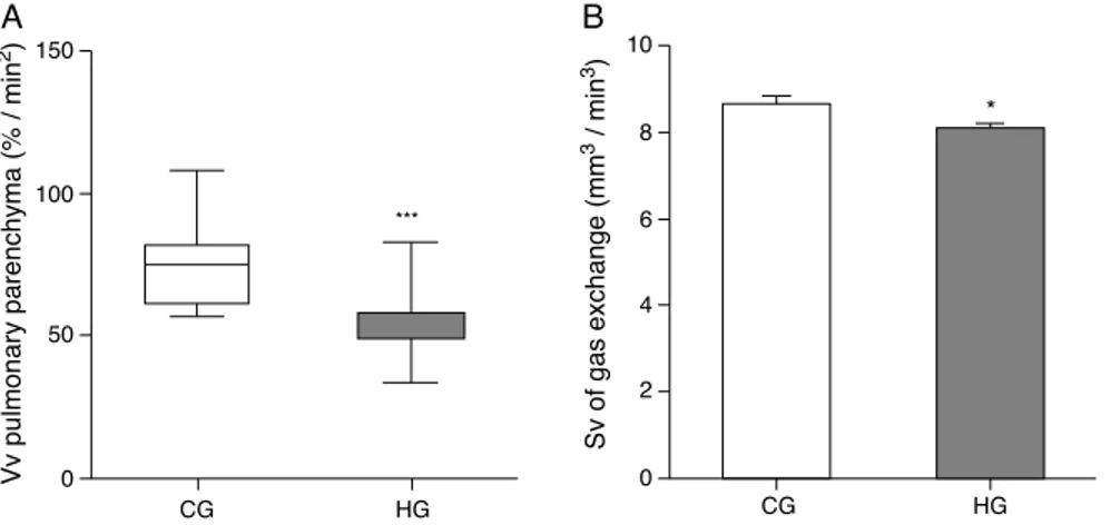
As for morphometric analyses, a decrease in
the Vv of lung parenchyma was observed in the
HG = 54.7/33.5/83.5%/mm2, CG = 75/56.7/107.9%/mm2,
p < 0.0001) (Fig. 2A), and the Sv of gas exchange of HG
(HG = 8.08±0.12 mm2/mm3; CG = 8.65
±0.20 mm2/mm3,
p = 0.0193 (Fig. 2B).
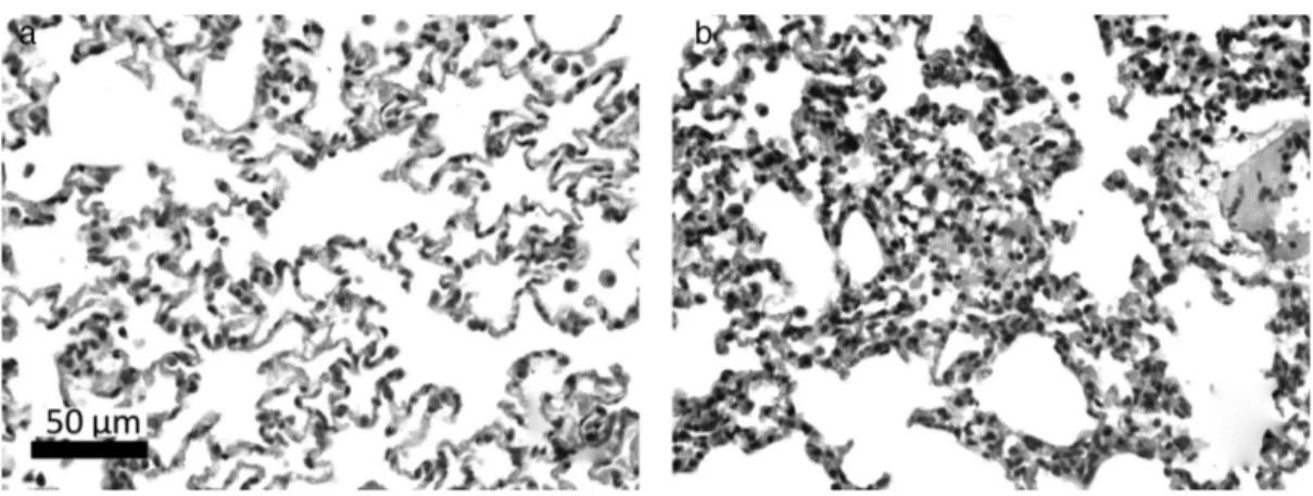
Histologically, the CG was characterized by presence of lung parenchyma of normal aspect (Fig. 3A). The HG showed diffuse parenchymal abnormalities, with varying degrees of intensity (from mild to severe). The pres-ence of areas of atelectasis and the prespres-ence of red blood cells in the alveolar lumen, as shown in Fig. 3B, were the most frequent alterations and were significantly
increased in HG (HG = 17.5/11.3/38.4 atelectasis/mm2,
CG = 14/6.1/24.4 atelectasis/mm2, p = 0.0166) when
com-pared to the CG (Fig. 4).
Discussion
The present study analyzed the effects of exposure to high concentrations of oxygen on lung histological patterns of neonatal Balb/c mice.
It was observed that hyperoxia induced a decrease in the number of alveolar macrophages, modified the lung histoar-chitecture, and increased the number of red cells in the air spaces.
The HG showed a decrease of macrophages in the alve-olar space after 24 h of exposure. In their studies, Petrache
et al.12 demonstrated in vitro (after 24 hour-exposure to
O2> 95%) andin vivo(after the mice were exposed for 72 h
to 100% O2) that alveolar macrophages undergo apoptosis
when compared to macrophages in normoxia. It was also demonstrated that, in the first 30 minutes of hyperoxia, the increased activity of ERK (extracellular signal-regulated kinase) protected alveolar macrophages, decreasing the rate of apoptosis. However, the same did not occur in the period from 8 to 24 h, because ERK activity returned to nor-mal values.
Nyunoya et al.13 observed that the decreased activity of
phosphatases during hyperoxia, including PP2A and MKP-3, is related to ERK inhibition, which decreases macrophage survival. Similar results were observed in the present study, as there was a significant decrease in alveolar macrophages in the lung of newborn animals exposed to hyperoxia for 24 h. In another study, macrophages cultures exposed to 95%
150
100
50
0
10
8
6
4
2
0
CG HG CG HG
*
Vv pulmonary parenchyma (% / min
2)
Sv of gas exchange (mm
3 / min 3)
***
A
B
Figure 2 Volume density (Vv) and surface density (Sv) in the pulmonary parenchyma in neonatal mice exposed to ambient air and
hyperoxia for 24 h.
CG = control group (animals exposed to ambient air); HG = hyperoxia group, (animals exposed to 100% oxygen for 24 hours). *** Means difference between CG and HG, with p-value < 0.0001 at Mann-Whitney test.
Figure 3 Photomicrographs representative of histological sections of lung parenchyma of neonatal mice exposed to ambient air and hyperoxia for 24 h. A Vvdecrease can be observed in the lung parenchyma, gas exchange Sv, and atelectasis increase in HG (b)
compared with the CG (a). Hematoxylin and eosin (H & E).
oxygen (hyperoxia) showed reduced proliferation when com-pared to cultures exposed to 21% oxygen (normoxia). This reduction was probably mediated by actin polymerization induced by oxidative stress, which altered the phagocytic
capacity to pathogens.14 Thus, it is suggested that
hyper-oxia may influence both the increase in apoptosis and the decrease in proliferation of alveolar macrophages.
Another study by Thébaud et al.15 demonstrated that
exposure to oxygen therapy at high concentrations inter-feres with the development of lung parenchyma, as newborn rats had a lower expression of vascular endothelial growth factor (VEGF) and, consequently, a decrease in the num-ber of blood capillaries, which resulted in increased air
spaces. Mascaretti et al.16 also reported this decrease in
the number of alveoli in an experimental model of expo-sure to hyperoxia in preterm rabbits of the New Zealand
50
40
30
20
10
0
CG HG
*
Atelectasis / mm
2
Figure 4 Mean number of alveoli with atelectasis per micro-scopic field in the lung parenchyma of neonatal mice exposed to ambient air and hyperoxia for 24 h.
CG, control group (animals exposed to ambient air); HG, hyper-oxia group, (animals exposed to 100% oxygen for 24 h). * Means the difference between CG and HG with p-value = 0.0166 in Mann-Whitney test.
lineage. Animal models have demonstrated structural lung
abnormalities resulting from exposure to hyperoxia.17,18
Neonates are subject to alterations caused by oxygen exposure, since their antioxidant system develops later. These alterations make the neonate vulnerable to such lesions, including parenchymatous lesions, which may be
irreversible.19 Dauger et al.20 studied mice exposed to
hyperoxia at 65% over a period of 28 days after birth, demon-strating a smaller number of alveoli, albeit with increased alveolar lumen. The alterations lasted for seven months after exposure, evidencing that hyperoxia causes perma-nent alterations in lung structure. Neonatal mice are at the saccular stage of lung development, and decreased
alveo-larization is a prevalent characteristic.21 This pattern was
demonstrated in the present study. However, exposure to hyperoxia exacerbated the decrease in volume density of the lung parenchyma and gas exchange surface area, com-pared to animals exposed to ambient air.
In clinical practice, atelectasis is often found during general anesthesia, especially in the postoperative period
and/or during mechanical ventilation.6The present results
indicate that exposure to hyperoxia for 24 h resulted in an increase in areas of pulmonary atelectasis. This can be explained by the induction of atelectasis by resorption, a mechanism responsible for impairment of gas exchange and
of structural lung parenchyma.22 Loewen et al.23 studied
rabbits of the New Zealand lineage and demonstrated the beneficial effect of supplementation of exogenous surfac-tant in lungs exposed to hyperoxia. In their study, animals exposed to hyperoxia at 100% associated with surfactant supplementation presented a decrease in areas of atelec-tasis, when compared to animals exposed to hyperoxia alone. This suggests that reduction in surfactant production induced by high doses of oxygen promotes increased areas of
atelectasis, which was also confirmed by Buonocore et al.24
Studies in experimental animals have demonstrated that hyperoxia promotes increased capillary permeability, extravasation of plasma proteins into the interstitium and alveolar space, and later, after a prolonged exposure,
fibro-sis in the alveolar wall.25 Tokieda et al.26 studied mice
with deficiency of surfactant protein B (SP-B) exposed to hyperoxia at 95%, and observed a susceptibility to
demonstrated the protective effects against damage caused by exposure to oxygen therapy. In that study, transgenic animals overexpressing signal transducer and activator of transcription 3(Stat3C) and, consequently, with an increased production of SP-B protein, presented resistance to
alve-olar hemorrhage. Alvealve-olar hemorrhage,20 non-cardiogenic
pulmonary edema,28 and damage to type I pneumocytes27
and type II pneumocyte hyperplasia have been mentioned as alterations resulting from high oxygen concentrations in
clinical practice.29
The stages of lung structural development are similar in humans and mice. In the mouse, after the ninth day of gestational development, lung formation begins, character-ized by embryonic events that depend on the interaction between epithelial and mesenchymal cells. The 12-hour postnatal period, which was chosen for the start of the present intervention, is described as crucial for the devel-opment of histological and biochemical alterations that can be evaluated in this experimental model. Moreover, at this time, the animals are at the intermediate saccular period of lung development and lung structures are significantly
formed.30 The objective of the present study was to
inves-tigate how the developing lung would be able to respond to exposure to oxygen at high concentrations, considering that in clinical practice newborns receive such treatment (supplemental oxygen therapy) in intensive care units.
The limitation to mimic the time and intensity of oxygen administration in experimental models is due to the lack of clinical studies that indicate the mean time and mean fraction of inspired oxygen used by newborns during the hospitalization period. However, this study stimulates the development of other clinical and experimental findings, for example, feasibility studies and specific markers of apopto-sis for alveolar macrophages, all with the aim of achieving therapeutic alternatives for the treatment of medical expo-sure to oxygen at high concentrations.
Conflicts of interest
The authors declare no conflicts of interest.
Acknowledgements
To FAPERJ for the scientific initiation grant to undergraduate student Renata Reis and to FAPEMIG for the postdoctoral grant to Professor Frank Silva Bezerra DECBI/UFOP through the project approved by Edict 15/2010 ‘‘Programa Primeiros Projetos’’.
References
1. Duke T. Neonatal pneumonia in developing countries. Arch Dis Child Fetal Neonatal Ed. 2005;90:F211---9.
2. Frey B, Shann F. Oxygen administration in infants. Arch Dis Child Fetal Neonatal Ed. 2003;88:F84---8.
3. Duke T, Graham SM, Cherian MN, Ginsburg AS, English M, Howie S, et al. Oxygen is an essential medicine: a call for international action. Int J Tuberc Lung Dis. 2010;14:1362---8.
4. O’Reilly MA. DNA damage and cell cycle checkpoints in hyper-oxic lung injury: braking to facilitate repair. Am J Physiol Lung Cell Mol Physiol. 2001;281:L291---305.
5. Usen S, Webert M. Clinical signs of hypoxaemia in children with acute lower respiratory infection: indicators of oxygen therapy. Int J Tuberc Lung Dis. 2001;5:505---10.
6. Reissmann H, Böhm SH, Suárez-Sipmann F, Tusman G, Buschmann C, Maisch S, et al. Suctioning through a double-lumen endotracheal tube helps to prevent alveolar collapse and to preserve ventilation. Intensive Care Med. 2005;31: 431---40.
7. Hay Jr WW, Bell EF. Oxygen therapy, oxygen toxicity, and the STOP-ROP trial. Pediatrics. 2000;105:424---5.
8. Kim Y, Kim H, Yoo HY, Kang JS, Kim SJ, Kim JK, et al. Suppression of CFTR-mediated Cl secretion of airway epithe-lium in vitamin C-deficient mice. J Korean Med Sci. 2011;26: 317---24.
9. Minamino T, Komuro I. Regeneration of the endothelium as a novel therapeutic strategy for acute lung injury. J Clin Invest. 2006;116:2316---9.
10. Nagato AC, Bezerra FS, Lanzetti M, Lopes AA, Silva MA, Porto LC, et al. Time course of inflammation, oxidative stress and tissue damage induced by hyperoxia in mouse lungs. Int J Exp Pathol. 2012;93:269---78.
11. Mandarim-de-Lacerda CA. Stereological tools in biomedical research. An Acad Bras Cienc. 2003;75:469---86.
12. Petrache I, Choi ME, Otterbein LE, Chin BY, Mantell LL, Horowitz S, et al. Mitogen-activated protein kinase pathway mediates hyperoxia-induced apoptosis in cultured macrophage cells. Am J Physiol. 1999;277:L589---95.
13. Nyunoya T, Monick MM, Powers LS, Yarovinsky TO, Hunning-hake GW. Macrophages survive hyperoxia via prolonged ERK activation due to phosphatase down-regulation. J Biol Chem. 2005;280:26295---302.
14. O’Reilly PJ, Hickman-Davis JM, Davis IC, Matalon S. Hyperoxia impairs antibacterial function of macrophages through effects on actin. Am J Respir Cell Mol Biol. 2003;28:443---50.
15. Thébaud B, Ladha F, Michelakis ED, Sawicka M, Thurston G, Eaton F, et al. Vascular endothelial growth factor gene therapy increases survival, promotes lung angiogenesis, and prevents alveolar damage in hyperoxia-induced lung injury: evidence that angiogenesis participates in alveolarization. Circulation. 2005;112:2477---86.
16. Mascaretti RS, Mataloun MM, Dolhnikoff M, Rebello CM. Lung morphometry, collagen and elastin content: changes after hyperoxic exposure in preterm rabbits. Clinics (Sao Paulo). 2009;64:1099---104.
17. Wispe JR, Roberts RJ. Molecular basis of pulmonary oxygen tox-icity. Clin Perinatol. 1987;14:651---66.
18. Crapo JD. Morphologic changes in pulmonary oxygen toxicity. Annu Rev Physiol. 1986;48:721---31.
19. Monte LF, Silva Filho LV, Miyoshi MH, Rozov T. Displasia bron-copulmonar. J Pediatr (Rio J). 2005;81:99---110.
20. Dauger S, Ferkdadji L, Saumon G, Vardon G, Peuchmaur M, Gaultier C, et al. Neonatal exposure to 65% oxygen durably impairs lung architecture and breathing pattern in adult mice. Chest. 2003;123:530---8.
21. Rogers LK, Tipple TE, Nelin LD, Welty SE. Differential responses in the lungs of newborn mouse pups exposed to 85% or > 95% oxygen. Pediatr Res. 2009;65:33---8.
22. Carvalho CR, de Paula Pinto Schettino G, Maranhão B, Beth-lem EP. Hyperoxia and lung disease. Curr Opin Pulm Med. 1998;4:300---4.
23. Loewen GM, Holm BA, Milanowski L, Wild LM, Notter RH, Mat-alon S. Alveolar hyperoxic injury in rabbits receiving exogenous surfactant. J Appl Physiol. 1989;66:1087---92.
24. Buonocore G, Perrone S, Tataranno ML. Oxygen toxicity: chem-istry and biology of reactive oxygen species. Semin Fetal Neonatal Med. 2010;15:186---90.
lung injury in mice. Am J Physiol Lung Cell Mol Physiol. 2004;287:L1042---7.
26. Tokieda K, Iwamoto HS, Bachurski C, Wert SE, Hull WM, Ikeda K, et al. Surfactant protein-B-deficient mice are sus-ceptible to hyperoxic lung injury. Am J Respir Cell Mol Biol. 1999;21:463---72.
27. Lian X, Qin Y, Hossain SA, Yang L, White A, Xu H, et al. Overexpression of Stat3C in pulmonary epithelium pro-tects against hyperoxic lung injury. J Immunol. 2005;174: 7250---6.
28. Song Y, Fukuda N, Bai C, Ma T, Matthay MA, Verkman AS. Role of aquaporins in alveolar fluid clearance in neonatal and adult lung, and in oedema formation following acute lung injury: studies in transgenic aquaporin null mice. J Physiol. 2000;525:771---9.
29. Babu PB, Chidekel A, Shaffer TH. Hyperoxia-induced changes in human airway epithelial cells: the protective effect of perflu-bron. Pediatr Crit Care Med. 2005;6:188---94.
www.jped.com.br
ERRATUM
Erratum of ‘‘Alterations in the pulmonary histoarchitecture of
neonatal mice exposed to hyperoxia’’
Renata B. Reis
a, Akinori C. Nagato
b, Clarissa R. Nardeli
c, Isadora C.P.
Matias
c, Wanderson G. Lima
d, Frank S. Bezerra
e,∗aAcadêmica, Curso de Fisioterapia, Laboratório de Biomorfologia e Patologia Experimental, Centro de Ciências da Saúde,
Universidade Severino Sombra (USS), Vassouras, RJ, Brasil
bMestre em Ciências, Professor Assistente, Laboratório de Biomorfologia e Patologia Experimental, Centro de Ciências da Saúde,
Universidade Severino Sombra (USS), Vassouras, RJ, Brasil
cAcadêmica, Curso de Biomedicina, Laboratório de Biomorfologia e Patologia Experimental, Centro de Ciências da Saúde,
Universidade Severino Sombra (USS), Vassouras, RJ, Brasil
dDoutor em Patologia, Professor Adjunto, Laboratório de Imunopatologia, Departamento de Ciências Biológicas, Universidade
Federal de Ouro Preto (UFOP), Ouro Preto, MG, Brasil
eDoutor em Ciências Morfológicas, Professor Adjunto, Laboratório de Bioquímica Metabólica, Departamento de Ciências
Biológicas, Universidade Federal de Ouro Preto (UFOP), Ouro Preto, MG, Brasil
In the original article ‘‘Alterations in the pulmonary histoarchitecture of neonatal mice exposed to hyperoxia’’ (J Pediatr (Rio J). 2013;89(3):300−306), in affiliation ‘e’, where it readsLaboratório de Medicina Metabólica, it should readLaboratório de Bioquímica Metabólica.
Doi: 10.1016/j.jpedp.2012.10.010
DOI of refers to article: http://dx.doi.org/10.1016/j.jpedp.2012.10.010
∗Corresponding author.
E-mail:frank@iceb.ufop.br (F.S. Bezerra).