the concept of collateral ventilation (in press). The concept of collateral ventilation is not new, having been described in 1930.(1) In normal
venti-Introduction
In a previous study, we proposed a protocol to test a new surgical treatment for patients with diffuse pulmonary emphysema, based on
Therapeutic application of collateral ventilation with
pulmonary drainage in the treatment of diffuse
emphysema: report of the first three cases*
Aplicação terapêutica da ventilação colateral com drenagem pulmonar no tratamento do enfisema pulmonar difuso: relato dos três primeiros casos
Roberto Saad Junior, Vicente Dorgan Neto, Marcio Botter, Roberto Stirbulov, Jorge Henrique Rivaben, Roberto Gonçalves
Abstract
Objective: To report the results obtained in three patients with diffuse pulmonary emphysema during the pre- and post-operative periods following a new surgical technique: collateral ventilation with lung parenchyma drainage. Methods: Patients suffering from pulmonary failure and disabling dyspnea, despite having received the gold standard treatment, including pulmonary rehabilitation, were selected for the evaluation of pulmonary drainage. During the pre- and post-operative periods, patients were submitted to plethysmography and six-minute walk tests, as well as completing the following quality of life questionnaires: Medical Outcomes Study 36-item Short-Form Health Survey, Saint George’s Respiratory Questionnaire, Eastern Cooperative Oncology Group Performance Status and Medical Research Council Scale. In all three cases, the post-operative follow-up period was at least 300 days. The tests were performed at the following time points: during the pre-operative period; between post-operative days 30 and 40; and on post-post-operative day 300. Data were analyzed using profile plots of the means. Results: When the results obtained in the pre-operative period were compared to those obtained at the two post-operative time points evaluated, improvements were observed in all parameters studied. Conclusions: The results suggest that the surgical technique proposed for the treatment of patients suffering from severe diffuse emphysema successfully reduces the debilitating symptoms of these patients, improving their quality of life considerably. Keywords: Pulmonary emphysema; Lung diseases, obstructive; Lung, hyperlucent.
Resumo
Objetivo: Relatar os resultados obtidos no pré- e pós-operatório de três pacientes portadores de enfisema pulmonar difuso grave, empregando uma nova técnica: ventilação colateral com drenagem do parênquima pulmonar. Métodos: Para a avaliação da drenagem pulmonar, foram selecionados pacientes que já haviam sido submetidos à terapêutica clínica máxima, incluindo a reabilitação pulmonar, e que ainda assim sofriam de falência respiratória com dispnéia incapacitante. Os pacientes foram submetidos, no pré- e no pós operatório, à pletismografia e ao teste da caminhada de seis minutos, assim como responderam aos seguintes questionários de qualidade de vida: Medical Outcomes Study 36-item Short-Form Health Survey, Saint George’s Respiratory Questionnaire, Eastern Cooperative Oncology Group Performance Status e Medical Research Council Scale. Todos os pacientes tiveram seguimento de no mínimo 300 dias de pós-operatório. Os testes foram realizados no pré-operatório, entre 30 e 40 dias de pós-operatório e após 300 dias de pós-operatório. Os dados foram analisados pelo método de gráficos de perfis de médias. Resultados: Quando comparados os resultados do pré-operatório com os do pós-operatório nos dois momentos, verificou-se que houve melhora em todos os parâmetros estudados. Conclusões: Os resultados sugerem que a técnica operatória proposta para o tratamento de doentes portadores de enfisema pulmonar difuso grave foi capaz de diminuir os sintomas debilitantes destes pacientes, tornando sua qualidade de vida muito melhor. Descritores: Enfisema pulmonar; Pneumopatias obstrutivas; Pulmão hipertransparente.
* Study carried out at the Santa Casa School of Medical Sciences in São Paulo, São Paulo, Brazil.
Correspondence to: Roberto Saad Junior. Rua dos Ingleses, 524, apto. 05, Morro dos Ingleses, CEP 01329-000, São Paulo, SP, Brasil. Tel 55 11 4330-1511. E-mail: [email protected]
Financial support: None.
Methods
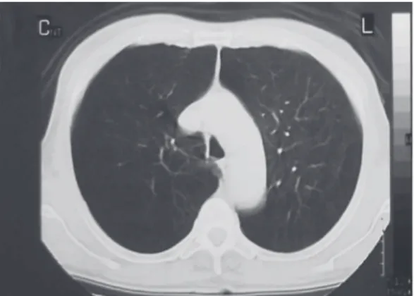
Patients were included in or excluded from the study based on the criteria detailed below. The inclusion criteria were as follows: having been diagnosed with emphysema, with chest X-ray (inhalation and exhalation) and HRCT evidence of homogeneous or heterogeneous diffuse emphysema showing pulmonary hype-rinflation (Figure 1); being less than 75 years of age; being incapacitated despite having received the gold standard treatment (pulmonary rehabilitation) for at least 6 months; postbron-chodilator FEV1 < 30% of predicted; residual volume ≥ 150% of predicted; total lung capa-city > 100% of predicted; smoking cessation at least 6 months prior to the procedure; and being considered a candidate for lung transplantation or lung volume reduction surgery.
We elected the following exclusion criteria: having had a myocardial infarction in the prece-ding six months; presenting a ventricular ejection fraction < 45%; interstitial or pleural disease which could impede the procedure; clinically significant bronchiectasis; pulmonary nodule; giant lung bullae (>1/3 of the lung volume); pulmonary hypertension (= 35 mmHg); comorbi-dities presenting five-year mortality rates > 50%; abnormal weight loss (< 70% or > 130% of ideal body weight); and evidence of systemic disease / neoplasia expected to impair survival.
This study protocol was approved by the Ethics in Human Research Committee of the Santa Casa School of Medical Sciences in São Paulo.
Prior to, during and after the procedure investigated, enrolled individuals were provided with full care in relation to the clinical evalua-lation, the inhaled and exhaled air flows through
the respiratory airways, since there is no obstruc-tion to this flow. However, in the patient with emphysema, the airways are obstructed, impeding expiratory flow. Consequently, there is an increase in the intra-alveolar pressure, forcing the air flow to the neighboring alveoli through passages or channels (Khon, Lambert and Martin pores) which divert it from the natural airway.(2,3) The air circulating among the alveolar air sacs causes pulmonary hyperinflation and remains trapped in the parenchyma, the consequences of which are all well known. In 1978, a proposal, based on this concept, was made.(4) The proposal, which had potential clinical applications, involved the crea-tion of artificial airways (spiracles) from the lung to the exterior, that is, outside the chest wall, which would allow the liberation of (an alternative exit for) the trapped air, decreasing hyperinflation and improving the respiratory mechanics.
The term airway bypass was proposed for the first time in 2003.(5) The term was applied to proce-dures which allowed communication between this trapped air (collateral ventilation) and the main respiratory airway in an extra-anatomical manner, allowing the trapped air to escape from the lung, thus decreasing the hyperinflation.
In a study conducted in 2007,(6) patients with severe emphysema were submitted to airway bypass through the endoscopic application of paclitaxel-eluting stents. The patients were submitted to general anesthesia. Subsequently, with the aid of fiberoptic bronchoscopy and Doppler ultrasound, a site in a segmental bronchus, free of blood vessels, was elected. Subsequently, an opening was made in the bronchial wall and the stent was put into place, creating a new passage between the respiratory tree and the adjacent pulmo-nary tissue. It was concluded that this procedure reduces hyperinflation, improving pulmonary function and reducing dyspnea in selected patients with severe pulmonary emphysema.
Based on these concepts,(7) the Santa Casa in São Paulo Group proposed an alternative proce-dure for the treatment of patients with severe pulmonary emphysema: drainage of the lung parenchyma, creating communication between the lung and the environment (spiracle). We initiated this pilot study 15 months ago.
The clinical and biochemical evolution of three patients submitted to this procedure is described with the objective of showing the results obtained in this period.
patient, awake, was maintained in the postanes-thesia care unit for at least 1 h and subsequently taken to the room.
The results for the three first patients submitted to this procedure are described below.
The statistical analysis of the data was performed using profile plots of the means.(8,9)
Results
All patients were hospitalized for 3 days. Chest tubes were removed after 24 h.
Patient 1: male, 47 years old, diagnosed with COPD, lung transplantation was indicated.
This patient engaged in no social activi-ties. He had stopped working, and was unable to ascend gentle slopes. He presented various attacks of dyspnea, which required treatment in the emergency room. He was enrolled in a pulmonary rehabilitation program and was monitored by a pulmonologist, receiving gold standard treatment.
He underwent surgery on 21 December 2006. At this writing, he had been in post-operative follow-up for 363 days. No serious complica-tions were reported. He reported resuming work (pushing an ice-cream cart). As a minor compli-cation, he reported pain at the drainage site for a period of 3 months.
Patient 2: male, 62 years old, diagnosed with severe COPD, lung transplantation was indicated.
He reported having had dyspnea for 7 years, with worsening of the symptoms for 3 years. He displayed respiratory arrest 2 years before, having tion and diagnosis of the disease (emphysema
with clinical symptoms of dyspnea). Individuals were fully informed regarding the procedure and the purpose of the study. All participating patients gave written informed consent. The following data were obtained from all of the participants using forms for subsequent analysis: written informed consent; identification; age; date; documentation showing that the indivi-dual met all of the inclusion criteria; anamnesis; significant physical examination findings; and pre-operative evaluation data. The selected patients were submitted to plethysmography and six-minute walk tests, in addition to completing quality of life questionnaires (Medical Outcomes Study 36-item Short-Form Health Survey; Saint George’s Respiratory Questionnaire; Eastern Cooperative Oncology Group Performance Status; and Medical Research Council Scale). For all of the patients, the post-operative follow-up period was at least 300 days. The tests were performed at the following time points: during the pre-operative period (T0), between post-operative days 30 and 40 (T1) and on post-operative day 300 (T2).
Surgical technique
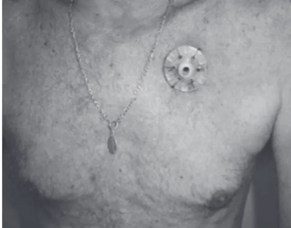
Patients were taken to the operating room, maintained in the supine position and submitted to local anesthesia, in the sixth intercostal space and extending for 2 cm, with the objective of performing conventional chest tube drainage in the hemithorax chosen for the performance of the pulmonary drainage. The choice of the side of the surgery was based on chest X-rays and on HRCT scans of the chest. Subsequently, a 4-cm incision was made in the selected hemithorax, under local anesthesia, in the third intercostal space, in the midclavicular line. Careful layer-by-layer dissection was performed until the pleural cavity was reached. Once the pleural cavity had been accessed, the lung parenchyma was secured with forceps in order to be opened safely. Four cardinal sutures were made in order to attach the lung to the parietal pleura. A 2- to 3-cm opening in the lung was sufficient. Through this opening, a chest tube was introduced (5 to 7 cm into the lung parenchyma) and affixed to the lung. A fenestrated 30 F silicone chest tube was used. The use of water-seal drainage was not required, since a patient can lead a normal life with the tube exposed to the environment (Figure 2). The chest tube is designed to be left in place for the lifetime of the patient. After the surgery, the
She was enrolled in a pulmonary rehabilitation program and was monitored by a pulmonologist, receiving gold standard treatment.
She underwent surgery on 20 January 2007. At this writing, she had been in post-operative follow-up for 310 days. No serious complications were reported. She reported improvement in her disposition, and she was sleeping all night without the use of oxygen therapy. There was no longer any need for emergency room visits due to attacks of dyspnea, and she became more socially active. As a minor complication, she presented cervical emphysema, which regressed a week after surgery.
For all three patients, the results obtained in the examinations carried out at the various time points are listed in Table 1.
In relation to the profile plots of the means of the analyzed variables, the differences between T0 and the other two time points were, in general, greater than those observed between T1 and T2.
The statistical analysis of the results involved only the three patients described. Due to the small sample size, the conclusions have a merely indicative (descriptive) character.
Discussion
In a recently published experimental study,(7) explanted lungs from lung transplant patients were evaluated. In the explanted native lung, a been admitted to the intensive care unit and
submitted to mechanical ventilation. He reported having been hospitalized, due to frequent infec-tions, at least once a month. He was unable to live alone, since he could not perform even the simplest of tasks, such as showering and eating. Therefore, he lived with his daughter. He was receiving oxygen therapy 18 h per day. He was unable to sleep at night without oxygen therapy. He was enrolled in a pulmonary rehabilitation program and was monitored by a pulmonolo-gist, receiving gold standard treatment.
He underwent surgery on 19 January 2007. At this writing, he had been in post-operative follow-up for 310 days. No serious complica-tions were reported. He was again able to live alone, since he was now able to perform all tasks with rarely any symptoms.
As a minor complication, a hematoma deve-loped in the chest wall.
Patient 3: female, 53 years old, diagnosed with severe COPD, lung transplantation was indicated.
She reported having dyspnea since 2003 (for 4 years). She used oxygen therapy 15 h per day, with a flow of 3 L/min, with the following schedule: 3 h in the morning, 3 h in the afternoon and 9 h at night. She reported experiencing extreme shor-tness of breath when bathing or ascending a flight of stairs. She had to stop working and was unable to walk. She suffered from constant panic attacks.
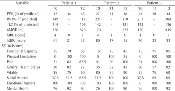
Table 1 - Results of the plethysmography, six-minute walk tests, and quality of life questionnaires, during the preoperative and post-operative periods.
Variable Patient 1 Patient 2 Patient 3
T0 T1 T2 T0 T1 T2 T0 T1 T2
FEV1 (% of predicted) 32 34 45 27 41 38 24 28 34
RV (% of predicted) 149 - 117 231 - 118 257 - 204
TLC (% of predicted) 114 - 108 142 - 121 143 - 136
6MWD (m) 320 - 570 170 - 225 150 - 435
MRC (score) 4 0 1 4 1 0 4 0 1
SGRQ (score) 61 21 37 80 17 15 70 23 33
SF-36 (scores)
Functional Capacity 15 70 55 15 75 45 15 75 85
Physical Limitation 0 100 100 0 100 25 0 100 100
Pain 31 62 87.5 41 90 100 31 100 100
General Health Status 30 82 72 35 82 42 30 57 82
Vitality 35 75 60 80 95 90 35 75 60
Social Aspects 37.5 62.5 62.5 37.5 100 100 37.5 65 65
Emotional Aspects 100 100 100 100 100 100 0 100 100
Mental Health 76 92 92 76 100 90 56 100 92
necessary, translating to longer hospital stays and a risk of severe bleeding.(5,6,11,12)
Two other therapeutic methods currently used for the treatment of severe pulmonary emphysema are lung volume reduction surgery and lung transplantation.
In both methods, the use of general anes-thesia, admission to the intensive care unit and long hospital stays are necessary. These methods present high morbidity and mortality, as well as high costs.
In the National Emphysema Treatment Trial,(13) mortality within 90 days after lung volume reduction surgery was found to be 7.9%. Lung volume reduction surgery is contrain-dicated for high-risk patients (those with an FEV1 < 20% of predicted). In selected patients submitted to this procedure, the following results were described(14): post-operative clinical and functional improvement; a 30% decrease in residual volume; a 50% increase in FEV1; and the discontinuation of supplementary oxygen in 80% of the patients. However, 2 years after the intervention, all patients presented values equal to those observed in the pre-operative period.
It is known that the pre-operative mortality is 6.2% among lung transplant patients, whereas the survival rate after bilateral transplantation is 86.4% in 1 year and 66.7% in 5 years.(15,16) The complications most frequently observed in the first year after the procedure are as follows: infection, in 80% of the cases; pulmonary hyper-tension, in 51.1%; renal dysfunction, in 25.7%; diabetes, in 21.5%; dyslipidemia, in 17.7%; severe acute rejection, in 10.5%; primary graft failure, in 10.2%; bronchiolitis obliterans, in 8.8%; and malignant neoplasia, in 3.9%.
However, since 2000, the Thoracic Surgery Group of the Santa Casa School of Medical Sciences in São Paulo has been improving techni-ques for the treatment of giant lung bullae using local anesthesia and drainage.(17) Therefore, in the 7 years after that first publication, we have accu-mulated considerable experience and confidence in this technique,(18,19) and we can now propose this new type of procedure not only for patients with giant lung bullae but also for patients with diffuse lung emphysema, as proposed in 1978,(4) and as carried out in the explanted native lungs of patients submitted to lung transplantation. (7) Therefore, the results obtained in the post-operative period, using drainage of the lung parenchyma, in these three patients with severe diffuse pulmonary disease constitute the first small opening was made and a silicone tube was
inserted in order to create a spiracle. Lungs were then ventilated through the bronchial tree or through transpleural communications. The flow-volume curve and the flow-volume of air recovered from the lungs by the spiracles were determined. Magnetic resonance imaging was employed in order to study the spatial distribution of helium administered through the natural airway or through the spiracles. The authors concluded that, due to the presence of collateral ventilation in the lung with emphysema, the direct commu-nication between the lung parenchyma and the environment improved the respiratory mechanics and, consequently, the ventilation. This study was the final result of many such studies conducted since 1930.(1) Therefore, 78 years after the study in which the concept was first defined, the existence of collateral ventilation in healthy lungs has been proven. In 1969, one group of authors measured the resistance of the interalveolar channels and demonstrated that, in the setting of hyperten-sion (emphysema), collateral ventilation becomes fundamentally important for the redistribution of air in the parenchyma, resulting in the develop-ment of the pulmonary hyperinflation and all of its consequences.(2) In a study conducted in 1978, it was suggested that a new procedure, based on the concept of collateral ventilation, be used for the treatment of patients with emphysema. (4) The author of that study stated the following: “Although it seems ridiculous at first, the creation of spiracles, allowing direct air exchanges between the pulmonary parenchyma and the environment, bypassing the normal airways (which are diseased), can produce significant improvement in the venti-lation and respiratory mechanics.” In addition to these aspects, some authors defend the hypothesis that there are air passages among the pulmonary lobes, so that, when there is severe emphysema, the consequent collateral ventilation maintains direct communication among the alveoli of the pulmo-nary lobes.(3,4,10) Therefore, in theory, a procedure enabling the communication of the upper lobe, for instance, with the environment, would allow the drainage not only of this upper lobe but also of the lower lobe of the same side.
About the authors
Roberto Saad Junior
Full Professor. Santa Casa School of Medical Sciences in São Paulo, São Paulo, Brazil.
Vicente Dorgan Neto
Adjunct Professor of the Thoracic Surgery Department. Santa Casa School of Medical Sciences in São Paulo, São Paulo, Brazil.
Marcio Botter
Instructor. Santa Casa School of Medical Sciences in São Paulo, São Paulo, Brazil.
Roberto Stirbulov
Head of the Pulmonology Department. Santa Casa School of Medical Sciences in São Paulo, São Paulo, Brazil.
Jorge Henrique Rivaben
Graduate Student in the Department of Surgery. Santa Casa School of Medical Sciences in São Paulo, São Paulo, Brazil.
Roberto Gonçalves
Full Professor in the Department of Thoracic Surgery. Santa Casa School of Medical Sciences in São Paulo, São Paulo, Brazil. report of the therapeutic application of collateral
ventilation in human beings.
Although the results obtained show that there was clinical improvement of the patients in the post-operative period, we cannot conclude that the surgical method proposed truly bring benefits. However, the findings of this pilot study convinced us to obtain further data from a larger sample of patients.
References
1. Van Allen C, Lindskog G, Richter HT. Gaseous interchange between adjacent lung lobules. Yale J Biol Med. 1930;2:297-300.
2. Hogg JC, Macklem PT, Thurlbeck WM. The resistance of collateral channels in excised human lungs. The Journal of Clinical Investigation. 1969;48(3):421-31.
3. Terry PB, Traystman RJ, Newball HH, Batra G, Menkes HA. Collateral ventilation in man. N Engl J Med. 1978;298(1):10-5.
4. Macklem PT. Collateral ventilation. N Engl J Med. 1978;298(1):49-50.
5. Rendina EA, De Giacomo T, Venuta F, Coloni GF, Meyers BF, Patterson GA, et al. Feasibility and safety of the airway bypass procedure for patients with emphysema. J Thorac Cardiovasc Surg. 2003;125(6):1294-9.
6. Cardoso PF, Snell GI, Hopkins P, Sybrecht GW, Stamatis G, Ng AW, et al. Clinical application of airway bypass with paclitaxel-eluting stents: early results. Thorac Cardiovasc Surg. 2007;134(4):974-81.
7. Choong CK, Macklem PT, Pierce JA, Lefrak SS, Woods JC, Conradi MS, et al. Transpleural ventilation of explanted human lungs. Thorax. 2007;62(7):623-30.
8. Neter J, Kutner MH, Nachtshein CJ, Wasserman W, editors. Applied Linear Statistical Models. Chigago: Irwin; 1996.
9. Bussab, WO, Morettin, PA, editors. Estatística Básica. São Paulo: Saraiva, 2005.
10. Higuchi T, Reed A, Oto T, Holsworth L, Ellis S, Bailey MJ, et al. Relation of interlobar collaterals to radiological heterogeneity in severe emphysema. Thorax. 2006;61(5):409-13.
11. Lunn WW. Endoscopic lung volume reduction surgery: cart before the horse? Chest. 2006;129(3):504-6. 12. Wan IY, Toma TP, Geddes DM, Snell G, Williams T,
Venuta F, et al. Bronchoscopic lung volume reduction for end-stage emphysema: report on the first 98 patients. Chest. 2006;129(3):518-26.
13. Saad Jr R, Mansano MD, Giannini JA, Dorgan Neto V. Tratamento operatório de bolhas de enfisema bolhoso: uma simples drenagem. J Pneumol. 2000;26(3):113-8. 14. Saad Jr R, Ethel Filho J, Stirbulov R. Enfisema pulmonar
difuso: proposta de tratamento. In: Saad Jr R, Carvalho WR, Ximenes Netto M, Forte V, editors. Cirurgia Torácica Geral. São Paulo: Atheneu; 2005. p.351-3.
15. Botter M, Saad Jr R, Botter DA, Rivabem JH, Gonçalves R, Dorgan Neto V. Tratamento operatório das bolhas pulmonares gigantes. Rev Assoc Med Bras. 2007;53(3):217-22.
16. Fishman A, Martinez F, Naunheim K, Piantadosi S, Wise R, Ries A, et al. A randomized trial comparing lung-volume-reduction surgery with medical therapy for severe emphysema. N Engl J Med. 2003;348(21):2059-73. 17. McKenna RJ Jr, Brenner M, Fischel RJ, Gelb AF. Should
lung volume reduction for emphysema be unilateral or bilateral? J Thorac Cardiovasc Surg. 1996;112(5):1331-8; discussion 1338-9.
18. Cassivi SD, Meyers BF, Battafarano RJ, Guthrie TJ, Trulock EP, Lynch JP, et al. Thirteen-year experience in lung transplantation for emphysema. Ann Thorac Surg. 2002;74(5):1663-9; discussion 1669-70.