* Study carried out at the Universidade do Estado do Rio de Janeiro – UERJ, Rio de Janeiro State University – Pedro Ernesto University Hospital, Rio de Janeiro, Brazil.
1. Chief of the Department of Respiratory Function at the Universidade do Estado do Rio de Janeiro – UERJ, Rio de Janeiro State University – Pedro Ernesto University Hospital, Rio de Janeiro, Brazil.
2. Adjunct Professor at the Universidade do Estado do Rio de Janeiro – UERJ, Rio de Janeiro State University – School of Medical Sciences, Rio de Janeiro, Brazil. 3. Physician in training in the Department of Radiology and Diagnostic Imaging of the Universidade do Estado do Rio de Janeiro – UERJ, Rio de Janeiro State University – Pedro Ernesto University Hospital, Rio de Janeiro, Brazil.
4. Adjunct Professor at the Universidade do Estado do Rio de Janeiro – UERJ, Rio de Janeiro State University – Institute of Biology, Rio de Janeiro, Brazil. 5. Full Professor at the Universidade do Estado do Rio de Janeiro – UERJ, Rio de Janeiro State University – School of Medical Sciences, Rio de Janeiro, Brazil. Correspondence to: Agnaldo José Lopes. Rua José do Patrocínio, 290/405, Grajaú, CEP 20560-160, Rio de Janeiro, RJ, Brazil.
Tel 55 21 2576 2030. E-mail: [email protected]
Submitted: 4 February 2007. Accepted, after review: 7 August 2007.
Tomografia computadorizada de alta resolução na silicose: correlação com radiografia e testes de função pulmonar
Agnaldo José Lopes1, Roberto Mogami2, Domenico Capone2, Bernardo Tessarollo3, Pedro Lopes De Melo4, José Manoel Jansen5
Abstract
Objective: To correlate tomographic findings with pulmonary function findings, as well as to compare chest X-ray findings with high-resolution computed tomography (HRCT) findings, in patients with silicosis. Methods: A cross-sectional study was conducted in 44 non-smoking patients without a history of tuberculosis. Chest X-ray findings were classified according to the International Labour Organization recommendations. Using a semiquantitative system, the following HRCT findings were measured: the full extent of pulmonary involvement; parenchymal opacities; and emphysema. Spirometry and forced oscillation were performed. Pulmonary volumes were evaluated using the helium dilution method, and diffusing capacity of the lung for carbon monoxide was assessed. Results: Of the 44 patients studied, 41 were male. The mean age was 48.4 years. There were 4 patients who were classified as category 0 based on X-ray findings and as category 1 based on HRCT findings. Using HRCT scans, we identified progressive massive fibrosis in 33 patients, compared with only 23 patients when X-rays were used. Opacity score was found to correlate most closely with airflow, DLCO and compliance. Emphysema score correlated inversely with volume, DLCO and airflow. In this sample of patients presenting a predominance of large opacities (75% of the individuals), the deterioration of pulmonary function was associated with the extent of structural changes. Conclusions: In the early detection of silicosis and the identification of progressive massive fibrosis, HRCT scans are superior to X-rays.
Keywords: Silicosis; Occupational diseases; Tomography, x-ray computed; Respiratory function tests.
Resumo
Objetivo: Correlacionar os parâmetros tomográficos com os de função pulmonar em portadores de silicose, bem como comparar os resultados da tomografia computadorizada de alta resolução (TCAR) com os da radiografia de tórax. Métodos: Foi realizado um estudo de corte trans-versal, em que foram avaliados 44 pacientes não-tabagistas e sem história pregressa de tuberculose. As radiografias foram classificadas de acordo com a proposta da Organização Internacional do Trabalho. Utilizando um sistema de escore semiquantitativo, os seguintes achados na TCAR foram quantificados: extensão total do envolvimento pulmonar, opacidades parenquimatosas e enfisema. Foram realizados espiro-metria, oscilações forçadas, avaliação dos volumes pulmonares pela técnica de diluição com hélio e medida da diffusing capacity of the lung for carbon monoxide (DLCO, capacidade de difusão do monóxido de carbono). Resultados: Dos 44 pacientes estudados, 41 eram homens, com média de idade de 48,4 anos. Na análise comparativa, 4 pacientes classificados na categoria 0 pela radiografia foram reclassificados na categoria 1 pela TCAR e, enquanto a radiografia diagnosticou 23 casos de fibrose maciça progressiva, a TCAR estabeleceu esse diagnóstico em 33 doentes. Para o escore de opacidades, as maiores correlações foram observadas com as medidas de fluxo, DLCO e complacência. Já o enfisema correlacionou-se negativamente com as medidas de volume, DLCO e fluxo. Nesta amostra com predomínio de grandes opacidades (75% dos indivíduos), a deterioração da função pulmonar associou-se com a extensão das alterações estruturais. Conclusões: Na silicose, a TCAR é superior à radiografia tanto na detecção precoce da doença quanto na identificação de fibrose maciça progressiva.
All tests were performed within one month of each and in the following order: pulmonary func-tion test, chest X-ray and HRCT.
The chest X-ray was performed in the antero-posterior position using a Siemens X-ray unit (model LX30; Siemens AG, Erlangen, Germany). The following technique was used: distance of 180 cm between focal plane and the film; device at 80 kVp; exposure time of 0.04 ms. Readings were performed independently by three specialists certi-fied by the Jorge Duprat Figueiredo Foundation for Occupational Safety and Medicine and by the Brazilian Ministry of Labor and Employment. For the sake of simplification, the classification of the profusion of small opacities was summarized: 0/−, 0/0 or 0/1 = 0; 1/0, 1/1 or 1/2 = 1/2/1; 2/2 or 2/3 = 2; 3/2; and 3/3 or 3/+ = 3. Large opacities were classified as types A, B or C, according to the standards provided by the ILO.(1) The results were
summarized as the medians of the readings. The pulmonary function tests consisted of the forced oscillation technique (FOT), spirometry, evaluation of lung volumes using the helium dilu-tion method and assessment of diffusing capacity of the lung for carbon monoxide (DLCO). The FOT was performed using an impedance analyzer, and the following parameters were evaluated: total respiratory resistance; airway resistance; and dynamic respiratory compliance (Crs,dyn). The remaining tests were performed using the Collins Plus Pulmonary Function Testing System (Warren E. Collins, Inc., Braintree, MA, USA), standardized and interpreted in accordance with the guide-lines established by the Brazilian Thoracic Society.
(11) Equations devised by Pereira (for spirometry)
and Neder (for static lung volumes and DLCO) were used in the interpretation of the following parameters(12-14):
• spirometry: forced vital capacity (FVC); forced expiratory volume in one second (FEV1); FEV1/ FVC ratio; forced expiratory flow between 25 and 75% of FVC (FEF25-75%); and FEF25-75%/
FVC ratio
• helium dilution method: residual volume; total lung capacity; and the residual volume/ total lung capacity ratio
• determination of single-breath DLCO
The HRCT was performed using a GE HiSpeed scanner (General Electric Medical Systems, Milwaukee, WI, USA).The following technique was used: 1-mm
Introduction
According to the current guidelines of the International Labour Organization (ILO),(1) the
prin-cipal method of diagnosing silicosis is the analysis of chest X-rays and of the history of occupational exposure to free silica. However, high-resolution computed tomography (HRCT) has increasingly taken a prominent position in the evaluation of sili-cosis, offering relevant additional information for the early detection of small opacities and emphysema and identification of complications.(2-4) Pulmonary
function tests, although not used as diagnostic tools, are widely employed in longitudinally studies of individuals with silicosis.(3,4)
Since imaging and function tests are the most widely used diagnostic resources in the follow-up evaluation of individuals with silicosis, it is funda-mental to establish the correlation between these two methods. Due to its higher sensitivity, HRCT has been the imaging test most often used for this purpose. However, in studies correlating imaging tests with respiratory function tests, quantifica-tion of the severity of the disease by funcquantifica-tion tests is affected by history of smoking and associated pulmonary emphysema, a fact highlighted by most investigators.(5-10)
The objective of the present study was to correlate tomographic parameters with respiratory function parameters in nonsmokers with silicosis and to compare the HRCT findings with the chest X-ray findings.
Methods
A descriptive study evaluating 53 nonsmokers with history of exposure to silica was carried out. Participants were informed regarding the objec-tive of the study, after which they all gave written informed consent. The protocol was approved by the ethics in research committee of the institution.
vessels; aortic arch; carina; confluence of pulmo-nary veins; and 1 cm above the right diaphragm.
(18) Using a semiquantitative evaluation system, each
of these levels (right and left, separately, totaling 10 levels) was analyzed as to the following aspects:
• score of total interstitial disease (TID, including nodules, masses, emphysema and other paren-chymal alterations):
0) no alteration
1) pulmonary involvement ≤5% of the area 2) pulmonary involvement from >5 to ≤25%
of the area
3) pulmonary involvement from >25 to 49% of the area
4) pulmonary involvement from 50 to 75% of the area
5) pulmonary involvement >75% of the area • score of total extent of parenchymal opacities,
including nodules and masses: 0) no parenchymal opacities
1) parenchymal opacities involving ≤5% of the area
2) parenchymal opacities involving >5 to ≤25% of the area
3) parenchymal opacities involving >25 to 49% of the area
slices, at 1.5-s intervals and increased by 10 mm; image reconstruction with a 512 × 512 pixel matrix, using a high-resolution algorithm; 1000 Hounsfield unit (HU)-width window; −700 HU medium window level. The tomographic findings were interpreted by consensus among four radiologists who had been previously trained for one month in how to apply the classification system adopted. Small opacities were classified into four categories, according to profusion, and in a manner similar to that used to classify the chest X-ray findings:
0) absence of micronodules
1) a small number of micronodules without vascular blurring
2) a large number of micronodules, with or without vascular blurring but with no confluence
3) confluence of nodules <10 mm, typi-cally accompanied by pronounced vascular blurring
Large opacities were classified as type A or B (respectively, one or more opacities > 10 mm having the sum of the diameters ≤50 mm, and one or more opacities having the sum of the diameters <50 mm).(9,10,15-17) Subsequently, HRCT was
evalu-ated as to the extent of pulmonary involvement, considering five cut-off levels: origin of major
a b
functional indices and tomographic scores were studied using Pearson’s parametric test. Values of p ≤ 0.05 were considered significant.
Results
Of the 44 patients studied, 41 were male. The mean age was 48.4 years. The mean duration of silica exposure and the mean time since withdrawal were 16.1 and 16.9 years, respectively. Sandblasting and stone cutting were the professional activities most often cited (45.5% and 34.1%, respectively). Other professional activities cited were marble quar-rying (6.8%), rock quarquar-rying (4.5%), foundry work (4.5%), dental prosthesis design (2.3%) and grinding (2.3%).
In the evaluation of the chest X-ray findings, 19 tests had technical quality level 1 and 25 tests had technical quality level 2. The distribution of indi-viduals in the in the ILO radiological categories for small opacities was as follows: 4 (9.1%) in category 0; 22 (50%) in category 1; 14 (31.8%) in category 2; and 4 (9.1%) in category 3. Regarding the form and size of the small opacities, the chest X-ray find-ings were classified as follows: q/q = 14; r/q = 7; p/q = 5; q/r = 4; p/p = 3; q/p = 3; r/r = 3; or q/t = 1. Type A large opacities were observed in 4 patients (9.1%), type B in 12 (27.3%) and type C in 7 (15.9%), whereas 21 patients (47.7%) did not present large opacities on chest X-rays. The classifications most often observed were as follows: ax (coalescence of small opacities: 22 cases); cn (calcification in small opacities: 14 cases); co (abnormalities in the shape or size of the heart: 14 cases); and di (distortion of the thoracic contents) Analysis of the inter-rater reliability for the profusion of small opacities (cate-4) parenchymal opacities involving 50 to 75%
of the area
5) parenchymal opacities involving >75 of the area
• score of extent of the emphysema: 0) no emphysema
1) emphysema affecting ≤5% of the area 2) emphysema affecting >5 to ≤25% of the
area
3) emphysema affecting >25 to 49% of the area
4) emphysema affecting 50 to 75% of the area
5) emphysema affecting >75% of the area For the analysis of the scores, pulmonary involvement was estimated using an influence factor (weight) to correct the differences in volume at each level: origin of major vessels (0.129); aortic arch (0.190); carina (0.222); confluence of pulmo-nary veins (0.228); 1 cm above the right diaphragm (0.230).(19) In the end, the TID, parenchymal
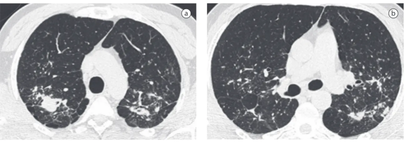
opaci-ties and emphysema values were obtained by adding the scores for each level (highest possible score for each patient, 10). An example of how these scores were obtained is shown in Figure 1
The statistical programs used were Epi Info 6.04 and Statistica 5.01b (StatSoft, Inc., Tulsa, OK, USA). Means, standard deviations and frequencies were used to describe the data. The calculation of the coefficient of concordance was used to compare chest X-ray readings. Analysis of variance was used to compare the means of function parameters among the various HRCT categories. All continuous variables were analyzed in order to determine the distribution of normality in accordance with the Kolmogorov-Smirnov test. Correlations between
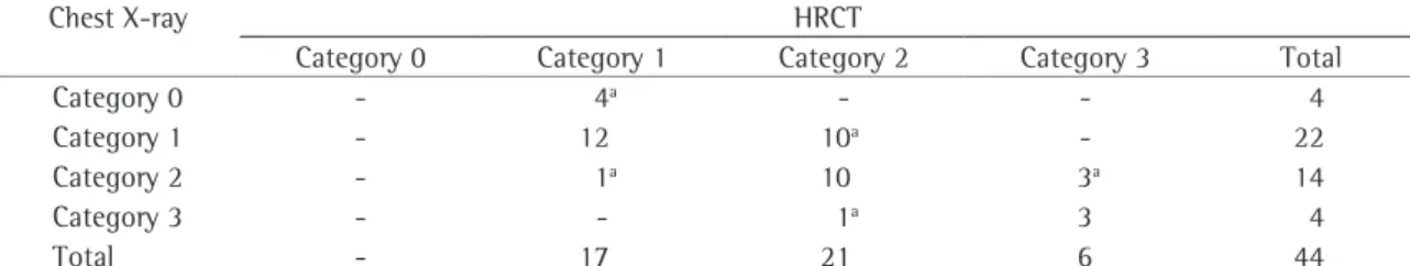
Table 1 - Distribution of individuals in the radiological and tomographic categories, according to the profusion of small opacities.
Chest X-ray HRCT
Category 0 Category 1 Category 2 Category 3 Total
Category 0 - 4a - - 4
Category 1 - 12 10a - 22
Category 2 - 1a 10 3a 14
Category 3 - - 1a 3 4
Total - 17 21 6 44
HRCT: high-resolution computed tomography. aCases of discrepancy between the HRCT classification and the chest X-ray
in 13 cases and moderate in 1); 13 had mixed respiratory disorder (mild in 8 cases, moderate in 3 and severe in 2); 11 had restrictive defect (mild in 8 cases, moderate in 2 and severe in 1); and 6 presented spirometric indices and static volumes that were within the normal ranges. In this study, 29 individuals presented a DLCO reduction, which was characterized as mild in 15 cases, moderate in 12 and severe in 2.
In relation to the HRCT scores, the mean values of TID, parenchymal opacities and emphysema were 3.7, 3.3 and 1.5, respectively. The coefficients of the correlations between functional parameters and values obtained in the HRCT analysis are given in Table 4. The opacities score correlated most strongly with airflow, DLCO and Crs,dyn. However, emphysema correlated inversely with lung volume, DLCO and airflow.
Discussion
In any study of morphofunctional correlations, it is fundamental that the investigators eliminate the influence of any factors other than the one analyzed. Most previously studies have included smokers and former smokers, as well as patients with a history of tuberculosis.(5,8-10) However, in the present study,
we were careful to exclude smokers, former smokers and individuals with a history of tuberculosis. Consequently, it was necessary to recruit patients from various referral centers for silicosis in order to reach the ideal sample size.
Conventionally, silicosis-related lesions are assessed radiologically according to the ILO clas-sification of pneumoconiosis, which was created for epidemiological purposes.(1) However, its usefulness
in clinical diagnosis is debatable, particularly in rela-tion to two aspects: the high inter-rater variability in chest X-ray analysis, especially to low profusion categories; and the fact that it can underestimate the presence of pulmonary disease.(16) In the present
study, greater discrepancy among specialists was observed for the small opacity category. Inter-rater variability was lower for large opacities than for small opacities. This finding is in accordance with those of other studies, which also showed higher kappa values for large opacities.(4,9,16-17) Despite these
limitations, chest X-ray is still the most efficient tool for the follow-up evaluation of workers exposed gories 0, 1, 2 and 3) and of large opacity types (0,
A, B and C), respectively, revealed kappa coefficients of 0.31 (p < 0.001) and 0.75 (p < 0.001).
The HRCT findings observed were micron-odules in 44 cases (100%), with identification of confluences in six tests; large opacities consistent with progressive massive fibrosis (PMF) in 33 cases (75%); emphysema in 27 cases (61.4%) - 21 cases of scar-related emphysema and 6 cases of panac-inar emphysema - 24 of which were concomitant to PMF; localized pleural thickening in 14 cases (31.8%), from which 10 of which were concomitant to PMF; and intrathoracic lymph node enlargement in 28 cases (63.6%).
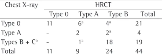
The comparisons between the chest X-ray and the HRCT classifications are shown in Tables 1 and 2 (for small and large opacities, respectively). For small opacities, there was concordance between the two methods in 56.8% of the cases. In this sample, 4 patients classified as category 0 based on the chest X-ray findings were reclassified as category 1 based on the HRCT findings. For large opacities, there was concordance between the two methods in 70.5% of the cases. The diagnosis of PMF was made based on chest X-ray findings in 23 cases, compared with 33 cases for HRCT findings.
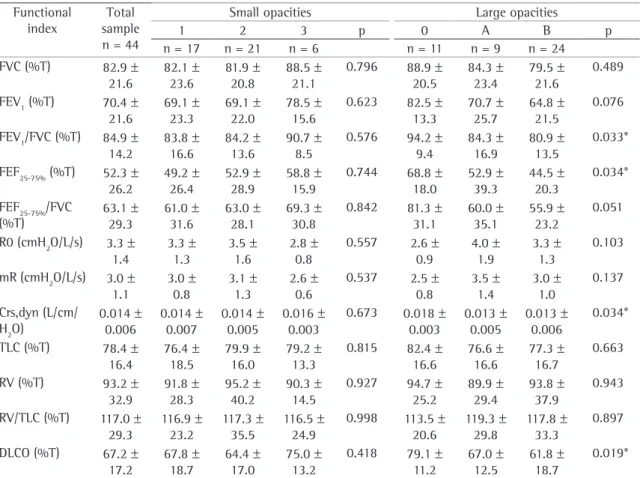
The means and standard deviations for the pulmo-nary function indices, as well as the distribution of those values according to the HRCT classification, are given in Table 3. For large opacities, statistically significant differences were observed in terms of the values obtained for airflow, Crs,dyn and DLCO.
In relation to the functional pattern, 14 indi-viduals had obstructive ventilatory defect (mild
Table 2 - Distribution of individuals in the radiological categories and distribution of individuals in the tomographic categories, according to the type of large opacity.
Chest X-ray HRCT
Type 0 Type A Type B Total Type 0 11 6a 4a 21
Type A - 2 2a 4
Types B + Cb - 1a 18 19
Total 11 9 24 44
HRCT: high-resolution computed tomography. aCases of
discrep-ancy between the HRCT classification and the chest X-ray classification. bCases of type B or C large opacities seen on chest
based on the HRCT findings, underscoring the value of the latter exam.
In terms of the capacity to detect large opaci-ties, the superiority of HRCT over chest X-ray is well established. In a study assessing the HRCT results of 76 patients, 10 cases of PMF were observed among the 26 classified as simple sili-cosis based on chest X-rays. In the present study, 10 new cases of PMF were diagnosed based on the HRCT findings, confirming the higher sensitivity of the method for detecting accelerated silicosis. Another interesting finding was the detection of emphysema, which was diagnosed in 27 patients (61.4%). Considering the fact that the sample consisted exclusively of nonsmokers, the detection of emphysema, in this study, can only be attrib-to silica, since it is an inexpensive procedure and
exposes subjects to only a low dose of radiation.(2)
Many studies have been carried out to establish the importance of HRCT in the early detection of small opacities. One study showed that 13 out of 32 individuals with a history of exposure to silica and normal chest X-ray results presented evidence of silicosis on HRCT scans.(16) In a study carried out
in Brazil and including 68 former miners, concord-ance of chest X-ray findings and HRCT findings was observed in 55 cases. Divergence was observed in 13 cases; HRCT diagnosed 5 new cases of sili-cosis and ruled out 8 cases.(17) In the present study,
4 patients classified as category 0 based on the chest X-ray findings were reclassified as category 1
Table 3 - Functional indices according to the tomographic categories of small and large opacities.a
Functional index
Total sample n = 44
Small opacities Large opacities
1 2 3 p 0 A B p
n = 17 n = 21 n = 6 n = 11 n = 9 n = 24 FVC (%T) 82.9 ±
21.6
82.1 ± 23.6
81.9 ± 20.8
88.5 ± 21.1
0.796 88.9 ± 20.5
84.3 ± 23.4
79.5 ± 21.6
0.489
FEV1 (%T) 70.4 ± 21.6
69.1 ± 23.3
69.1 ± 22.0
78.5 ± 15.6
0.623 82.5 ± 13.3
70.7 ± 25.7
64.8 ± 21.5
0.076
FEV1/FVC (%T) 84.9 ± 14.2
83.8 ± 16.6
84.2 ± 13.6
90.7 ± 8.5
0.576 94.2 ± 9.4
84.3 ± 16.9
80.9 ± 13.5
0.033*
FEF25-75% (%T) 52.3 ± 26.2
49.2 ± 26.4
52.9 ± 28.9
58.8 ± 15.9
0.744 68.8 ± 18.0
52.9 ± 39.3
44.5 ± 20.3
0.034*
FEF25-75%/FVC (%T)
63.1 ± 29.3
61.0 ± 31.6
63.0 ± 28.1
69.3 ± 30.8
0.842 81.3 ± 31.1
60.0 ± 35.1
55.9 ± 23.2
0.051
R0 (cmH2O/L/s) 3.3 ± 1.4
3.3 ± 1.3
3.5 ± 1.6
2.8 ± 0.8
0.557 2.6 ± 0.9
4.0 ± 1.9
3.3 ± 1.3
0.103
mR (cmH2O/L/s) 3.0 ± 1.1
3.0 ± 0.8
3.1 ± 1.3
2.6 ± 0.6
0.537 2.5 ± 0.8
3.5 ± 1.4
3.0 ± 1.0
0.137
Crs,dyn (L/cm/ H2O)
0.014 ± 0.006
0.014 ± 0.007
0.014 ± 0.005
0.016 ± 0.003
0.673 0.018 ± 0.003
0.013 ± 0.005
0.013 ± 0.006
0.034*
TLC (%T) 78.4 ± 16.4
76.4 ± 18.5
79.9 ± 16.0
79.2 ± 13.3
0.815 82.4 ± 16.6
76.6 ± 16.6
77.3 ± 16.7
0.663
RV (%T) 93.2 ± 32.9
91.8 ± 28.3
95.2 ± 40.2
90.3 ± 14.5
0.927 94.7 ± 25.2
89.9 ± 29.4
93.8 ± 37.9
0.943
RV/TLC (%T) 117.0 ± 29.3
116.9 ± 23.2
117.3 ± 35.5
116.5 ± 24.9
0.998 113.5 ± 20.6
119.3 ± 29.8
117.8 ± 33.3
0.897
DLCO (%T) 67.2 ± 17.2
67.8 ± 18.7
64.4 ± 17.0
75.0 ± 13.2
0.418 79.1 ± 11.2
67.0 ± 12.5
61.8 ± 18.7
0.019*
responsible for the obstructive phenomenon, since 75% of the patients presented PMF in the HRCT results. These masses of PMF, composed of clusters of fibrotic nodules, can distort the lung parenchyma and cause an irregular increase in the area of the adjacent air spaces, ultimately limiting airflow.(10,22,23,24)
Comparing the means of the pulmonary func-tion parameters between the different HRCT categories, we observed that, for small opacities, there was no significant difference in any isolated functional parameter for any HRCT category evalu-ated. This finding is in accordance with those of a previous study in which the micronodules detected on the HRCT scan, per se, were found to have no influence on functional deterioration.(15) However,
in the present study, we observed that, in patients presenting large opacities, there was a progressive decrease in DLCO, Crs,dyn and airflow decreased in parallel with increases in the extent of damage (HRCT classification). These findings are in accord-ance with those of other studies also showing that functional damage correlates more strongly with the extent of large opacities than with that of small opacities.(5,9-10,23) Therefore, an HRCT finding of
large opacities can be an important indicator of the severity of silicosis.(9,10,25)
In our study, as well as in others, parenchymal opacity correlated negatively with a reduction in DLCO.(3,5,10,23) Some investigators have attributed this
correlation to the presence of PMF, which tends to diminish as the disease advances. That results in the appearance of scar-related emphysema and a decrease in the surface area available for gas exchange, which reduces DLCO.
The analysis of the association obtained between parenchymal opacity and Crs,dyn is interesting, since, to our knowledge, there have been no studies correlating compliance values with HRCT findings. In practice, Crs,dyn represents how easily the respi-ratory system, including the lungs and chest wall, reaches total lung capacity.(26) In accelerated silicosis,
the intensification of pleuropulmonary involvement causes a reduction in Crs,dyn. Therefore, since the method of measurement is noninvasive, Crs,dyn might eventually become another functional param-eter evaluated in the follow-up treatment of patients with silicosis. In addition, our results indicate that, in nonsmoking silicosis patients presenting large opacities (as in 75% of our sample), deterioration uted to the presence of PMF or of the very silica
dust itself.
On the HRCT scans, we observed localized pleural thickening in 31.8% of the cases, compared with the 58.2% found in another study in which individuals with a history of tuberculosis were included (31% of the sample).(20) In that study,
most of the pleural thickening was diffuse rather than localized. Despite the fact that diffuse pleural thickening can cause restrictive functional damage, the authors of that same study concluded that the clinical relevance of this finding in silicosis remains uncertain.(20)
In relation to pulmonary function, we observed that ventilatory defect (obstructive or mixed) was quite common, being diagnosed in 61.4% of the cases. In silicosis, airflow limitation is caused by many factors, such as bronchial stenosis secondary to peribronchiolar fibrosis, as well as lymph node enlargement and centrilobular emphysema.(10,21)
However, in the present study, it is possible that the large opacities and the accompanying scar-related emphysema be the principal mechanisms
Table 4 - Pearson’s correlation coefficients for the correlations between functional indices values and results of the scores obtained with high-resolution computed tomography in 44 individuals evaluated.
Functional index Total score
pacO score
Emphysema score FVC (%T) −0.42** −0.33* −0.44**
FEV1 (%T) −0.61** −0.55** −0.44**
FEV1/FVC (%T) −0.46** −0.51** −0.10
FEF25-75% (%T) −0.59** −0.58** −0.35*
FEF25-75%/FVC (%T) −0.41** −0.46** −0.12
R0 (cmH2O/L/s) +0.20 +0.19 +0.10 Rm (cmH2O/L/s) +0.13 +0.11 +0.13 Crs,dyn (L/cm/H2O) −0.48** −0.45** −0.25
TLC (%T) −0.42** −0.32* −0.51** RV (%T) −0.06 −0.01 −0.30* RV/TLC (%T) +0.31* +0.28 +0.06 DLCO (%T) −0.63** −0.57** −0.43**
miners with long exposure to silica dust. Occup Environ Med. 1994;51(8):557-63.
8. Bergin CJ, Müller NL, Vedal S, Chan-Yeung M. CT in silicosis: correlation with plain films and pulmonary function tests. AJR Am J Roentgenol. 1986;146(3):477-83.
9. Talini D, Paggiaro PL, Falaschi F, Battolla L, Carrara M, Petrozzino M, et al. Chest radiography and high resolution computed tomography in the evaluation of workers exposed to silica dust: relation with functional findings. Occup Environ Med. 1995;52(4):262-7.
10. Ooi GC, Tsang KW, Cheung TF, Khong PL, Ho IW, Ip MS, et al. Silicosis in 76 men: qualitative and quantitative CT evaluation--clinical-radiologic correlation study. Radiology. 2003;228(3):816-25.
11. Sociedade Brasileira de Pneumologia e Tisiologia. Diretrizes para Testes de Função Pulmonar. J Pneumol. 2002;28(Supl 3):S1-S238.
12. Pereira CAC, Barreto SP, Simões JG, Pereira FWL, Gerstler JG, Nakatani J. Valores de referência para espirometria em uma amostra da população brasileira adulta. J Pneumol. 1992;18(1):10-22.
13. Neder JA, Andreoni S, Castelo-Filho A, Nery LE. Reference values for lung function tests. I. Static volumes. Braz J Med Biol Res. 1999;32(6):703-17.
14. Neder JA, Andreoni S, Peres C, Nery LE. Reference values for lung function tests. III. Carbon monoxide diffusing capacity (transfer factor). Braz J Med Biol Res. 1999;32(6):729-37. 15. Gevenois PA, Sergent G, De Maertelaer V, Gouat F, Yernault
JC, De Vuyst P. Micronodules and emphysema in coal mine dust or silica exposure: relation with lung function. Eur Respir J. 1998;12(5):1020-4.
16. Bégin R, Ostiguy G, Fillion R, Colman N. Computed tomography scan in the early detection of silicosis. Am Rev Respir Dis. 1991;144(3 Pt 1):697-705.
17. Carneiro APS, Siqueira AL, Algranti E, Ferreira CS, Kavakama JI, Bernardes ML, et al. Estudo comparativo entre tomografia computadorizada de alta resolução e radiografia de tórax no diagnóstico da silicose em casos incipientes. J Pneumol. 2001;27(4):199-205.
18. Copley SJ, Wells AU, Sivakumaran P, Rubens MB, Lee YC, Desai SR, et al. Asbestosis and idiopathic pulmonary fibrosis: comparison of thin-section CT features. Radiology. 2003;229(3):731-6.
19. Wells AU, Rubens MB, du Bois RM, Hansell DM. Serial CT in fibrosing alveolitis: prognostic significance of the initial pattern. AJR Am J Roentgenol. 1993;161(6):1159-65. 20. Arakawa H, Honma K, Saito Y, Shida H, Morikubo H, Suganuma
N, et al. Pleural disease in silicosis: pleural thickening, effusion, and invagination. Radiology. 2005;236(2):685-93. 21. Terra-Filho M, Santos UP. Silicosis. J Bras Pneumol.
2006;32(Supl 2):S59-S65.
22. Bégin R, Ostiguy G, Cantin A, Bergeron D. Lung function in silica-exposed workers. A relationship to disease severity assessed by CT scan. Chest. 1988;94(3):539-45.
23. Arakawa H, Gevenois PA, Saito Y, Shida H, De Maertelaer V, Morikubo H, et al. Silicosis: expiratory thin-section CT assessment of airway obstruction. Radiology. 2005;236(3):1059-66.
24. Leung CC, Chang KC, Law WS, Yew WW, Tam CM, Chan CK, et al. Determinants of spirometric abnormalities among silicotic patients in Hong Kong. Occup Med (Lond). 2005;55(6):490-3.
of pulmonary function correlates with the extent of the structural alterations.
As has been reported in previous studies, we found that emphysema presented significant correla-tions with airflow, lung volume and DLCO. However, the correlation coefficients were slightly inferior to those obtained in studies that included smokers in their samples. Therefore, again, the difference can be attributed to smoking, which is known to reduce airflow and DLCO.
A critical analysis of the results of the present study and its limitations is called for. One poten-tial limitation of this study was that the dose and incidence of the occupational exposure were not quantified. Such data might have furthered the understanding of morphofunctional correlations. Another limitation is that the coefficient of concord-ance was not evaluated for the HRCT readings. Therefore, the interpretation of the tomographic findings by consensus implies possible acceptance of the analysis of the most experienced specialists by the less experienced specialists, which constitutes a methodological bias.
In conclusion, our study shows that, in the diag-nosis of silicosis HRCT scans are superior to chest X-rays, both for the early detection of the initial phases of the disease and for the identification of PMF.
References
1. Occupational Safety and Health. Guidelines for the use of the ILO International classification of radiographs of pneumoconiosis. 2nd ed. rev. Geneva: International Labour
Organization; 2002.
2. Capitani EM. Silicosis (still) among us. J Bras Pneumol. 2006;32(6):xxxiii-xxxv
3. Ferreira AS, Moreira VB, Ricardo HMV, Coutinho R, Gabetto JM, Marchiori E. Progressive massive fibrosis in silica-exposed workers. High-resolution computed tomography findings. J Bras Pneumol. 2006; 32(6): 523-8.
4. Antao VC, Pinheiro GA, Terra-Filho M, Kavakama J, Müller NL. High-resolution CT in silicosis: correlation with radiographic findings and functional impairment. J Comput Assist Tomogr. 2005;29(3):350-6.
5. Kinsella M, Müller N, Vedal S, Staples C, Abboud RT, Chan-Yeung M. Emphysema in silicosis. A comparison of smokers with nonsmokers using pulmonary function testing and computed tomography. Am Rev Respir Dis. 1990;141(6):1497-500.
6. Bégin R, Filion R, Ostiguy G. Emphysema in silica- and asbestos-exposed workers seeking compensation. A CT scan study. Chest. 1995;108(3):647-55.
27. Mesquita-Júnior JA, Lopes AJ, Jansen JM, Melo PL. Using the forced oscillation technique to evaluate respiratory resistance in individuals with silicosis. J Bras 2Pneumol. 2006;32(3):213-20.
25. Bégin R, Bergeron D, Samson L, Boctor M, Cantin A. CT assessment of silicosis in exposed workers. AJR Am J Roentgenol. 1987;148(3):509-14.