THE EFFECT OF ABIOTIC FACTORS ON THE HATCHING
OF Moina micrura KURZ, 1874 (CRUSTACEA: CLADOCERA)
EPHIPPIAL EGGS
ROJAS, N. E. T.,1 MARINS, M. A.2 and ROCHA, O.2
1Centro de Pesquisa em Reprodução e Larvicultura, Instituto de Pesca, São Paulo, SP, Brazil 2Departamento de Ecologia e Biologia Evolutiva, Universidade Federal de São Carlos, Rodovia Washington
Luiz, km 235, CEP 13565-905, São Carlos, SP, Brazil
Correspondence to: Nilton Eduardo Torres Rojas, Instituto de Pesca, Av. Francisco Matarazzo, 455, CEP 05001-900, São Paulo, SP, Brazil, e-mail: niltonrojas@uol.com.br
Received January 21, 2000 – Accepted March 8, 2000 – Distributed August 31, 2001 (With 2 figures)
ABSTRACT
The roles of some abiotic factors in controlling the hatching rates of Moina micruraephippial eggs were investigated. Determination of optimum hatching conditions would be important in developing the use of this species as a food for fish larvae in aquaculture. Ephippia were exposed to different treat-ments in the laboratory, and monitored for hatching over a period of seven days. Optimum hatching conditions were: pH 5-9, temperature 25oC, photoperiod eight or more hours light per day and light
intensity equal to or greater than 850 lux. Differences in water ionic concentrations (from deionized water to 880 mg.L–1 of selected salts) had no effect.
Key words: Moina micrura, ephippia, encysted embryos, abiotic factors, egg hatching.
RESUMO
Influência de fatores abióticos na eclosão dos ovos efipiais de Moina micrura
Kurz, 1874 (Crustacea: Cladocera)
Foram investigados os efeitos de alguns fatores abióticos na taxa de eclosão dos ovos efipiais de Moina micrura. O conhecimento das melhores condições para a eclosão desses ovos pode ser importante na utilização dessa espécie para a aqüicultura, como alimento para larvas de peixes. Os efípios foram expostos a diferentes experimentos em laboratório por um período de sete dias. As melhores condições de eclosão foram: pH de 5 a 9, temperatura de 25oC, fotoperíodo de oito ou mais horas de luz e
intensidade luminosa igual ou maior que 850 lux. Diferentes concentrações de íons na água (de água deionizada e até 880 mg.L–1 de sais selecionados) não influenciaram a eclosão.
Palavras-chave: Moina micrura, efípio, embriões encistados, fatores abióticos, eclosão de ovos.
INTRODUCTION
Many zooplankton taxa produce encysted em-bryos, usually resulting from sexual reproduction. Such resting stages form a key part of aquaculture, and the mastering of both acquisition and hatching techniques of such cysts and ephippia is crucial for larviculture of fish and crustaceans.
Moina micrura is a common species found throughout South America. Zaniboni Filho (1992)
inten-sity (environmental features found to effect the hatching rates of other crustaceans – Stross, 1966; Davison, 1969; Sorgeloos et al., 1986) and the hat-ching of Moina micruraephippial eggs, in an effort to define optimum hatching techniques. This would serve as a departure point for the culture and/or use of neonates of M. micrura for feeding fish larvae.
MATERIAL AND METHODS
For full details on the materials and methods employed, see Rojas (1995).
Cladocera were collected in fish ponds in Pindamonhangaba (22o55’55”S; 45º27’02”W), São
Paulo, Brazil.
From 1992 to 1994, populations were kept in laboratory aquaria with 30-40 L of water (maintained at a temperature of 25 ± 2ºC, with a photoperiod of 14 hours light at an intensity of 500 lux). Two-thirds of the water were renewed every week. The replacing water came from an outdoor fibro-cement tank (capacity 1,000 L), containing water plants (Elodea sp.) and fish (Poecilia sp.).
The water level of this tank was maintained mostly by rain water. Before being used in the cultures, the water was filtered through a 30 mm plankton net,
and aerated for 24 hours. The Moina were fed “ad libitum” twice a day, in the morning with Baker’s yeast (Saccharomyces cerevisiae) and, at the end of the afternoon, with algae (Chlorella homosphaera, Sce-nedesmus ecornis,Kirchneriella lunaris, Chlamy-domonas sp. and Nephrocylium lunatium), cultured in W.C. medium (Guillard & Lorenzen, 1972). The latter were harvested during the exponential growth phase, centrifuged and resuspended before being used.
Production of ephippia was apparently spon-taneous. Every week, the bottoms of the culture
containers were checked and, upon finding a suffi-cient number of ephippia, these were siphoned off and kept in water at a temperature of 5 ± 1oC.
Fol-lowing a minimum period of 15 days, the eggs were checked for the occurrence of hatching (loss of dormancy) and then used in the tests.
The experiments, lasting for 7 days, were carried out in beakers (100 ml) with 50 ml of water. A mixture of biologically filtered and distilled water (1:1), previously aerated for 12 hours, was used in experiments 1, 3, 4 and 5. For experiment 2, deionized water was used. During the expe-riments, the water was not replaced. pH and con-ductivity correction and/or monitoring were directly performed in each beaker.
In the first experiment, the eggs were exposed to pH values of 3, 4, 5, 6, 7, 8, 9 10, 11 and 12. To adjust the pH, sodium hydroxide (NaOH) and hydrochloric acid (HCl) solutions were used, drops being added twice a day (at 8:00 a.m. and 6:00 p.m.). In the second experiment, the eggs were kept at different concentrations of the main ions occurring in freshwater (APHA, 1989).
The salts used and their respective concen-trations are given in Table 1.
In the third experiment, carried out in a ger-mination chamber, the eggs were kept at tempe-ratures of 10, 15, 20, 25, 30, 35 and 40 ± 1oC. The light
intensity inside the chamber (650 lux with a fluores-cent lamp) was similar to that used in the laboratory during experiments 1 and 2.
In the fourth experiment, the eggs in the ger-mination chamber were submitted to different pho-toperiods: 0, 8, 12, 16 and 24 hours light.
The temperature was maintained at 25 ± 1oC
and the light intensity, except at 0 photoperiod, at 650 lux.
Concentration of salts used (mg.L–1) Water type
NaHCO3 CaSO4 · 2H2O MgSO4 KCl
Very soft 12.0 7.5 7.5 0.5
Soft 48.0 30.0 30.0 2.0
Moderately hard 96.0 60.0 60.0 4.0
Hard 192.0 120.0 120.0 8.0
Very hard 384.0 240.0 240.0 16.0
TABLE 1
In the fifth experiment, carried out in a ger-mination chamber, the eggs were submitted to dif-ferent light intensities: 0, 650, 850, 1,350 and 2,000 lux. The temperature was kept at 25 ± 1ºC and the photoperiod at 12 hours, except for the treatment in which the eggs were kept in the dark.
Each experiment was replicated, while each treatment of each experiment had ten repetitions, using ten eggs in each.
For each experimental replicate, all eggs used were produced by the same population and at the same period. The data were examined using Analysis of Variance (ANOVA), experimental replicates being combined; treatment means were compared with the Tukey complementary test (Pimentel-Gomes, 1985).
RESULTS
Ephippia had an average size of 458.11 (S.D. 27.79) ¥ 287.48 (S.D. 19.49) mm (n = 100).
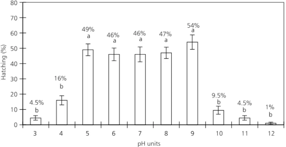
Ephippial egg hatching occurred within a wide range of pH values (Fig. 1). There were no significant differences (DTukey = 1,503; p < 0.05) in the range of
pH 5 to 9. Some hatching occurred at extreme pH values: 1% at pH 12 and 4.5% at pH 3.
With regard to the different ionic concentrations, there were no significant differences (p > 0.05) among the treatment’s. The pH variation between the six treatments was not great; however, conductivity showed considerable variation (Table 2).
Considering temperature (Fig. 2), hatching occurred within a wide range (15 to 40oC).
b b b a a a a a
b
b
3 4 5 6 7 8 9
80 70 60 50 40 30 20 10 0
11
10 12
pH units
Hatching (%)
4.5% 16%
49%
46%
46% 47%
54%
9.5% 4.5%
1%
Fig. 1 — Hatching rates (means and standard errors) of M. micrura ephippial eggs at different pH values. The letters a and b indicate means which did not differ significantly.
Water type
Total hatching
(%)
Standard errors
(±)
PH variation
Conductivity variation (µS.cm–1)
Deionized 32.5 3.23 6.63-8.56 3-15
Very soft 38.0 4.51 6.52-7.65 32-45
Soft 34.0 2.34 6.48-8.16 116-143
Moderately hard 29.5 3.73 6.84-8.56 150-354
Hard 33.0 3.71 7.30-8.67 243-617
Very hard 28.0 3.04 7.70-8.87 425-994
TABLE 2
Hatching rates (means and standard errors) of M. micrura ephippial eggs submitted to different water ionic concentrations (experiment 2). Ranges of pH and conductivity values during the
The 25oC treatment was significantly different
from the others (DTukey = 0.989; p < 0.05) and resulted
in the best hatching (76%). A reasonable result was also achieved at the temperature of 20oC (49% of
ha-tching).
Hatching was considerably reduced at the extreme temperature of 40oC (2% of hatching). There
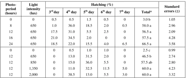
was no significant difference in the hatching of eggs exposed to different photoperiods varying from 8 to 24 hours or light intensities varying from 850 to 2,000
lux (Table 3). Hatching was almost nonexistent in the dark in both experiments.
DISCUSSION
With the procedure used in feeding M. mi-crura, we have tried to eliminate the possibility that the diet might be influencing the feasibility of the ephippia since, with the organisms being suffi-ciently well feed, they should have generate ener-getically rich eggs.
Hatching (%) Photo-period (hours) Light Intensity
(lux) 3rd day 4th day 5th day 6th day 7th day Total*
Standard errors (±)
0 8 12 16 24 0 650 650 650 650 0.5 1.0 17.5 21.0 18.5 0.5 36.0 31.0 34.5 22.0 1.5 18.5 5.5 2.0 15.5 0.5 2.0 2.5 0 4.0 0 0.5 0 0 6.5 3.0 b 58.0 a 56.5 a 57.5 a 66.5 a 1.05 2.96 2.09 4.28 3.58 0 12 12 12 12 0 650 850 1,350 2,000 0 0 0 0 0 0.5 13.0 15.0 11.0 38.5 1.0 31.5 36.0 32.5 13.0 1.0 2.0 5.5 11.5 5.5 0 0 0 5.0 3.0 2.5 c 46.5 b 57.5 ab 60.0 a 60.0 a 0.99 2.74 2.80 4.23 3.32
* Photoperiod: ∆Tukey = 1,188; p < 0.05.
Light intensity: ∆Tukey = 1,187; p < 0.05.
d c c a b c 0 d 100 90 80 70 60 50 40 30 20 10 0 Hat chi ng ( % ) Temperature (ºC)
10 15 20 25 30 35 40
22.5% 49% 76% 15.5% 14.5% 2% TABLE 3
Hatching rates (means and standard errors) of M. micrura ephippial eggs submitted to different photoperiods and light intensities (experiments 4 and 5). For each experiment, means which did
not differ significantly are indicated by the same letter.
Fig. 2 — Hatching rates (means and standard errors) of M. micrura ephippial eggs at different temperatures. The letters indicate
The production of ephippia by the M. micrura populations was apparently spontaneous and oc-curred synchronously for short periods of time. Dif-ferent egg production induction factors have been tested by us (water temperature, photoperiod, light intensity, quality and quantity of food, and parental stage), but the positive results were sporadic and it has not been possible to identify a single factor which might effectively induce the production of these eggs. In practice, the populations were kept in aquaria until they commenced intense and syn-chronous ephippial production. The period between one bout of ephippial production and another was never the same. Future work should be directed towards identifying the factors inducing the pro-duction of cladoceran ephippia in tropical regions. The lack of knowledge of such factors has been one of the major difficulties in carrying out the present study.
In none of the experiments did the hatching rate approach 100%, as has been found for other crustaceans (Davison, 1969; Sorgeloos et al., 1986). This fact might be related to an intrinsic characteristic of the species, but could also be due to differences in the hatching techniques employed.
The effect of pH
Since the water used in the experiments was not replaced during the seven days of experimen-tation, no abrupt pH and conductivity variations took place, thus exposing the eggs to pH values close to the ones planned for each treatment. The greatest differences among pH values in all con-tainers of a particular treatment were between 1.0 and 1.5 unit. Due to this variation, the term “egg exposition” instead of “egg maintenance” was used for the different pH values. The “maintenance” of eggs at a set pH value would require the use of an appropriate buffer, which would introduce other variations in the environment, such as in the salt content.
Sato (1967, apud Sorgeloos et al., 1986) pointed out that, after breaking through the Artemiacyst shell, the embryo enters in direct contact with the outside environment through the hatching mem-brane and, at this moment, the ionic composition of the hatching environment should be similar to that of sea water, with pH between 8 and 9 for a hatching around 100%. Thus, it seems that the pH tolerance of M. micruraembryos (maximum hatching
bet-ween pH values of 5 and 9) is greater than that seen for Artemia.
The hatching of the M. micrura eggs at extre-me pH values shows that they might be capable, when present in the sediment of fish ponds, of resisting the liming processes, usually used in aquaculture, and which cause great pH variations, and the large pH fluctuations which can occur on a daily basis in such ponds, thereby remaining viable and producing young to recolonize the en-vironment.
Ionic concentration
As it was found in experiment 1 that M. mi-cruraeggs satisfactorily hatched in the pH ran-ge of 5 to 9, the differences among pH values experienced in the present experiment probably did not influence the results achieved. Similarly, in relation to the conductivity variation, it may be seen that the ephippial eggs hatched in a wide range of this factor (Table 2).
During attempts by us at cultivating M. micrura, some culture media recommended for Cladocera (D’Agostino & Provasoli, 1970; Conklin & Provasoli, 1977) were used. These procedures have not given satisfactory results, probably because they were originally developed for populations from temperate regions in the northern hemisphere where, in general, waters have greater ionic concentrations. It was therefore of interest to examine the effects of different ionic concentrations on the hatching success of ephippial eggs of a warm-water cladoceran population. Despite the absence of significant differences among the treatment results, the organisms which hatched in very soft and soft waters showed greater activity. In the treatments with hard and very hard waters, the organisms presented, following birth, an apparently normal behavior but, when left in the hatching con-tainer, they died after a few hours. Further research on the sensitivity of M. micrura to total ionic con-centration and the concon-centration of particular ions in the water would be of interest.
For the experiments with temperature, photope-riod and light intensity, the pH was near neutrality (6 to 8), and conductivity was similar to the treatments of very soft and soft waters as defined in experiment 2 (32 to 143 mS.cm–1). These pH and conductivity
Temperature
Davison (1969) obtained 100% hatching of de-capsulated eggs of Daphnia pulexat temperatures of 24oC or lower, whereas at 30oC, only 20% of the
embryos completed their development. For Artemia sp., Sorgeloos et al. (1986) recommended that the temperature for egg hatching should be kept constant between 25 to 30oC, since below this range hatching
is slower and, above it, energy losses occur due to the embryo’s accelerated metabolism. In the present study, egg hatching was sensitive to temperature variation, especially at the highest values, where a noticeable reduction in hatching took place. However, the hatching rate of 2% at a temperature of 40oC shows the capacity
of these eggs to resist the high temperatures, which can occur in fish ponds in tropical regions.
Light
For Daphnia,Stross (1966, 1969) noted that the methodology used for stocking eggs (low tem-peratures for a few weeks) was more important for obtaining better hatching results than the use of different photoperiods and light intensities. Stross (1971) used, for D. pulexeggs, lighting pulses at different hatching stages, and noted that the best results occurred with an initial 16-hour light pulse, followed by a period of darkness (72% of hatching), or with two 2-hour pulses: an initial one and another from the 14th to the 16th hour of the
expe-riment (63.6% of hatching). Davison (1969) stated that D. pulexephippia require a light intensity of 418 lux for 100% activation, due to the “shell” protection provided to the embryo. For Artemiasp. cysts, Sorgeloos et al. (1986) compared the hatching of four lots of eggs from different regions at light intensities of 20, 100, 1,000 and 2,000 lux; they found that a minimum light intensity of 2,000 lux, using either natural or artificial light, was essential to obtain maximum hatching (100%), at least in the first hours of cyst hydration.
In the present study, the hatching of M. mi-cruraephippia was strongly affected both by the photoperiod and the light intensity. In relation to the number of days required for hatching, it was not possible to correlate photoperiod with the length of time for the greatest hatching (Table 3). However, with regard to light intensity, it was found that, in the treatment of 2,000 lux, there was a tendency for egg hatching to occur in a shorter period of time. The use of light intensities greater than 2,000 lux might offer even better hatching results specially if combined with a photoperiod of 24 hours light.
In summary, optimum conditions for the ha-tching of ephippial eggs of M. micrura were found to involve a water pH of around neutrality (5-9), and a temperature of 25oC. Photoperiod and light
intensity with a minimum of 8 hours and 850 lux, respectively, were necessary.
Acknowledgments — We thank Dr. Kennedy Francis Roche
for his comments on the manuscript and linguistic impro-vements.
REFERENCES
AMERICAN PUBLIC HEALTH ASSOCIATION, 1989,
Standard methods for the examination of water and wastewate. 17th ed. Ed. American Public Health Associa-tion, New York, pp. 8-16.
CONKLIN, D. E. & PROVASOLI, L., 1977, Nutritional requirements of the water flea Moina macrocopa. Biol. Bull., 152: 337-350.
D’AGOSTINO, A. & PROVASOLI, L., 1970, Dixenic culture of Daphnia magna Straus. Biol. Bull., 139: 485-494. DAVISON, J., 1969, Activation of the ephippial egg of
Daphnia pulex. J. Gen. Physiol., 53: 562-575.
FREGADOLLI, C. H., 1993, Food selection of pacu Pia-ractus mesopotamicus Holmberg, 1887 and tambaqui Colossoma macropomum Cuvier, 1818 larvae in the
laboratory. B. Tec. CEPTA,6(1): 1-50 (in Portuguese).
GUILLARD, R. R. L. & LORENZEN, C. J., 1972, Yellow-green algae with cholorophyllid-c. J. Phycol., 8: 10-14.
PIMENTEL-GOMES, F., 1985, Experimental statistics. Ed. Nobel, Piracicaba, SP, 466p. (in Portuguese). ROJAS, N. E. T., 1995, Hatching of Brachionus
calyciflorus Pallas, 1776 (Monogononta: Ploima) and
Moina micrura Kurz, 1874 (Crustacea: Cladocera) resting eggs induced by abiotic factors. Master’s Thesis, UFSCar, São Carlos, 147p. (in Portuguese). SORGELOOS, P., LAVENS, P., LÉGER, P., TACKAERT, W. & VERSICHELE, D., 1986, Manual on the cultivation and use of Artemia in aquacultur. Ed. United Nations Organization for Agriculture and Nutrition, Aquila Project Doc. 10, 301p. (in Spanish).
STROSS, R. G., 1966, Light and temperature requirements for diapause development and release in Daphnia.
Ecology, 47: 368-374.
STROSS, R. G., 1969, Photoperiod control of diapause in
Daphnia. III Two stimulus control of long-day,
short-day induction. Biol. Bull., 137: 359-374.
STROSS, R. G., 1971, Photoperiod control of diapause in Daphnia. IV Light and CO2 – sensitive phases within the cycle of activation. Biol. Bull., 140: 137-155.
ZANIBONI FILHO, E., 1992, Incubation, larviculture and fingerling production of tambaqui (Colossoma macropomum Cuvier, 1818). Ph.D. Thesis, UFSCar,