ABSTRACT: Zucchini yellow mosaic virus (ZYMV) causes substantial economic losses in cucurbit crops. Although ZYMV has been present in Brazil for more than 20 years, there is little informa-tion about the biological and molecular characteristics of the isolates found in the country. This study aimed to characterize the experimental hosts, pathotypes and genetic diversity of a col-lection of eleven Brazilian ZYMV isolates within the coat protein gene. For biological analysis, plant species from Amaranthaceae, Chenopodiaceae, Cucurbitaceae, Fabaceae, Solanaceae, and Pedaliaceae were mechanically inoculated and pathotypes were identified based on the reaction of a resistant Cucumis melo, accession PI414723. All of the cucurbit species/varieties and Sesamum indicum were systemically infected with all isolates. The nucleotide sequence variability of the coat protein gene ranged from 82 % to 99 % compared to the corresponding sequences of ZYMV isolates from different geographical locations. No recombination event was detected in the coat protein gene of the isolates.
Keywords: Potyvirus, nucleotide sequence, genetic diversity
Introduction
Zucchini yellow mosaic virus (ZYMV) belongs to the genus Potyvirus, family Potyviridae; this positive sense single-stranded RNA virus has a genome of approxi-mately 9,600 nucleotides contained in elongated and flexuous particles that are approximately 750 nm long and 12 nm in diameter. The viral RNA encodes a poly-protein, which generates eleven proteins, after cleavage by virus-encoded proteases. The coat protein (CP) has a molecular weight of 36 kDa (Lisa and Lecoq, 1984; Adams et al., 2012). ZYMV is experimentally transmit-ted by 26 species of aphids in a non-persistent manner (Katis et al., 2006).
ZYMV affects all cultivated cucurbit species [zuc-chini squash (Cucurbita pepo L. cv. Caserta, squash ( Cu-curbita maxima Duch. Ex Lam.), pumpkin (Cucurbita moschata Duch. Ex Poir.), cucumber (Cucumis sativus L.), melon (Cucumis melo L.), and watermelon (Citrullus lanatus (Thunb.) Matsum. & Nakai.)] worldwide. In Bra-zil, ZYMV was primarily detected in the states of São Paulo and Santa Catarina in the early 1990s (Vega et al., 1995; Kurozawa et al., 2005). It is likely that ZYMV is currently present in all cucurbit-growing regions of Bra-zil. Infected plants exhibit severe leaf mosaic, yellow-ing and eventually "shoestryellow-ing" symptoms in the leaves. The fruits are stunted, twisted and deformed, resulting in reduced yield making the plants unmarketable, espe-cially zucchini squash. In addition to cultivated species, ZYMV can systemically infect certain wild cucurbit spe-cies (Desbiez and Lecoq, 1997). The experimental evalu-ation of yield losses caused by ZYMV and the potyvirus Papaya ringspot virus - type W (PRSV - W) in zucchini
squash showed that plants that were infected at 5, 15, and 20 days after emergence did not produce marketable fruits (Pereira et al., 2007).
Although ZYMV has been present in Brazil for over 20 years and is associated with significant yield losses in cucurbit crops, little is known about some of the biological and molecular traits of Brazilian isolates. Such information may be critical for the understanding of disease epidemiology and breeding for resistant vari-eties and hybrids. Therefore, this study aimed to charac-terize eleven Brazilian isolates of ZYMV on experimen-tal hosts, the pathotype and genetic diversity of the coat protein gene.
Materials and Methods
Virus isolates
ZYMV isolates were obtained from cucurbits grown in the states of Espírito Santo (ZYMV-ES), Pará (ZYMV-PA), Rio Grande do Norte (ZYMV-RN), Paraná (ZYMV-PR), Rio Grande do Sul (ZYMV-RS), São Paulo (ZYMV-RI and ZYMV-P), Mato Grosso (ZYMV-MT), and the Federal District (ZYMV-DF and ZYMV-Fe). A mild strain, named ZYMV-M, which was isolated by Rabelo and Rezende (2004), was also included in this study as a reference. All but the ZYMV-RI isolate, which was main-tained in vivo for several months, were originally stored in dehydrated tissue at -20 °C. During the course of the host range experiments they were mechanically trans-mitted two or three times to zucchini squash plants. The isolates were maintained on zucchini squash plants by mechanical transmission. For mechanical inoculation, infected leaves were ground in a 0.02 M phosphate buf-1University of São Paulo/ESALQ – Dept. of Plant Pathology
and Nematology, C.P. 09 – 13418-900 – Piracicaba, SP – Brazil.
2Embrapa Vegetables, Rod. BR-060, km 09 (Brasília/ Anápolis), C.P. 218 – 70351-970 – Brasília, DF – Brazil. *Corresponding author <[email protected]>
Edited by: Emerson Medeiros Del Ponte
Biological and molecular characterization of Brazilian isolates of
Zucchini yellow
David Marques de Almeida Spadotti1, Débora Targino Wassano1, Jorge Alberto Marques Rezende1*, Luis Eduardo Aranha
Camargo1, Alice Kazuko Inoue-Nagata2
Received June 03, 2014 Accepted August 08, 2014
fer (pH 7.0) containing 0.02 M sodium sulfite and diluted 1:10 (w/v). The diluted inoculum was rubbed onto leaves that were dusted with carborundum.
Host range
The ZYMV isolates were mechanically inoculated onto two to six plants of various cucurbit species and other hosts listed in Table 1. The infection of test-plants was determined by examining the occurrence of local and/or systemic symptoms at 7, 14, and 21 days after inoculation. Infection was confirmed by plate trapped antigen – enzyme linked immunosorbent assay (PTA-ELISA) and reverse transcription – polymerase chain re-action (RT-PCR) analyses as described below. Whenever needed, the viral infection was also confirmed by back mechanical inoculation to C. pepo cv. Caserta.
Pathotype identification
The pathotype was determined based on the reac-tion of the ZYMV resistant C. melo accessionPI414723 that was mechanically inoculated with ten severe iso-lates of ZYMV, as proposed by Pitrat and Lecoq (1984). C. pepo cv. Caserta plants were also inoculated as con-trols for infectivity of each isolate. The infection was as-sessed through symptom evaluation and by PTA-ELISA.
PTA-ELISA
The test plants were analyzed by plate-trapped antigen (PTA)-enzyme-linked immunosorbent assay (ELISA) (Mowat and Dawson, 1987) using polyclonal
antiserum against purified virion of ZYMV. Appropriate negative and positive controls were included. The reac-tion was considered positive when the OD405nm reading was at least three times that of the healthy sample.
RNA isolation and RT-PCR
Total RNA was extracted from leaf tissue according to Toth et al. (2002) and submitted to one-step RT-PCR in a thermocycler using the following parameters: 42 ºC for 30 min; 94 ºC for 2 min; 35 cycles of 94 ºC for 30 s, 65 ºC for 30 s, and 72 ºC for 60 s; and a final elongation step at 72 ºC for 10 min. RT-PCR was carried out using the prim-ers ZY2 [5'-GCTCCATACATAGCTGAGACAGC-3'], which was derived from the NIb gene, and ZY3 [5'- TAGGCTT-GCAAACGGAGTCTAATC-3'], which anneals to the 3' un-translated region (Thomson, 1995); to amplify a fragment of 1186 bp containing part of the NIb gene, the complete coat protein gene and most of the 3’ untranslated region.
The RT-PCR reaction mixture contained 12.5 µL of PCR Master Mix 2X (Fermentas), 1 µM of each primer, 1 unit of reverse transcriptase AMV (Avian myeloblastosis virus, Promega), 5.0 µL of RNA, and RNase-free water in a 25 µL final volume. The RT-PCR products were ana-lyzed on a 0.8 % agarose gel that was stained with SYBR Safe DNA Gel Stain (Invitrogen) diluted 1:10,000 in a TBE buffer using the 1 Kb Plus DNA Ladder (Invitrogen) as the molecular size marker.
Nucleotide sequence analysis
The RT-PCR amplicons of each ZYMV isolate were purified using the Wizard SV Gel and PCR Clean-Up
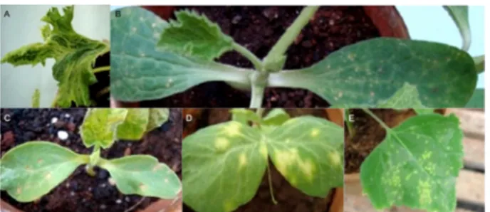
Sys-Table 1 – Host range reaction of several species mechanically inoculated with 11 Brazilian ZYMV isolates and detection of the virus by PTA-ELISA.
Plants ZYMV isolates
ES PA RN PR RS RI P DF MT Fe M
Cucurbita pepo cv. Caserta M (+) NLL, M (+) M (+) M (+) M (+) M (+) M (+) NLL, M (+) M (+) M (+) A (+) C. moschata cv. Menina
Brasileira M (nt) M (+) M (nt) M (nt) M (nt) M (nt) M (nt) M (nt) nt (nt) nt (nt) M (nt)
C. maxima cv. Exposição M (+) M (+) M (+) M (+) M (+) M (+) M (+) M (+) M (+) nt (nt) M (+) Cucumis sativus cv. Safira M (+) LLN, M (+) M (+) M (+) M (+) M (+) LLN, M (+) M (+) M (+) nt (nt) nt (nt) C. sativus cv. Aodai M (+) M (+) M (+) A (-) M (+) M (+) M (+) M (+) M (+) M (+) M (+) Citrullus lanatus cv. Crimson
Sweet M (+) M (+) M (+) M (+) M (+) M (+) M (+) M (+) M (+) nt (nt) M (+)
C. lanatus cv. Fair fax M (+) M (+) M (+) M (+) M (+) M (+) M (+) M (+) M (+) M (+) M (+) Cucumis melo cv. Ouro A (-) M (+) LLN, M (+) M (+) M (+) M (+) M (+) M (+) M (+) M (+) A (+) C. melo cv. Caipira M (+) M (+) LLN, M (+) M (+) LLN, M (+) M (+) M (+) M (+) M (+) LLN, M (+) LLN, M (+)
C. anguria M (+) M (+) M (+) M (+) M (+) M (+) M (+) M (+) M (+) M (+) M (+)
Luffa cylindrica M (+) M (+) CLL, M (+) A (-) M (+) M (+) M (+) M (+) M (+) M (+) M (+)
Chenopodium amaranticolor CLL (nt) CLL (nt) CLL (nt) CLL (nt) CLL (nt) CLL (nt) CLL (nt) A (nt) nt (nt) nt (nt) A (nt) C. quinoa CLL (nt) CLL (nt) CLL (nt) CLL (nt) CLL (nt) CLL (nt) CLL (nt) A (+) nt (nt) nt (nt) A (nt) Gomphrena globosa A (-) NLL (il+) CLL (il+) A (-) A (-) CLL (il+) A (-) A (-) A (il+) nt (nt) A (il+) Pisum sativum A(il+) CLL (il+) A (il+) A (-) A (-) A (il+) CLL (il+) A (-) A (il+) nt (nt) A (-) Phaseolus vulgaris Nt (nt) A (-) nt (nt) A (-) A (-) A (-) A (-) A (-) nt (nt) nt (nt) nt (nt) Nicotiana benthamiana A (-) A (-) A (il+) A (-) M (+) A (il+) A (+) A (-) X (nt) nt (nt) A (-)
N. glutinosa A (-) X (nt) A (-) A (-) A (-) A (-) A (-) A (-) A (-) nt (nt) A (-)
Sesamum indicum A(+) A (+) A (+) A (+) A (+) A (+) A (-) A (-) M (+) A (-) A (-)
tem and then directly sequenced in both directions using the same PCR amplification primers at Macrogen Inc., Seoul, Korea. The resulting nucleotide sequences of the coat protein gene were assembled using Electrophero-gram Quality Analysis software (http://asparagin.cenar-gen.embrapa.br/phph/) and then aligned using Clustal W (Thompson et al., 1994). The deduced amino acid sequences of the coat protein gene were obtained with the ExPASy proteomics server program (http://ca.expasy. org/tools/dna.html). The nucleotide and deduced amino acid sequences were compared with the corresponding sequences of 26 ZYMV isolates from different geograph-ical regions that were deposited in the GenBank.
A phylogenetic analysis was conducted using the molecular evolutionary genetics analysis (MEGA) soft-ware version 5.0 (Tamura et al., 2011), and a phylogenet-ic tree was constructed using the neighbor-joining meth-od and a maximum composite likelihometh-od mmeth-odel with bootstrap analysis with 1000 repetitions. The nucleotide sequences of the coat protein gene of Soybean mosaic vi-rus (SMV) and Papaya ringspot virus (PRSV) were used as out-groups. The nucleotide sequences of the coat protein gene of the Brazilian isolates of ZYMV were submitted to recombination analysis using the methods included in the RDP 3.0 package with default settings (Martin et al., 2010).
Results
Eleven ZYMV (ten severe and one mild) isolates that were collected from seven states in Brazil were me-chanically inoculated into a range of 19 plant species belonging to Amaranthaceae, Chenopodiaceae, Cucurbita-ceae, FabaCucurbita-ceae, SolanaCucurbita-ceae, and Pedaliaceae (Table 1). In-oculated plants were maintained in a greenhouse with-out environmental control.
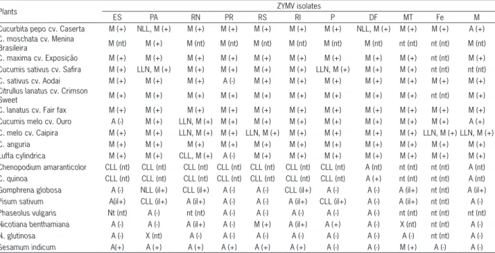
The majority of cucurbit species was systemically infected with all isolates and showed the typical mosa-ic symptoms, ranging from mild to severe in intensity (Table 1). The symptoms also included, in some cases, leaf narrowing, blistering, wilting, systemic necrosis and plant death. The ZYMV-M isolate caused mild symp-toms, as expected, on all of the cucurbit species tested and caused an asymptomatic infection of C. pepo cv. Caserta and C. melo cv. Ouro. The local reaction of the cucurbit cotyledons after mechanical inoculation varied according to the species/variety and the virus isolate (Table 1; Figure 1A-C).
The ZYMV isolates ES, PA, RN, PR, RS, RI and P induced chlorotic local lesions on the inoculated leaves of Chenopodium amaranticolor (Figure 1E) and Chenopo-dium quinoa, whereas the mild isolate M did not induce symptoms. ZYMV-DF was asymptomatic on C. quinoa, but the virus could be serologically detected (Table 1). The isolates ZYMV- ES, PA, RN, RI, P, and MT infected Pisum sativum, but the infection was confined to the in-oculated leaves as evidenced by chlorotic local lesions only for the isolates P (Figure 1D) and PA.
ZYMV-PA induced necrotic local lesions on Gomphrena globosa, whereas ZYMV-RN and RI induced chlorotic local le-sions.
The isolates ZYMV-M and MT did not induce any symptoms in G. globosa, but the virus was detected in the inoculated leaves using PTA-ELISA and by back inoculation test. S. indicum plants were systemically infected by seven isolates of ZYMV, although only those inoculated with ZYMV-MT were symptomatic. Nicotiana benthamiana plants were systemically in-fected by ZYMV-RS and P, but only ZYMV-RS induced symptoms. ZYMV-RN and RI were asymptomatic in N. benthamiana. Phaseolus vulgaris and Nicotiana glutinosa
plants were not infected by any ZYMV isolate. The ten severe ZYMV isolates did not infect C. melo PI414723; therefore, these isolates were classified as belonging to pathotype 0.
The ZY2 and ZY3 primers amplified a fragment of approximately 1186 bp for all of the Brazilian isolates containing the complete coat protein gene of the virus. The PCR products were directly sequenced, and the electropherograms were clear with a single peak with high confidence for all of the samples (data not shown). The coat protein gene of all of the isolates comprised 837 nucleotides, encoding a 279 amino acids protein. The nucleotide sequences were compared pairwise, and the nucleotide identity of the coat protein gene ranged from 93 % to 100 % among the Brazilian isolates and from 92 % to 94 % with isolate TW-TN3 (NC_003224) used as reference.
The coat protein sequence from the eleven Brazil-ian isolates and 26 isolates from different parts of the world were aligned, and a phylogenetic tree was con-structed (Figure 2). The ZYMV isolates formed three dis-tinct groups, named A, B, and C; group A was divided into four subgroups, as proposed by Coutts et al. (2011). The Brazilian isolates were all grouped within subgroups I and II of group A. No recombination event was de-tected in the coat protein gene of any of the Brazilian ZYMV isolates using seven independent RDP3 methods.
1989), showing only localized infection on the inoculat-ed leaves for some Brazilian isolates of the virus. These results suggested that cucurbits are the most important virus source in the field, which is relevant in Brazil due to the numerous cucurbit species present in cultivated and non-cultivated areas. S. indicum could be a potential source of the virus, but further testing is required to test this hypothesis.
The nucleotide sequence identity of the coat pro-tein gene among the Brazilian isolates of ZYMV ranged from 93 % to 100 %, and the deduced amino acid se-quence identity ranged from 97 % to 100 %. Compared to the corresponding nucleotide sequences of ZYMV isolates from different geographic origins, the identity varied from 82 % to 99 %, whereas that of the deduced amino acid sequences varied from 87 % to 99 %.
The phylogenetic tree (Figure 2) constructed using the nucleotide sequences of the coat protein gene of Bra-zilian and 26 other ZYMV isolates from different regions of the world showed three well-supported groups (A, B and C) and four subgroups within group A, as proposed by Coutts et al. (2011). Clearly, the Brazilian isolates be-longed to group A, subgroups I and II. The ZYMV-PR, Fe, RN, MT, PA, and RS isolates clustered in group A, subgroup I, along with a Brazilian isolate previously col-lected in the state of São Paulo State (GU586790) and with isolates from Asia and Europe, whereas isolates ZYMV-DF, M, P, and RI were in group A, subgroup II, with isolates from North America and Europe.
The isolate ZYMV-ES from the state of Espírito Santo is distantly related to subgroup I. The results sug-gest that ZYMV isolates may have been introduced into Brazil on at least three occasions, potentially with infect-ed seinfect-eds from abroad. ZYMV-infectinfect-ed seinfect-eds may act as effective viral reservoirs and partially accounting for the current geographic distribution of the virus (Simmons et al., 2011). Coutts et al. (2011) cautiously suggested that absence of local lesion in C. quinoa might be a way to assign ZYMV isolates to A-II group. In the present study, of the four A-II isolates (DF, M, P, and RI), ZYMV-P and RI induced chlorotic local lesions in this species, that did not concur with previous reports (Coutts et al., 2011). The results also suggested that the coat protein gene sequence might not be associated with symptom-atology in this host.
The C. melo PI414723 accession is the only source of resistance to ZYMV (Daning-Poleg et al., 2002) and is widely used in breeding programs not only because of its resistance to this virus but also because of its resis-tance to Papaya ringspot virus, the aphid Aphis gossypii, and the powdery mildew fungus Podosphaera xanthii (Pi-trat and Lecoq, 1983; McCreight et al., 1992). Resistance to ZYMV is controlled by the Zym-1 gene (Périn et al., 2002) and possibly by two additional genes of minor ge-netic effects (Danin-Poleg et al., 1997). The reaction of PI414723 to ZYMV, however, is not uniform which led Lecoq and Pitrat (1984) to classify isolates of the virus into pathotypes based on three possible reactions of this
Discussion
The cucurbit species tested in this study were sys-temically infected with the ten severe isolates of ZYMV and exhibited symptoms of severe mosaic, followed by growth reduction, foliar malformation, and reduced plant growth. Interestingly, C. melo cv. Ouro was not in-fected by ZYMV-ES, and Cucumis sativus cv. Aodai and Luffa cylindrica were not infected by ZYMV-PR. A large variety of symptoms have been reported in plants in-oculated with the ZYMV isolates collected from France (Lisa and Lecoq, 1984), USA (Provvidenti et al., 1984), and Australia (Coutts et al., 2011), similar to the results of our study. The ZYMV-M isolate caused only mild or asymptomatic infections of cucurbit species, confirming its potential for cross protection.
Among the non-cucurbit plants, S. indicum was the only species that was systemically infected with differ-ent ZYMV isolates. In Sudan, a ZYMV isolate causing severe mosaic and leaf deformation in S. indicum was reported in 1997 (Mahgoub et al., 1997). This is the first record of ZYMV infection of S. indicum under ex-perimental conditions in Brazil. This infection, however, was mostly asymptomatic, except for isolate ZYMV-MT. P. sativum was systemically infected by ZYMV isolates from Lebanon and Israel, although the plants were as-ymptomatic (Lesemann et al., 1983; Antignus et al.,
accession: no symptoms (pathotype 0), systemic necro-sis (pathotype 1), and yellowing, mosaic and deforma-tion of the leaves and wilting (pathotype 2). As the ten severe ZYMV isolates used in this study were assigned to pathotype 0, it is concluded that muskmelon hybrids carrying the Zym-1 resistance gene of this wild acces-sion may be effective in controlling ZYMV in different melon-producing regions of Brazil.
References
Adams, M.J.; Zerbini, F.M.; French, R.; Rabenstein, F.; Stenger, D.C.; Valkonen, J.P.T. 2012. Family Potyviridae. p. 1069-1089. In: King, A.M.Q.; Adams, M.J.; Carstens, E.B; Lefkowitz, E.J., eds. Virus taxonomy: classification and nomenclature of viruses; Ninth Report of the International Committee on Taxonomy of Viruses. Elsevier Academic Press, San Diego, CA, USA.
Antignus, Y.; Raccah, B.; Gal-On, A.; Cohen, S. 1989. Biological and serological characterization of zucchini yellow mosaic and watermelon mosaic virus - 2 isolates in Israel. Phytoparasitica 17: 289-298.
Coutts, B.A.; Kehoe, M.A.; Webster, C.G.; Wylie, S.J.; Jones, R.A.C. 2011. Zucchini yellow mosaic virus: biological properties, detection procedures and comparison of coat protein gene sequences. Archives of Virology 156: 2119-2131.
Danin-Poleg, Y.; Paris, H.S.; Cohen, S.; Rabinowitch, H.D.; Karchi, Z. 1997. Oligogenic inheritance of resistance to Zucchini yellow mosaic virus in melons. Euphytica 93: 331-337.
Danin-Poleg, Y.; Tadmor, Y.; Tzuri, G.; Reis, N.; Hirschberg, J.; Katzir, N. 2002. Construction of a genetic map of melon with molecular markers and horticultural traits, and localization of genes associated with ZYMV resistance. Euphytica 125: 373-384. Desbiez, C.; Lecoq, H. 1997. Zucchini yellow mosaic virus. Plant
Pathology 46: 809-829.
Katis, N.I.; Tsitsipis, J.A.; Lykouressis, D.P.; Papapanayotou, A.; Margaritopoulos, J.T.; Kokinis, G.M.; Perdikis, D.C.; Manoussopoulos, I.N. 2006. Transmission of Zucchini yellow mosaic virus by colonizing and non-colonizing aphids in Greece and new aphid species vectors of the virus. Journal of Phytopathology 154: 293-302.
Kurozawa, C.; Pavan, M.A.; Rezende, J.A.M. 2005. Cucurbit diseases. = Doenças das cucurbitáceas. p. 293-302. In: Kimati, H.; Amorim, L.; Rezende, J.A.M.; Bergamin Filho, A.; Camargo, L.E.A., eds. Plant pathology = Manual de fitopatologia. Agronômica Ceres, São Paulo, SP, Brazil (in Portuguese). Lecoq, H; Pitrat, M. 1984. Strains of Zucchini yellow mosaic virus
in muskmelon (Cucumis melo L.). Journal of Phytopathology 111: 165-173.
Lesemann, D.E.; Makkouk, K.M.; Koenig, R.; Natafji Samman, E. 1983. Natural infection of cucumbers by Zucchini yellow mosaic virus in Lebanon. Phytopathologische 108: 304-313.
Lisa, V.; Lecoq, H. 1984. Zucchini Yellow Mosaic Virus. CMI/AAB, Kew, UK. (Descriptions of Plants Viruses, 282).
Mahgoub, H.A.; Desbiez, C.; Wipfscheibel, C.; Dafalla, G.; Lecoq, H. 1997. Characterization and occurrence of Zucchini yellow mosaic virus in Sudan. Plant Pathology 46: 800-805.
Martin, D.P.; Lemey, P.; Lott, M.; Moulton, V.; Posada, D.; Lefeuvre, P. 2010. RDP3: a flexible and fast computer program for analyzing recombination. Bioinformatics 26: 2462-2463. McCreight, J.D.; Bohn, W.; Kishaba, A.N. 1992. Pedigree of PI
414723 melon. Cucurbit Genetics Cooperative Report 15: 51-52.
Mowat, W.P.; Dawson, S. 1987. Detection of plant viruses by
ELISA using crude sap extracts and unfractionated
antisera. Journal of Virological Methods 15: 233-247.
Pereira, M.J.Z.; Sussel, A.A.B.; Silva, R.F.; Kuhn, O.J.; Domingues, F.; Rezende, J.A.M. 2007. Evaluation of yield losses caused by ZYMV and PRSV-W in zucchini squash. Summa Phytopathologica 33: 192-194 (in Portuguese, with abstract in English).
Périn, C.; Hagen, L.S.; De Conto, V.; Katzir, N.; Danin-Poleg, Y.; Portnoy, V.; Baudracco-Arnas, S.; Chadoeuf, J.; Dogimont, C.; Pitrat, M. 2002. A reference map of Cucumis melo based on two recombinant inbred lines populations. Theoretical and Applied Genetics 104: 1017-1034.
Pitrat, M.; Lecoq, H. 1983. Two alleles for watermelon mosaic virus 1 resistance in melon. Cucurbit Genetics Cooperative Report 6: 52–53.
Pitrat, M.; Lecoq, H. 1984. Inheritance of Zucchini yellow mosaic virus resistance in Cucumis melo L. Euphytica 33: 57-61. Provvidenti, R.; Gonsalves, D.; Humaydan, H.S. 1984. Occurrence
of Zucchini yellow mosaic virus in cucurbits from Connecticut, New York, Florida, and California. Plant Disease 68: 443-446. Simmons, H.E.; Holmes, E.C.; Gildow, F.E.; Bothe-Goralczyk,
M.A.; Stephenson, A.G. 2011. Experimental verification of seed transmission of Zucchini yellow mosaic virus. Plant Disease 95: 751-754.
Tamura, K.; Peterson, D.; Peterson, N.; Stecher, G.; Nei, M.; Kumar, S. 2011. MEGA5: molecular evolutionary genetics analysis using maximum likelihood, evolutionary distance, and maximum parsimony methods. Molecular Biology and Evolution 28: 2731-273.
Thompson, J.D.; Higgins, D.G.; Gibson, T.J. 1994. CLUSTAL W: improving the sensitivity of progressive multiple sequence alignment through sequence weighting, position specific gap penalties and weight matrix choice. Nucleic Acids Research 22: 4673-4680.
Thomson, K.G.; Dietzgen, R.G.; Gibbs, A.J.; Tang, Y.C.; Liesack, W.; Teakle, D.S.; Stackebrandt, E. 1995. Identification of
Zucchini yellow mosaic potyvirus by RT-PCR and analysis of sequence variability. Journal of Virological Methods 55: 83-96. Toth, I.K.; Hyman, L.J.; Berteau, Y.; Fréchon, D. 2002. Methods for
the Detection and Quantification of Erwinia carotovora subsp.
atroseptica (Pectobacterium carotovorum subsb. atrosepticum) on Potatoes: A Laboratory Manual. Scottish Crop Research Institute, Dundee, Scotland.