High frequency of mutation G377S
in Brazilian type 3 Gaucher disease
patients
1Departamento de Genética e Biologia Evolutiva, Instituto de Biociências, Universidade de São Paulo, São Paulo, SP, Brasil
2Hospital Evangélico de Londrina, Londrina, PR, Brasil 3Hospital de Base do Distrito Federal, Brasília, DF, Brasil 4Departamento de Pediatria, Escola Paulista de Medicina,
Universidade Federal do Estado de São Paulo, São Paulo, SP, Brasil 5Serviço de Hemoterapia, HEMORIO, Rio de Janeiro, RJ, Brasil 6Serviço de Hemato-Oncologia, Faculdade de Ciências Médicas, Santa Casa de São Paulo, São Paulo, SP, Brasil
R. Rozenberg1, F.T. Araújo1, D.C. Fox1, P. Aranda2, A. Nonino3, C. Micheletti4, A.M. Martins4, R. Cravo5, E. Sobreira6 and L.V. Pereira1
Abstract
Gaucher disease (GD), the most prevalent lysosome storage disorder, presents an autosomal recessive mode of inheritance. It is a paradigm for therapeutic intervention in medical genetics due to the existence of effective enzyme replacement therapy. We report here the analysis of GD in 262 unrelated Brazilian patients, carried out in order to establish the frequency of the most common mutations and to provide prognostic information based on genotype-phenotype correlations. Among 247 type 1 GD patients, mutation N370S was detected in 47% of all the alleles, but N370S/N370S homozygosity was found in only 10% of the patients, a much lower frequency than expected, suggesting that most individuals presenting this genotype may not receive medical attention. Recombinant alleles were detected at a high frequency: 44% of the chromosomes bearing mutation L444P had other mutations derived from the pseudogene sequence, present in 25% of patients. Three neuronopathic type 2 patients were homozygous for L444P, all present-ing additional mutations (E326K or recombinant alleles) that probably lead to the more severe phenotypes. Six children, classified as type 1 GD patients, had a L444P/L444P genotype, showing that neuronopathic symptoms may only manifest later in life. This would indicate the need for a higher treatment dose during enzyme replacement therapy. Finally, mutation G377S was present in 4 homozygous type 1 patients and also in compound heterozygosity in 5 (42%) type 3 patients. These findings indicate that G377S cannot be unambiguously classified as mild and suggest an allele-dose effect for this mutation.
Correspondence
R. Rozenberg
Departamento de Genética e Biologia Evolutiva Instituto de Biociências, USP R. do Matão, 277, Sala 350 05508-900 São Paulo, SP Brasil
Fax: +55-11-30917583 E-mail: [email protected]
Research supported by FAPESP, CNPq, and Genzyme do Brasil.
Received December 13, 2005 Accepted July 20, 2006
Key words
•Gaucher disease •GBA gene •Allele dose-effect
Introduction
Gaucher disease (GD), the most preva-lent lysosome storage disorder, presents an autosomal recessive mode of inheritance.
sever-ity of neurologic symptoms define the three types of GD. Approximately 95% of patients present type 1 GD (MIM# 230800), without neurologic involvement and with onset dur-ing childhood or adulthood (1). Neurono-pathic variants are type 2 GD (MIM# 230900), characterized by early onset and survival of up to 2 years of age, and type 3 GD (MIM# 231000) of infantile or juvenile onset and with a less ominous course with survival into adulthood.
GD is caused by mutations in the GBA gene (1q21). This is a 7-kb gene containing 11 exons, which codes for the acid ß-glu-cosidase enzyme, also known as glucocer-ebrosidase (2). The absence of this enzyme leads to the accumulation of glucocerebroside in the cells of the reticuloendothelial system and ultimately to the clinical manifestation of GD. More than 200 GD-causing muta-tions have been identified so far and the most frequent ones are [c.1226A>G;N370S] and [c.1448T>C;L444P], herein called N370S and L444P, respectively. The clini-cal diagnosis of GD may be confirmed by the biochemical assay of acid ß-glucosidase activity. Unlike enzymatic diagnosis, muta-tion analysis has a considerable, although not absolute, predictive value with respect to disease prognosis (1). Opportunely, an ex-tensive genotype-phenotype correlation is present for the most frequent mutations. Generally, GD-causing mutations are classi-fied as mild, severe or null alleles (3). The presence of a mild mutation leads to type 1 GD even in combination with a second se-vere mutation or a null allele. The neurono-pathic variants are due to homozygosity of severe mutations, or the combination of a severe mutation and a null allele. Homozy-gosity of null alleles is not found, suggesting that this genotype is incompatible with life. Despite the genotype-phenotype correlation, a variable expression of clinical symptoms is present in patients with the same geno-type, indicating the existence of other fac-tors, genetic and/or environmental,
influ-encing the phenotype (4).
GD is the first lysosome storage disease for which an efficient enzyme replacement therapy (ERT) was developed (Imiglucerase) and became a paradigm for therapeutic in-tervention in human genetics (5). ERT is the standard treatment for type 1 and 3 GD patients, and efficiently ameliorates non-neu-rological symptoms. The enzyme dose in-fused during ERT is established for each patient according to body weight and clini-cal manifestations. Dose individualization is subject to medical decision including the initial dose and its reduction during the life-long treatment. The high cost of treatment dictates that ERT should be administered at the most efficient possible dose. In Brazil, the patients’ therapy is subsidized by the public health system. Thus, the disease may be of low incidence but has a significant economic impact. There are no estimates of GD prevalence in Brazil, but there are ap-proximately 400 diagnosed cases (6), the third largest patient population undergoing ERT in the world.
In this study the frequency of the most common mutations in a sample of 262 unre-lated Brazilian GD patients was determined. Several genotype-phenotype correlations were identified and their implications for the diagnosis and treatment of GD are discussed.
Patients and Methods
Gaucher disease patients and relatives
collected by physicians or health profes-sionals participating in the treatment of the patients and sent to the laboratory in 2-mL Eppendorf tubes. Participants signed in-formed consent forms and the project was approved by the Brazilian National Com-mittee of Ethics in Research (CONEP).
Mutation screening, for statistical pur-poses was performed in one case from each family, for a total of 262 cases, 247 of which were classified by their physicians as type 1, 3 as type 2, and 12 as type 3 GD patients. Since not all of these patients were evaluated by one of the authors, we could not confirm the classification in every case. Most of these patients are undergoing ERT, except type 2 cases, which are not treated and have a reduced life expectancy. The genotype was analyzed separately for type 2 and 3 patients in order to avoid bias in the evalua-tion of allelic and genotypic frequencies.
Molecular diagnosis
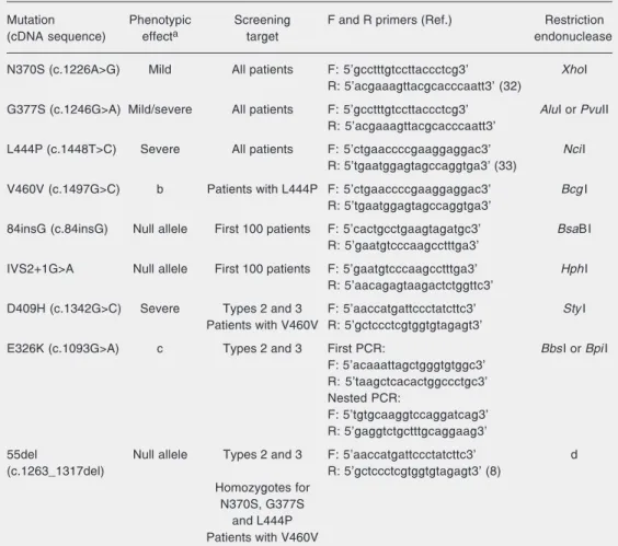
DNA was extracted from mouth mucus by the method of Richards et al. (7). Muta-tions N370S, L444P, G377S, c.84insG (84insG), IVS2+1G>A, c.1263_1317del (55del), V460V, D409H, and E326K were analyzed on the basis of restriction fragment length polymorphism. The enzymatic effect of these mutations, their screening strategy, the primers (Invitrogen, Carlsbad, CA, USA), and restriction endonucleases (New Eng-land Biolabs, Ipswich, MA, USA or from Fermentas, Vilnius, Lithuania) used for de-tection and the reference method, when pre-viously established, are described in Table 1. After endonuclease digestion, fragments were submitted to 12% polyacrylamide gel electrophoresis (mutations N370S, G377S, 84insG, and IVS2+1G>A) or 2% agarose gel electrophoresis (mutations L444P, V460V, D409H, 55del, and E326K).
All patients were screened for mutations N370S and L444P, the most frequent muta-tions worldwide. Mutation G377S is the third
most frequent mutation in Portugal and was therefore also included in the initial analy-sis. In the absence of these three mutations in one of the alleles, the first one hundred patients were screened for mutations 84insG and IVS2+1G>A. Patients homozygous for N370S, G377S or L444P were investigated for the presence of mutation 55del, present in the pseudogene (psGBA) contiguous to the GBA gene. This deletion, if present in GBA, would be localized at the annealing site of primers used for detecting mutations N370S, G377S and L444P (8).
Because mutation L444P may be a part of a recombinant allele (REC), with DNA sequence derived from the highly homolo-gous psGBA, all patients presenting this mutation were tested for V460V (9). When V460V was detected, indicating the pres-ence of a REC, mutations 55del and D409H were screened in order to determine the ex-tent of the recombination sequence. More-over, when a REC was detected, the patient’s parents were tested, when possible, to deter-mine if the mutations were in the same al-lele. The most frequent RECs are RecNciI (L444P, A456P and V460V) and RecTL (D409H, L444P, A456P, and V460V). Due to the absence of analysis of A456P, we opted to present RECs by the mutations actually detected. Finally, the 15 patients classified as types 2 and 3 were also tested for 55del, D409H and E326K due to the high frequency of these mutations among them.
Results
The sample analyzed was composed of 247 (94%) type 1, 3 (1%) type 2 and 12 (5%) type 3 GD patients. Type 2 GD patients may be under-represented since they are not treated by ERT and have a short life expect-ancy. Nevertheless, the distribution was simi-lar to that of other studies in distinct popula-tions (1).
the first one hundred patients analyzed, only one allele harboring mutation 84insG and none with IVS2+1G>A were detected. There-after, screening for these mutations was halted and they were considered to be un-identified. Almost half of the patients were compound heterozygotes presenting the N370S/L444P genotype. Combination of N370S with a second unidentified mutation was the second most frequent genotype, pres-ent in approximately 30% of the alleles. The remaining patients had many other geno-types.
Molecular analysis of 6 children with a
clinical diagnosis of type 1 GD showed a genotype characteristic of type 3 GD (ho-mozygous for L444P). For statistical pur-poses these children were considered to be type 1 patients according to their clinical characterization, although their genotypes suggest that neuronopathic symptoms may be expected to occur later in life.
The mutation frequencies, independent-ly of their genotypic distribution, are pre-sented as the first column of Table 3. While almost half of the patients’ alleles carry N370S (47%), the other half is divided be-tween L444P (27%), G377S (2%) and an
Table 1. Screening target, primers and restriction enzymes used for the detection of some mutations that cause Gaucher disease.
Mutation Phenotypic Screening F and R primers (Ref.) Restriction (cDNA sequence) effecta target endonuclease
N370S (c.1226A>G) Mild All patients F: 5’gcctttgtccttaccctcg3' XhoI R: 5’acgaaagttacgcacccaatt3' (32)
G377S (c.1246G>A) Mild/severe All patients F: 5’gcctttgtccttaccctcg3' AluI or PvuII R: 5’acgaaagttacgcacccaatt3'
L444P (c.1448T>C) Severe All patients F: 5’ctgaaccccgaaggaggac3' NciI R: 5’tgaatggagtagccaggtga3' (33)
V460V (c.1497G>C) b Patients with L444P F: 5’ctgaaccccgaaggaggac3' BcgI R: 5’tgaatggagtagccaggtga3'
84insG (c.84insG) Null allele First 100 patients F: 5’cactgcctgaagtagatgc3' BsaBI R: 5’gaatgtcccaagcctttga3'
IVS2+1G>A Null allele First 100 patients F: 5’gaatgtcccaagcctttga3' HphI R: 5’aacagagtaagactctggttc3'
D409H (c.1342G>C) Severe Types 2 and 3 F: 5’aaccatgattccctatcttc3' StyI Patients with V460V R: 5’gctccctcgtggtgtagagt3'
E326K (c.1093G>A) c Types 2 and 3 First PCR: BbsIor BpiI F: 5’acaaattagctgggtgtggc3'
R: 5’taagctcacactggccctgc3' Nested PCR:
F: 5’tgtgcaaggtccaggatcag3' R: 5’gaggtctgctttgcaggaag3'
55del Null allele Types 2 and 3 F: 5’aaccatgattccctatcttc3' d (c.1263_1317del) R: 5’gctccctcgtggtgtagagt3' (8)
Homozygotes for N370S, G377S
and L444P Patients with V460V
aPhenotypic effect of mutations according to Ref. 3. Mutation G377S has been detected with both mild and
severe effects (see text). bMutation V460V is a silent alteration used to indicate the presence of a recombinant
unidentified mutation (24%).
Since mutation L444P may be present alone or as part of a REC, this possibility was always examined. Among type 1 GD pa-tients, 46% (62/134) of alleles with L444P were found to be RECs with L444P+V460V. Testing for D409H followed the detection of V460V and only 2% (3/134) of alleles with L444P were positive, disclosing a REC with L444P+V460V+D409H. Mutation 55del was not detected in any of the RECs, suggesting that they probably had pseudogene-derived mutations restricted to exons 9 and 10. How-ever, other recombination events upstream from 55del may be present. When including type 2 and 3 patients in the statistical analy-sis, RECs were identified in 44% (66/150) of the chromosomes bearing mutation L444P and in 25% (66/262) of all GD patients.
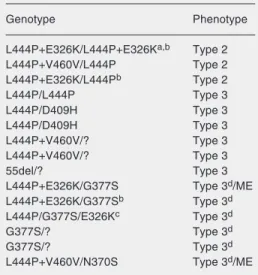
The 15 patients with type 2 and 3 GD were screened for mutations N370S, G377S, L444P, V460V, 55del, D409H, and E326K. Table 4 presents the genotypes found. We detected a homozygote patient whose par-ents are first cousins presenting E326K in combination with L444P in both alleles with a type 2 clinical phenotype. The only com-pound heterozygote presenting L444P in combination with a REC also had type 2 GD. Among the type 3 patients, the presence of mutations N370S in one case and G377S in 5 cases was unexpected since these are gen-erally considered to be mild mutations (3).
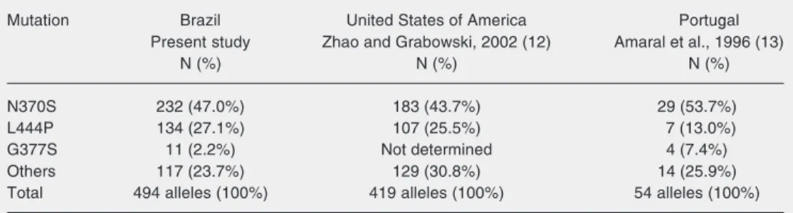
Table 3. Frequency of the most common mutations in Brazilian, Portuguese and non-Jewish North American type 1 Gaucher disease patients.
Mutation Brazil United States of America Portugal Present study Zhao and Grabowski, 2002 (12) Amaral et al., 1996 (13)
N (%) N (%) N (%) N370S 232 (47.0%) 183 (43.7%) 29 (53.7%) L444P 134 (27.1%) 107 (25.5%) 7 (13.0%) G377S 11 (2.2%) Not determined 4 (7.4%) Others 117 (23.7%) 129 (30.8%) 14 (25.9%) Total 494 alleles (100%) 419 alleles (100%) 54 alleles (100%) Statistical analysis comparing the present study with the North American sample considered G377S to belong to the “others” category.
Discussion
Frequency of the most common mutations
Although 47% of all GD-causing mu-tations are N370S (Table 3), only ~10% of the patients are N370S/N370S homozy-gotes (Table 2), a frequency much lower than the 22% expected (0.472). These data
probably reflect the fact that many N370S homozygotes are not obtaining medical attention due to the absence or mildness of symptoms. The 24 homozygotes herein detected indicate a penetrance of 45% for this genotype (0.10/0.22), a value that is close to that described for the Ashkenazi Jewish population (10) and for the Portu-guese population (11). These cases
indi-Table 2. Genotypic frequency observed in 247 Brazilian patients with type 1 Gaucher disease. Genotype Number of patients (frequency) N370S/L444P 112 (45.3%)
cate that despite its Mendelian mode of in-heritance, GD presents complex phenotypes. The frequency of the two most common GD-causing mutations among Brazilian pa-tients is similar to that described for other populations, where N370S and L444P rep-resent roughly half and one quarter of the alleles, respectively (1). Mutations 84insG and IVS2+1G>A were found to be too rare. In fact, these mutations have a high fre-quency among Jewish patients but not in the general population of most countries (1). The low detection rate of the 84insG and IVS2+1G>A in the present study may re-flect the low frequency of the Jewish popu-lation (<0.1%) in Brazil.
From Table 3 it is possible to observe that the frequency of the most common mu-tations in Brazilian type 1 GD patients de-tected in the present study is similar to that found in a sample of non-Jewish North Am-ericans (12) (P = 0.263). However, it is
significantly different (P = 0.028) when com-pared to that of Portuguese cases (13), ac-cording to chi-square analysis. The genetic background of the Brazilian population in-cludes important African and Indigenous components, in addition to the Portuguese ancestry, that may explain the greater simi-larity to the North American sample.
Mutation G377S is the third most frequent among GD patients in Portugal and Spain (13). In Brazil, it also appeared at a consider-able frequency (2.2%), indicating that it is important in preliminary screening. A corre-spondence in mutations causing another lyso-some storage disease has also been described for Tay-Sachs disease and points to a common origin of the mutated alleles prior to the Portu-guese colonization of Brazil (14).
Among the alleles with L444P, 44% (66/ 150) were shown to be RECs, present in 25.2% (66/262) of patients, corresponding to 12.6% (66/524) of the alleles studied. Our approach was limited by the lack of analysis of the A456P mutation, present in the RECs. There-fore, we have opted to present RECs by the mutations actually diagnosed. Since some pa-tients may have A456P and lack the silent mutation in V460V, the frequency of RECs in this sample may be even higher. The high frequency of RECs was also described in other countries (9,15). It is generally observed that the RECs have a more severe impact on clini-cal manifestation than L444P alone (9,16-18). In agreement with this observation the three homozygotes for L444P additionally carrying E326K or RECs had type 2 GD, while those with genotype L444P/L444P but lacking com-plex alleles had milder phenotypes. It is pos-sible that these patients’ more severe pheno-types reflect a modifier effect of E326K and RECs leading to a lower residual enzyme activity, although other loci may also have influenced the severity of the phenotype (4,19). The type 2 patient with the L444P+E326K/ L444P+E326K genotype did not present the severe perinatal-lethal phenotype, downplay-ing this genotype-phenotype correlation (20).
Table 4. Genotypes of 15 neuronopathic Brazilian patients with Gaucher disease.
Genotype Phenotype L444P+E326K/L444P+E326Ka,b Type 2
L444P+V460V/L444P Type 2 L444P+E326K/L444Pb Type 2
L444P/L444P Type 3 L444P/D409H Type 3 L444P/D409H Type 3 L444P+V460V/? Type 3 L444P+V460V/? Type 3 55del/? Type 3 L444P+E326K/G377S Type 3d/ME
L444P+E326K/G377Sb Type 3d
L444P/G377S/E326Kc Type 3d
G377S/? Type 3d
G377S/? Type 3d
L444P+V460V/N370S Type 3d/ME
ME = myoclonic epilepsy. aParents are first
cous-ins. bThese patients have siblings not included in
this study, presenting the same genotype and phenotype. cE326K phase was not determined,
i.e., it was cis or trans to L444P. dNeuronopathic
Type 1 pre-neuronopathic affected children
Among type 3 GD patients, neurological manifestations appear after 10 years of age in 50% of the cases (1). Therefore, it is always possible that children under this age, diag-nosed as type 1, may actually be type 3 pa-tients that are in a pre-neuronopathic phase. Molecular diagnosis may help to identify these patients and this was the case for 6 children in this study, aged 5 to 6 years, in which mutation L444P was detected in homozygosity. It is possible that other children clinically classi-fied as type 1 patients, with unidenticlassi-fied muta-tions and without a mild mutation, are in the same situation. Although only ~5% of GD patients are type 3 cases, the onset of GD is generally during infancy, which makes the possibility of a pre-neuronopathic patient a frequent doubt. The anticipation of the diag-nosis of a neuronopathic GD variant by DNA testing is due to the need to inform the family, since these patients are likely to manifest de-velopmental and language problems (4,21). More important is the impact on ERT. One possibility is that a high-dose treatment may impact the neuronopathic symptoms (21,22) but conclusive evidence is still lacking for GD. The European consensus for the treat-ment of neuronopathic GD has established that the ERT of type 3 patients, including the pre-neuronopathic ones identified by molecu-lar diagnosis, is to be performed at a higher dose that should not be reduced even after symptoms ameliorate (23).
Neuronopathic patients with mutations N370S and G377S
The evidence for considering G377S a mild mutation is based on its high residual enzyme activity (17.6%) according to meas-urement of acid ß-glucosidase activity (24). Moreover, all reports of G377S homozy-gotes involve type 1 GD patients (16,25-27). Accordingly, in the present study four G377S homozygotes had a type 1 phenotype.
How-ever, five compound heterozygotes carrying one G377S allele were type 3 patients. Other reports have also detected type 3 patients carrying one copy of G377S (3,28-30). These observations suggest the possibility of an allele-dose effect, whereby two copies of G377S would lead to an enzymatic residual activity sufficient to prevent neurological involvement, but one copy of G377S with a second severe mutation would lead to type 3 GD. An allele dose-effect has already been described for mutation K79N (31). In this model, the residual activity of mutant en-zymes produced by the two alleles should be considered in an additive manner. Some mutations, such as G377S, K79N, N188S, V394L, and R463C could be at the limit of residual activity that elicits neuropathic symptoms (3,16,31).
The patients reported here represent fur-ther in vivo evidence for a critical role of a GD-causing mutation in the development of neurological disease. However, the partici-pation of other modifiers cannot be excluded. In this study, another Brazilian family with two siblings (aged 5 and 15 years) present-ing the G377S/L444P genotype lacked any neurological involvement (Table 2). These siblings were shown not to carry the E326K mutation (data not shown), while 2 other patients with genotype G377S/L444P+ E326K presented neuronopathic symptoms (Table 4). This finding reinforces a possible negative modifier effect of E326K.
It is clear that DNA testing provides in-formation both on the diagnosis and the prognosis of GD and new insights on geno-type-phenotype correlations should be promptly integrated into medical practice. Moreover, since ERT is individualized and the treatment dose is subject to medical deci-sion, molecular diagnosis may be critical to establish a proper dose, as in the case of the L444P homozygote children still lacking neurological symptoms. Finally, our find-ings indicate that an allele dose-effect may be associated with mutation G377S. This mutation should be part of the screening panel for GD patients from Iberian coun-tries, including their former colonies. Fur-ther studies may be critical to establish its exact genotype-phenotype correlation.
Acknowledgments
The authors wish to thank the following persons for their valuable contributions: Dr.
Angela Morgante, Dr. Fernanda Teresa de Lima, Katia Maria da Rocha, Dr. Martha Lima, Dr. Paulo Alberto Otto, and Dr. Regina Celia Mingroni Neto from IB-USP; Dr. Gilda Porta from FM-USP; Dr. Ana Paula Cons-tancio, Andrea Callipo, Dr. Carlos Ruchaud, Daniella Jorge, Rodrigo Azambuja, Cristiana Petriz, and Dr. Rogerio Vivaldi from Genzyme do Brasil; Dr. Angela Zanette from HEMOBA; Dr. Cintia Xerez from HEMOAM; Dr. Maria Cristina Pereira, Dr. Maria Teresa M. de Paula, and Dr. Renata Cravo from HEMORIO; Dr. Silvia Thomas from HEMOMAT; Dr. Cristina Bortolheiro and Dr. Cristina Pozzi from Santa Casa de Misericórdia SP; Dr. Erlane Ribeiro from Hospital Albert Sabin CE; Dr. Eugenia Ribeiro Valadares from HC-BH; Lara Franco from UH; Dr. Mara A.D. Pianovski; Dr. Marcelo Kerstenetzki from Hospital de Restauração (PE), and Dr. Maria de Fatima Montoril from HEMOPA.
References
1. Beutler E, Grabowski GA. Gaucher disease. In: Scriver CR, Beaudet AL, Sly WS, Valle DV (Editors), The metabolic and molecular bases of inherited disease. 8th edn. New York: McGraw-Hill; 2001. p 3635-3668.
2. Sorge J, West C, Westwood B, Beutler E. Molecular cloning and nucleotide sequence of human glucocerebrosidase cDNA. Proc Natl Acad Sci U S A 1985; 82: 7289-7293.
3. Beutler E, Gelbart T, Scott CR. Hematologically important muta-tions: Gaucher disease. Blood Cells Mol Dis 2005; 35: 355-364. 4. Goker-Alpan O, Hruska KS, Orvisky E, Kishnani PS, Stubblefield
BK, Schiffmann R, et al. Divergent phenotypes in Gaucher disease implicate the role of modifiers. J Med Genet 2005; 42: e37. 5. Germain DP. Gaucher’s disease: a paradigm for interventional
ge-netics. Clin Genet 2004; 65: 77-86.
6. Michelin K, Wajner A, de Souza FT, de Mello AS, Burin MG, Pereira ML, et al. Application of a comprehensive protocol for the identifica-tion of Gaucher disease in Brazil. Am J Med Genet A 2005; 136: 58-62.
7. Richards B, Skoletsky J, Shuber AP, Balfour R, Stern RC, Dorkin HL, et al. Multiplex PCR amplification from the CFTR gene using DNA prepared from buccal brushes/swabs. Hum Mol Genet 1993; 2: 159-163.
8. Tayebi N, Stern H, Dymarskaia I, Herman J, Sidransky E. 55-Base pair deletion in certain patients with Gaucher disease complicates screening for common Gaucher alleles. Am J Med Genet 1996; 66: 316-319.
9. Tayebi N, Stubblefield BK, Park JK, Orvisky E, Walker JM, Lamarca ME, et al. Reciprocal and nonreciprocal recombination at the glucocerebrosidase gene region: implications for complexity in Gaucher disease. Am J Hum Genet 2003; 72: 519-534.
10. Beutler E, Nguyen NJ, Henneberger MW, Smolec JM, McPherson RA, West C, et al. Gaucher disease: gene frequencies in the Ashkenazi Jewish population. Am J Hum Genet 1993; 52: 85-88. 11. Lacerda L, Amaral O, Pinto R, Oliveira P, Aerts J, Sa Miranda MC.
Gaucher disease: N370S glucocerebrosidase gene frequency in the Portuguese population. Clin Genet 1994; 45: 298-300.
12. Zhao H, Grabowski GA. Gaucher disease: Perspectives on a proto-type lysosomal disease. Cell Mol Life Sci 2002; 59: 694-707. 13. Amaral O, Pinto E, Fortuna M, Lacerda L, Sa Miranda MC. Type 1
Gaucher disease: identification of N396T and prevalence of glucocerebrosidase mutations in the Portuguese. Hum Mutat 1996; 8: 280-281.
14. Rozenberg R, Martins AM, Micheletti C, Mustacchi Z, Pereira LV. Tay-Sachs disease in Brazilian patients: prevalence of the IVS7+1g>c mutation. J Inherit Metab Dis 2004; 27: 109-110. 15. Cormand B, Harboe TL, Gort L, Campoy C, Blanco M, Chamoles N,
et al. Mutation analysis of Gaucher disease patients from Argentina: high prevalence of the RecNciI mutation. Am J Med Genet 1998; 80: 343-351.
1777-1786.
17. Stone DL, Tayebi N, Orvisky E, Stubblefield B, Madike V, Sidransky E. Glucocerebrosidase gene mutations in patients with type 2 Gaucher disease. Hum Mutat 2000; 15: 181-188.
18. Mignot C, Gelot A, Bessieres B, Daffos F, Voyer M, Menez F, et al. Perinatal-lethal Gaucher disease. Am J Med Genet A 2003; 120: 338-344.
19. Montfort M, Chabas A, Vilageliu L, Grinberg D. Functional analysis of 13 GBA mutant alleles identified in Gaucher disease patients: Pathogenic changes and “modifier” polymorphisms. Hum Mutat
2004; 23: 567-575.
20. Chabas A, Gort L, Diaz-Font A, Montfort M, Santamaria R, Cidras M, et al. Perinatal lethal phenotype with generalized ichthyosis in a type 2 Gaucher disease patient with the [L444P;E326K]/P182L geno-type: effect of the E326K change in neonatal and classic forms of the disease. Blood Cells Mol Dis 2005; 35: 253-258.
21. Erikson A, Astrom M, Mansson JE. Enzyme infusion therapy of the Norrbottnian (type 3) Gaucher disease. Neuropediatrics 1995; 26: 203-207.
22. Vogler C, Levy B, Grubb JH, Galvin N, Tan Y, Kakkis E, et al. Overcoming the blood-brain barrier with high-dose enzyme replace-ment therapy in murine mucopolysaccharidosis VII. Proc Natl Acad Sci U S A 2005; 102: 14777-14782.
23. Vellodi A, Bembi B, de Villemeur TB, Collin-Histed T, Erikson A, Mengel E, et al. Management of neuronopathic Gaucher disease: a European consensus. J Inherit Metab Dis 2001; 24: 319-327. 24. Amaral O, Marcao A, Sa MM, Desnick RJ, Grace ME. Gaucher
disease: expression and characterization of mild and severe acid beta-glucosidase mutations in Portuguese type 1 patients. Eur J Hum Genet 2000; 8: 95-102.
25. Amaral O, Lacerda L, Marcao A, Pinto E, Tamagnini G, Sa Miranda MC. Homozygosity for two mild glucocerebrosidase mutations of probable Iberian origin. Clin Genet 1999; 56: 100-102.
26. Bembi B, Zambito MS, Sidransky E, Ciana G, Carrozzi M, Zorzon M, et al. Gaucher’s disease with Parkinson’s disease: clinical and pathological aspects. Neurology 2003; 61: 99-101.
27. Giraldo P, Pocovi M, Perez-Calvo J, Rubio-Felix D, Giralt M. Report of the Spanish Gaucher’s disease registry: clinical and genetic characteristics. Haematologica 2000; 85: 792-799.
28. Germain DP, Kaneski CR, Brady RO. Mutation analysis of the acid beta-glucosidase gene in a patient with type 3 Gaucher disease and neutralizing antibody to alglucerase. Mutat Res 2001; 483: 89-94. 29. Park JK, Orvisky E, Tayebi N, Kaneski C, Lamarca ME, Stubblefield
BK, et al. Myoclonic epilepsy in Gaucher disease: genotype-pheno-type insights from a rare patient subgroup. Pediatr Res 2003; 53: 387-395.
30. Wong K, Sidransky E, Verma A, Mixon T, Sandberg GD, Wakefield LK, et al. Neuropathology provides clues to the pathophysiology of Gaucher disease. Mol Genet Metab 2004; 82: 192-207.
31. Zhao H, Bailey LA, Elsas LJ, Grinzaid KA, Grabowski GA. Gaucher disease: in vivo evidence for allele dose leading to neuronopathic and nonneuronopathic phenotypes. Am J Med Genet A 2003; 116: 52-56.
32. Beutler E, Gelbart T, West C. The facile detection of the nt 1226 mutation of glucocerebrosidase by ‘mismatched’ PCR. Clin Chim Acta 1990; 194: 161-166.
The David Rockefeller Center for Latin American Stud-ies, Harvard University, invites candidates for its “Visit-ing Scholars and Fellows Program”. The “Visit“Visit-ing Schol-ars and Fellows Program” is a program for academic and non-academic professionals interested in developing re-search for a determined period of time (one or two aca-demic semesters) while residing at Harvard University. The selection of professionals is made by an examination.
With the support of Mr. Jorge Paulo Lemann, the Center
recently created the “Lemann Visiting Fellowship”. This is a fellowship for a professional for the above mentioned program whose research project has Brazil as the study objective. This fellowship is awarded once each academic year (September-June) and covers the administrative ex-penses of the Center and airline tickets. The professional will also receive financial assistance of US$15,000 (fif-teen thousand American dollars, subject to income tax), regardless of the number of semesters for which he is accepted.
Candidates must include, in English:
- Curriculum vitae;
- One page explaining why the candidate wishes to study at Harvard;
- Three or four pages describing the proposed research project;
- Two letters of reference from persons who can prove the qualifications of the candidate and the importance of the proposed project.
For additional information, contact:
Visiting Scholars and Fellows Program Coordinator David Rockefeller Center for Latin American Studies Harvard University
61 Kirkland Street
Cambridge, MA 02138 USA
Tel: (617) 495-3366 Fax: (617) 496-2802 e-mail: [email protected]
Web site: http://www.fas.harvard.edu/~drclas
Latin American Studies
Harvard University
“Lemann Visiting Scholars and Fellows
Program“ Brasil
The Alexander Von Humboldt Foundation, which exists more than 130 years and whose actual structure was estab-lished a few years after WWII (1953), is offering fellow-ships for post-doctorate study and for specialized research projects in all areas of knowledge. There are 500 fellow-ships annually, distributed without limitation of national-ity, and 150 fellowships (Feodor Lynen Research Fellow-ships), only for German researchers and professors, to work as invited researchers of ex-fellows of all nationali-ties and their Universinationali-ties. The Alexander Von Humboldt Foundation Fellowships are open to all professors who meet the above-mentioned requisites and who are less than 40 years of age. There is also one specialized fellow-ship for candidates between 40 and 45 years of age. These fellowships are for 6 to 24 months, receiving 3400 to 4200 German marks monthly.
Interested candidates should contact:
Alexander Von Humboldt-Stiftung Jean Paul Strasse 12
D-53173 BONN (Germany) Telefax: 0049-228-833-199 Internet: http://www.avh.de
Ex-fellows of the Foundation are represented in Brazil by:
“Clube Humboldt do Brasil”
Av. Brig. Faria Lima, 1572 - 7o andar, sala 705 01463-900 São Paulo, SP
President Prof. Dr. Bruno Konig Junior
Department of Anatomy, ICB, USP, São Paulo, Brasil Tel: (011)818-7258