Rev Saúde Pública 2005;39(4) www.fsp.usp.br/rsp
Skin lesions in diabetic patients
N T Foss, D P Polon, M H Takada, M C Foss-Freitas and M C Foss
Departamento de Clínica Médica. Faculdade de Medicina de Ribeirão Preto. Universidade de São Paulo. Ribeirão Preto, SP, Brasil
Correspondence to:
Norma Tiraboschi Foss Hospital das Clínicas - FMRP/USP Av. Bandeirantes, 3900
14049-900 Ribeirão Preto, SP, Brasil E-mail: [email protected]
Received on 21/10/2004. Approved on 11/3/2005.
Keywords
Skin diseases. Dermatomycoses. Diabetes mellitus. Metabolic control.
Abstract
Objective
It is yet unknown the relationship between diabetes and determinants or triggering factors of skin lesions in diabetic patients. The purpose of the present study was to investigate the presence of unreported skin lesions in diabetic patients and their relationship with metabolic control of diabetes.
Methods
A total of 403 diabetic patients, 31% type 1 and 69% type 2, underwent dermatological examination in an outpatient clinic of a university hospital. The endocrine-metabolic evaluation was carried out by an endocrinologist followed by the dermatological evaluation by a dermatologist. The metabolic control of 136 patients was evaluated using glycated hemoglobin.
Results
High number of dermophytosis (82.6%) followed by different types of skin lesions such as acne and actinic degeneration (66.7%), pyoderma (5%), cutaneous tumors (3%) and necrobiosis lipoidic (1%) were found. Among the most common skin lesions in diabetic patients, confirmed by histopathology, there were seen necrobiosis lipoidic (2 cases, 0.4%), diabetic dermopathy (5 cases, 1.2%) and foot ulcerations (3 cases, 0.7%). Glycated hemoglobin was 7.2% in both type 1 and 2 patients with adequate metabolic control and 11.9% and 12.7% in type 1 and 2 diabetic patients, respectively, with inadequate metabolic controls. A higher prevalence of dermatophytoses was seen in the both groups with inadequate metabolic control.
Conclusions
The results showed a high prevalence of skin lesions in diabetic patients, especially dermatophytoses. Thus, poor metabolic control of diabetes increases patient’s susceptibility to skin infections.
INTRODUCTION
Diabetes mellitus (DM) is a clinical syndrome of chronic and degenerative course caused by a disor-der in insulin secretion and/or action which results in metabolic changes, especially high blood glucose.12
Based on its etiopatogenic and pathophysiological mechanisms, this condition is classified into type 1 DM and type 2 DM.
Type 1 DM is usually an autoimmune disorder char-acterized by the production of autoantibody against ß-cells of Langerhans islets, which in turn leads to reduced insulin production. DM develops in geneti-cally susceptible individuals and could be associ-ated to several environment factors.8 Conversely, type
Rev Saúde Pública 2005;39(4) www.fsp.usp.br/rsp
Skin lesions in diabetic patients Foss NT et al
of a university hospital in the city of Ribeirão Preto, Brazil, in 2000, were studied. Of them, 31% were type 1 and 69% were type 2 DM patients, most were white (70.3%) females (65.3%) and had a mean age of 19.9±2.3 and 63.1±3.4 years old respectively. The study patients were examined by an endocrinologist for a metabolic evaluation and then were seen by a dermatologist who conducted a dermatological evalu-ation. DM metabolic control was documented in 136 patients by measuring their levels of glycated hemoglobin using ion-exchange chromatography.18
Glycated hemoglobin below 8% was considered as an adequate control and values above 8% as an inad-equate control.
Statistical analyses were carried out using the para-metric Student’s t-test at 5% significance level.16
RESU LTS
The dermatological evaluations showed skin le-sions in most patients, though they hadn’t been re-ported before in the visits. There were identified 1,198 skin lesions, about three to four (mean=3.7) lesions per patient. The prevailing skin lesions were dermato-phytoses (82.6%), followed by a group of skin condi-tions such as acne (4.7%), actinic degeneration, which comprised actinic, solar and seborrheic keratoses, solar melanosis and poikiloderma (62.0%), pyoder-mites (5%), malign skin tumors (3%) and necrobiosis to the action of circulating insulin. Type 2 DM is
often associated to quantitative and qualitative defi-ciency of insulin secretion for maintaining normal blood glucose.4
It is described high incidence of infections in both DM types. Infections in DM patients have a more se-vere clinical course and are one of the most commonly seen chronic complications of DM.15 The causes of
increased infection susceptibility in these patients are not yet clear. Previous studies have suggested a possi-ble immune response abnormality specific to DM pa-tients5 but have also pointed out to the role of macro
and microangiopathy and/or diabetic neuropathy.13
Understanding the pathophysiological mechanisms involved in chronic DM complications is key as they constitute compromising factors to patients’ quality of life resulting in significant increased disease burden and mortality. Multicentric studies as the Diabetes Con-trol and Complication Trial (DCCT)6 and the United
Kingdom Prospective Diabetes Study (UKPDS)19,20 have
shown metabolic control as an important factor for pre-venting chronic complications.
It is known that chronic high blood glucose contrib-utes for the development of chronic complications by inducing non-enzymatic glycation of proteins.2 The
end-products are first reversible but, due to chronic high blood glucose, some proteins of vessel walls un-dergo significant changes compromising the local tis-sue.7 This process can involve, for instance,
endothe-lial and collagen proteins, leading to increased infec-tion susceptibility. The higher blood glucose, the greater the deposit of glycated metabolites.
Besides the metabolic change, other factors favoring increased infections in DM patients should be mentioned. Among them, chronic vascular and neurological complications, and impaired immune response mostly characterized by reduced neutrophil chemotaxis and phagocytosis in DM compared to non-DM individuals. Epithelial and mucosa cells of DM patients have also been described as having in-creased adhesion of pathogens such as Candida
al-bicans in oral and vaginal mucosa cells and Es-cherichia coli in urinary epithelial cells.17
Given DM patients’ susceptibility to infections, the present study aimed at investigating the occurrence of skin lesions in these patients, even if they did not specifically report any lesions.
M ETH O D S
A total of 403 patients seen at the outpatient clinic
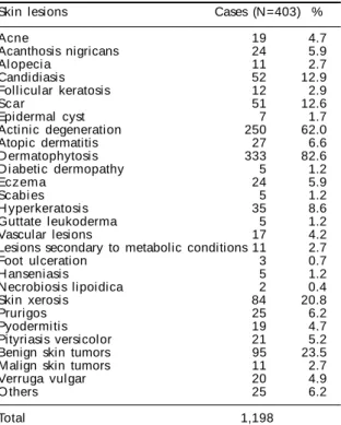
Table 1 - O ccurrence of skin lesions in diabetes mellitus pati ents seen at a uni versi ty hospi tal . Ri bei rão Preto, Brazi l , 2000.
Skin lesions Cases (N=403) %
Acne 19 4.7
Acanthosis nigricans 24 5.9
Al opeci a 11 2.7
Candidiasis 52 12.9
Follicular keratosis 12 2.9
Sc ar 51 12.6
Epidermal cyst 7 1.7
Actinic degeneration 250 62.0
Atopic dermatitis 27 6.6
D ermatophytosis 333 82.6
D iabetic dermopathy 5 1.2
Eczema 24 5.9
Scabi es 5 1.2
H yperkeratosi s 35 8.6
Guttate leukoderma 5 1.2
Vascular lesions 17 4.2
Lesions secondary to metabolic conditions 11 2.7
Foot ulceration 3 0.7
H anseniasis 5 1.2
Necrobiosis lipoidica 2 0.4
Skin xerosis 84 20.8
Prurigos 25 6.2
Pyodermitis 19 4.7
Pityriasis versicolor 21 5.2
Benign skin tumors 95 23.5
Malign skin tumors 11 2.7
Verruga vulgar 20 4.9
O thers 25 6.2
!
Rev Saúde Pública 2005;39(4) www.fsp.usp.br/rsp
Skin lesions in diabetic patients Foss NT et al
lipoidica (0.4%). Only 19% of the patients studied did not show any skin lesions, as shown in Table 1.
Of all dermatophytoses, 42.6% were onychomy-coses (n=172) and 29.2% were tinea pedis (n=118). The association of different tineas (tinea pedis and
cruris or tinea pedis, corporis and cruris) was seen in
30 patients, 9% of all cases of dermatophytoses. Interdigital candidiasis was found in 13% (n=52) and pityriasis versicolor in 5.2% (n=21). There were 19 cases (5%) of pyodermites with no previous report-ing, among them folliculites, furuncles, ecthyma and even two cases of erysipela, one of them in an early phase without treatment.
Both type 1 and type 2 DM patients had skin infec-tions such as pyodermites and superficial mycoses. Acne cases were exclusively found among type 1 DM patients. Skin tumors, such as basocellular epithe-lioma, were identified only in type 2 DM patients, whether or not associated with elastosis.
Acanthosis nigricans was identified in 6% (n=24) of cases. This is an interesting finding, since these le-sions had been unnoticed before. However, conditions more commonly seen in DM patients, such as diabetic dermopathy and necrobiosis lipoidica diabeticorum, were rare. Only two cases of necrobiosis (0.4%), five cases of diabetic dermopathy (1.2%) and three cases of foot ulcerations (0.7%) were confirmed in the histopa-thology exam. Another significant finding was that most DM patients had skin xerosis. Skin xerosis and dermatophytosis; seborrheic keratosis, onychomyco-sis and xeroonychomyco-sis; and juvenile acne and tinea pedis were the most common concomitant lesions.
Of 136 DM patients metabolically evaluated using glycated hemoglobin, 28 (20.6%) were type 1 and 108 (79.4%) type 2. Among type 1 DM patients, 14% had adequate metabolic control, while 17% of type 2 DM patients had glycated hemoglobin below 8% (Table 2). Among type 1 DM patients, mean age was 23.7 years in those with adequate metabolic control and 20.3 years in those with inadequate metabolic con-trol. Among type 2 DM patients, mean age was about 58 years in the whole group regardless of their meta-bolic control. Glycated hemoglobin was on average
7.2% in those with adequate metabolic control in both type 1 and 2 DM patients and 11.9% in type 1 and 12.7% in type 2 DM patients with inadequate metabolic control (Table 2). No statistical significant differences were found for age and DM course in both adequate and inadequate metabolic control groups for type 1 as well as type 2 DM patients.
However, when patients were grouped according to their metabolic control, those with adequate control had mostly xerosis (25%), followed by seborrheic keratosis (20.8%), solar elastosis (20.8%), dermato-phytosis (12.5%), seborrheic dermatitis (12.5%) and acanthosis nigricans (4.2%), while those with inad-equate metabolic control had mostly dermatophyto-sis (55.3%), followed by candidiadermatophyto-sis (12.5%), acne (7.2%), seborrheic keratosis (6.2%), acanthosis nigricans (5.4%), solar elastosis (5.4 %), seborrheic dermatitis (4.4%) and xerosis (3.6%) (Table 3).
D ISCU SSIO N
The study findings showed high occurrence (81%) of several different skin lesions in DM patients. Some of them were chronic well-defined lesions which had not been reported before and were only detected in the dermatological examination. The occurrence of more than one lesion per patient corroborates Bub & Olerud3 findings when they verified that almost all
DM patients had skin lesions. Hence, DM patients should undergo careful skin examination.
Among skin lesions identified, it has also been noted 82.6% of dermatophytoses and 42.6% fungal
ony-Table 3 - Distribution of skin lesions among diabetes mellitus patients with adequate and inadequate metabolic control. Ribeirão Preto, Brazil, 2000.
Skin lesions M etabolic control
Adequate Inadequate
% %
D ermatophytosis 12.5 55.3
Candidiasis 4.2 12.5
Skin xerosis 25 3.6
Solar elastosis 20.8 5.4
Seborrheic keratosis 20.8 6.2
Seborrheic dermatitis 12.5 4.4
Acanthosis nigricans 4.2 5.4
Juvenile acne N D 7.2
Total 100.0 (n=24) 100.0 (n=112)
N D : N ot detected
Table 2 - Clinical characteristics of diabetes mellitus (DM) patients according to metabolic control. Ribeirão Preto, Brazil, 2000.
Type 1 DM patients Type 2 DM patients
M etabolic control A IN A IN
No. patients (%) 4 (14%) 24 (86%) 20 (17.6%) 88 (82.4%)
Age (years) 23.7 20.3 58.5 58.3
Gender 3 F/1 M 14 F/10 M 16 F/4 M 60 F/28 M
D isease course (years) 12.7 9.6 11.9 13
Glycated hemoglobin (%) 7.2 11.9 7.2 12.7
" Rev Saúde Pública 2005;39(4) www.fsp.usp.br/rsp
Skin lesions in diabetic patients Foss NT et al
chomycoses. About 10% of all fungal lesions were asymptomatic cases of tinea corporis and cruris, which corroborates Lugo-Somolinos & Sanches11 findings.
Superficial mycoses (tineas) are known to cause pruri-tus17 and its absence could suggest a compromised
re-sponse in DM patients. It might be due to impaired su-perficial innervations caused by diabetic neuropathy, a condition that predisposes infections and traumas.1
Another contributing factor to skin lesions is the presence of macro and microangiopathy seen in DM patients. They cause vascular alterations with increased permeability and reduced vascular response to sympa-thetic nerve stimuli leading to a reduced ability to respond to thermal stress and/or local hypoxy.3
Besides, to colonize a keratin-covered skin, fungal agents need to cross a natural barrier created by cor-neal layer, which requires, among others, overcom-ing the fungistatic action of fatty acids produced by keratin cells.14 Thus, epidermis invasion by spores
occurs when this natural barrier is compromised and deeper epidermal layers are not able to activate the immune response against infections, as both mecha-nisms are impaired in the skin of DM patients.3
In addition to the significant number of dermato-phytoses, exfoliating, thickened dry skin – a condi-tion known as cutaneous xerosis – was the most fre-quent condition seen in the patients studied. Skin xero-sis was either an isolated condition (21%) or associ-ated to other skin lesions (64%). This condition is prob-ably derived from an increased production and accu-mulation of free radicals or advanced glycosylation end-products (AGE), in excess in the skin of DM pa-tients.3 In fact, high blood glucose or high levels of
other hexoses, pentoses and their phosphorilated de-rivatives seen in DM result in increased production of Amadori products which act as precursors in AGE pro-duction.2 The occurrence of skin xerosis can be
associ-ated to metabolic changes that lead to AGE produc-tion, and even to the metabolic control. High blood glucose leads to non-enzymatic glycosylation of these glycated products, which are directly associated to the level of DM metabolic control.
These findings were seen in type 1 DM patients with adequate metabolic control, of which 41% did not have any skin lesions. However, among those of same age and disease course but with inadequate
metabolic control, skin infections and acanthosis nigricans were more prevalent. In this group, only 6% of patients with inadequate metabolic control did not have any skin lesions.
It is also worth highlighting that, in type 1 DM patients, seborrheic keratosis (13%) was the skin le-sion most commonly seen among patients aged over 50. This suggests that disease progress associated to the production of glycation products would facili-tate the occurrence of these lesions.7
Among type 2 DM patients, skin infections, such as dermatophytosis and candidiasis, were more common compared to seborrheic and actinic keratosis. Simi-larly, those with inadequate metabolic control had considerably more dermatophytoses and skin candi-diasis than those with adequate metabolic control. This is in agreement to Gupta et al9 findings, showing a
26% frequency of onychomycoses in both type 1 and 2 DM patients, about one-third of all patients.
While no association between lesion frequency and DM course was found, a higher frequency of lesions, such as solar elastosis, were found to be associated to aging processes and skin deterioration in older age groups. Lesions such as acne were more common in younger age groups.
The study findings suggest that DM potentially enhances skin aging processes, as shown in Table 3. High solar elastosis and seborrheic keratosis were seen in type 2 DM patients and a significant frequency of seborrheic keratosis was found in type 1 DM patients, a group comprising mostly young patients (mean age: 20 years old), regardless of their metabolic control. Also, increased skin infections, both fungal and bac-terial, were detected among type 1 and type 2 DM patients with inadequate metabolic control. This find-ing indicates that inadequate metabolic control renders DM patients more susceptible to skin infec-tions. Furthermore, it could become more severe, ag-gravating these patients’ metabolic imbalance and compromising their general condition.10
#
Rev Saúde Pública 2005;39(4) www.fsp.usp.br/rsp
Skin lesions in diabetic patients Foss NT et al
REFEREN CES
1. Ansel JC, Armstrong CA, Song I, Quinlan KL, Olerud JE, Caughman SW, Bunnett NW. Interactions of the skin and nervous system. J Invest Dermatol Symp Proc 1997;2:23-6.
2. Beisswenger PJ, Moor LL, Curphey TJ. Relationship betw een glycemic control and colagen-linked advanced glycosylation end products in type I diabetes. Diabetes care 1993;16:689-94.
3. Bub JL, Olerud JE. Diabetes Mellitus. In: Freedberg IM, Elsen AZ, Wolff K, Austen KF, Goldsmith LA, Katz SI, M cGrow-Hill, editors. Chapter 168. Fitzpatrick’s dermatology in general medicine. N ew York: McGraw-Hill; 2003. p. 1651-61.
4. De Fronzo RA, Ferrannini E. Insulin resistance. A multifacetted syndrome responsible for type 2 diabetes mellitus, obesity, hypertention, dyslipidemia and atherosclerotic cardiovascular disease. D iabetes Care 1991;14:173-94.
5. Delamaire M, Maugendre D, Moreno M, Le Goff MC, Allannic H, Genetet B. Impaired leucocyte functions in diabetic patients. Diabet Med 1997;14:29-34.
6. Diabetes Control and Complications Trial Research Group. The effect of intensive treatment of diabetes on the development and progression of long-term complications in insulin-dependent diabetes mellitus. N Engl J Med 1993;329:977-86.
7. Dyer DG, Dunn JA, Thorpe SR, Bailie KE, Lyons TJ, McCance DR, Baynes JW. Accumulation of mailard reaction products in skin collagen in diabetes and aging. J Clin Invest 1993;91:2463-9.
8. Eisenbarth GS. Type 1 diabetes mellitus. A chronic autoimmune disease. N Engl J Med 1986;314:1360-8.
9. Gupta AK, Konnikov N, MacDonald P, Rich P, Rodger NW, Edmonds MW et al. Prevalence and
epidemiology of toenail onychomycosis in diabetic subjects: a multicentre survey. Br J Dermatol 1998;139:665-71.
10. Josh N, Caputo GM, Wettekamp MR, Karchmer AW. Infections in patients with diabetes melitus. N Engl J Med 1999;16:1906-12.
11. Lugo-Somolinos A, Sanchez JL. Prevalence of dermatophytosis in patients with diabetes. J Am Acad Dermatol 1992;26:408-10.
12. Marble A, Krall LP, Bradley RF, Christlieb AR, Soeldner JS, editors. Joslin’s diabetes mellitus. 12th ed.
Philadelphia: Lea-Febiger; 1985. p. 526-52.
13. McMahon MM, Bristian BR. Host defenses and susceptibility to infections in patients with diabetes mellitus. Infec Dis Clin of North Am 1995;9:1-9.
14. Nelson M M , M artin AG, Heffernan M P. Superficial fungal infections: dermatophytosis, onychomicosis, tinea Nigra, Piedra. Fungal diseases with cutaneous involvment. In: Freedberg IM, Elsen AZ, Wolff K, Austen KF, Goldsmith LA, Katz SI, editors. Fitzpatrick’s dermatology in general medicine. N ew York: McGraw Hill; 2003. Chapter 205, Section 29, p. 1989-2005.
15. Shah BR, Hux JE. Quantifying the risk of infectious disease for people with diabetes. Diabetes Care 2003;26:510-3.
16. Shott S. Statistics for health professionals. Philadelphia: W.B. Saunders; 1990.
17. Leonhardt JM, Heymann W R. Cutaneous manifestations of other endocrine diseases. In: Freedberg IM, Elsen AZ, Wolff K, Austen KF, Goldsmith LA, Katz SI, editors. Fitzpatrick’s dermatology in general medicine. N ew York: MacGraw-Hill; 2003. Chapter 169, p. 1662-70.
18. Trivelli LA, Ranney HM, Lai HT. Hemoglobin components in patients with diabetes mellitus. N Engl J Med 1971;284:353-7.
19. UK Prospective Diabetes Study Group (UKPDS). Intensive blood-glucose control with sulphonylureas or insulin compared with conventional treatment and risk of complications in patients with type 2 diabetes (UKPDS 33). Lancet 1998;352:837-53.
20. UK Prospective Diabetes Study Group (UKPDS). Effect of intensive blood-glucose control with metiformin on complications in overweight patients with type 2 diabetes (UKPDS 34). Lancet