MicroRNA-340-5p modulates cisplatin resistance
by targeting LPAAT
b
in osteosarcoma
L. Song
1, P. Duan
2, Y. Gan
1, P. Li
1, C. Zhao
1, J. Xu
1, Z. Zhang
1and Q. Zhou
1 1Department of Orthopedics, First Affiliated Hospital, Third Military Medical University, Chongqing, China 2Southwest Eye Hospital, First Af
filiated Hospital, Third Military Medical University, Chongqing, China
Abstract
MicroRNAs (miRNAs) play an important role in drug resistance and modulate the efficiency of chemotherapy. A recent study indicated that miR-340 functions as a tumor suppressor in various types of cancer. However, the role of miR-340 in chemotherapy has not been reported yet. In this study, we found that miR-340 enhanced cisplatin (CDDP)-induced cell death. Induction of miR-340-5p expression decreased the IC50of CDDP and increased the apoptosis of CDDP-resistant MG-63 and Saos-2 cells. Moreover, miR-340-5p decreased the accumulation of MRP1 and MDR1. We further explored the mechanism underlying the promoting effects of miR-340-5p on CDDP-induced cell death. We identified a potential target of miR-340 in the 30 untranslated region of
lysophosphatidic acid acyltransferase (LPAATb) using the online program Targetscan (www.microrna.org). Luciferase reporter assays showed that miR-340 binds to the 30UTR of LPAATb. Enforced expression of miR-340-5p decreased the accumulation of
LPAATbin both MG-63 and Saos-2 cells. Silencing LPAATbdecreased the IC50of CDDP and increased the apoptosis of CDDP-resistant MG-63 and Saos-2 cells, which is consistent with the effect of miR-340-5p on CDDP-induced cell death. Moreover, induced expression of LPAATbcompromised the effects of miR-340-5p on CDDP-induced cell death and accumulation of MRP1 and MDR1. Taken together, our data indicated that miR-340-5p enhanced the sensitivity to CDDP by targeting LPAATb.
Key words: Sensitivity to cisplatin; LPAATb; miR-340-5p; Osteosarcoma
Introduction
Osteosarcoma (OS) is an aggressive malignant neo-plasm that arises from primitive transformed cells of mes-enchymal origin, exhibits osteoblastic differentiation, and produces malignant osteoid. OS is ranked highest in mor-bidity among all primitive malignant tumors (1). Although chemotherapy is frequently used in OS, many factors lead to its failure. Drug resistance is the main factor affecting the efficiency of chemotherapy (1). Previous studies indi-cated that drug resistance is a compliindi-cated process involv-ing many genes, includinvolv-ing microRNAs (miRNAs) (2–4).
miRNAs are non-coding RNAs approximately 18–22 nucleotides in length (5). Recent studies demonstrated that miRNAs are key regulators of tumor initiation and progression (6–8). They typically modulate proliferation, migration, invasion and drug resistance of tumor cells by targeting oncogenes, tumor suppressor genes, transcrip-tion factors, and other regulators involved in cell death and survival (7,9–11). MicroRNA-340 (miR-340) wasfirst reported to be a suppressive miRNA in breast cancer (12). Subsequent studies of colorectal cancer (13), osteo-sarcoma (14), melanoma (15), and gastric cancer (16) confirmed that miR-340 plays an inhibitory role in the proliferation, migration, and invasion of cancer cells.
1-Acylglycerol-3-phosphate O-acyltransferase 2, also known as lysophosphatidic acid acyltransferaseb(LPAATb), is a member of the 1-acylglycerol-3-phosphate O -acyl-transferase family (17). The protein is located within the endoplasmic reticulum membrane and converts lysophos-phatidic acid to phoslysophos-phatidic acid, the second step in de novo phospholipid biosynthesis (17). Recent studies in ovarian cancer suggested that LPAATbplays a role in tumor progression (18,19). Rastegar et al. (20) reported that LPAATb promotes the tumor growth of human OS. It also functions as a downstream target of miRNA (21). MicroRNA-24 inhibits cell proliferation by targeting LPAATb
in OS (21). LPAATb is also involved in drug resistance (22). The LPAATb inhibitor CT-32615 triggers caspase-dependent apoptosis and can overcome resistance to conventional therapeutics (i.e., dexamethasone, doxoru-bicin, melphalan) in multiple myeloma cells (22).
In the current study, we analyzed the expression of miR-340-5p in OS and CDDP-resistant cells and exam-ined the effects of miR-340-5p on CDDP-induced cell death and expression of drug resistance-related genes. We also investigated the mechanism underlying the tran-scription regulation of miR-340-5p on LPAATb. Our results
Correspondence: Q. Zhou:<zhouqiangphd@sina.com>
provide novel insight into the CDDP resistance of OS, which may help to improve the efficacy of chemotherapy.
Material and Methods
Cell culture
OS cell lines MG-63 and Saos-2, and CDDP-resistant OS cells MG-63/CDDP and Saos-2/CDDP were obtained from Shanghai Cell Institute (China). MG-63, Saos-2, MG-63/CDDP, and Saos-2/CDDP cells were grown in 1640 medium containing 10% fetal bovine serum (Gibco/ BRL, USA) supplemented with 100 U/mL penicillin G and 100mg/mL streptomycin (Sigma-Aldrich, USA). Cells were maintained at 37°C in a humidified 5% CO2incubator.
Constructs
To create the luciferase reporter constructs contain-ing the wild-type 30 untranslated region of LPAATb, the full-length 30UTR of LPAATb was amplified and cloned into the pmirGLO vector (Promega, USA). The primers used to amplify the 30UTR of LPAATb are as follows: forward 50CTAGGCATGCAGACCACGGCAGGGCATG30 and reverse 50CCCAAGCTTTTGCCACTTCCAAGAGT GTG30. Luciferase reporter constructs containing mutat-ed binding sites were creatmutat-ed using the QuikChanges Site-Directed Mutagenesis Kit (Strategene, USA) with the wild-type 30UTR of LPAAT b as a template. The primers used to amplify the 30UTR of LPAATb were as follows: 50CACTGTACTCCGTTGCTGTTTTTATCTGA ACACACTCTTGGAAGTGGC30 and 50GCCACTTCCA AGAGTGTGTTCAGATAAAAACAGCAACGGAGTACAG TG30. The LPAATbexpression constructs were created by subcloning the coding region of LPAATbinto a pcDNA3.0-expressing vector (Invitrogen, USA). The primers used to amplify the 30UTR of LPAATbwere as follows: 50CG GGGTACCATGGAGCTGTGGCCGTGTC30 and 50GC CACTTCCAAGAGTGTGTTCAGATAAAAACAGCAAC GGAGTACAGTG30. All constructs were verified by sequencing.
Transfection of miRNAs and siRNAs
The mimics of miR-340-5p and siRNAs targeted to LPAATb were obtained from GenePharma Co., Ltd. (China). First, 2104OS cells were plated into 6-well plates the day before transfection. Next, 100 nM miRNAs or siRNAs were transfected into OS cells using Lipofectamine 2000 reagent (Invitrogen) according to the manufacturers’ instruction. A scramble sequence was used as the negative control (NC). The transfection efficiency of miRNAs and siRNAs was determined by quantitative real-time RT-PCR (qRT-PCR) and western blot analysis, respectively.
qRT-PCR
Total RNA was extracted using TRIZOL reagent (Ambion, USA) according to the manufacturer’s instructions.
cDNA used to examine the expression of LPAATb was synthesized using the PrimeScriptt RT reagent kit (TaKaRa, Japan) according to the manufacturer’s instruc-tions. Expression of LPAATbwas examined using SYBRs Premix Ex TaqtII (TaKaRa) and GAPDH served as inter-nal reference. All experiments were performed in dupli-cate and repeated twice. The results are represented as the fold-induction using the 2-DDCTmethod. Primers used to examine the expression of LPAATb were as follows: LPAATb, forward: 50-CCTTCCTCCACATCTCCAAG-30, reverse: 50-CCGGACAGAGTGGTATTTGG-30; miR-340-5p, forward: 50-GCGGTTATAAAGCAATGAGA-30, reverse: 50-GTGCGTGTCGTGGAGTCG-30; U6, forward: 50-CTCG CTTCGGCAGCACA-30, reverse: 50-AACGCTTCACGAAT TTGCGT-30.
Western blot analysis
Western blot analysis was performed according to standard procedures as previously described (23). Briefly, proteins were separated by 10% SDS-PAGE and then transferred to nitrocellulose membranes (Bio-Rad, USA). After blocking in 5% nonfat milk, the membranes were incubated with the following primary antibodies: rabbit anti-LPAATbpolyclonal antibody (ab62599; 1:500; Abcam, UK), rabbit anti-MRP1 polyclonal antibody (ab180960; 1:500; Abcam), rabbit anti-MDR1 polyclonal antibody (ab170904; 1:500; Abcam), and rabbit anti-GAPDH mAb (1:1,000; Abcam). The proteins were visualized using enhanced chemiluminescence reagents (Pierce, USA).
Proliferation assays
Cell Counting Kit-8 (CCK8) was used to evaluate the growth of OS cells treated with CDDP according to the manufacturer’s protocol. Briefly, 104cells/well were plated
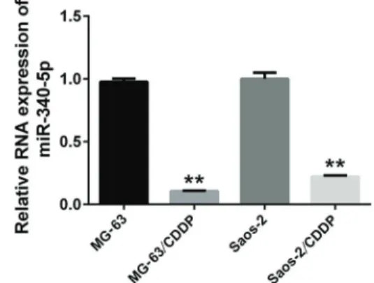
Figure 1. miR-340-5p is down-regulated in cisplatin (CDDP)-resistant osteosarcoma cells. The expression of miR-340-5p was examined in MG-63, Saos-2, MG-63/CDDP, and Saos-2/CDDP cells. U6 served as an internal reference. The relative expres-sion of miR-340-5p was calculated using the 2-DDCT method. All experiments were repeated three times. Data are reported as means±SD. **Po0.05 compared to its respective control
in triplicate in 96-well plates. CCK8 solution was added to each well at a 1:10 dilution. Cells were incubated for 4 h, and then the absorbance at 450 nm was measured using a multi-well plate reader. The IC50 value was calculated using SPSS software (USA).
Apoptosis assays
An Annexin V-FITC apoptosis detection kit (Multi-sciences, China) was used to detect apoptosis in OS cells.
According to the manufacturer’s instructions, the cells were digested with trypsin and centrifuged at 300 g for 5 min at 4°C. After collection, the cells were washed twice with PBS and centrifuged at 300 g for 5 min at 4°C, and 3105cells were collected and suspended in 500mL binding buffer. Next, 5mL Anexin V-FITC and 5 mL propidium iodide were added and mixed at room temperature in the dark for 15 min. Within 1 h, the cells were detected byflow cytometry.
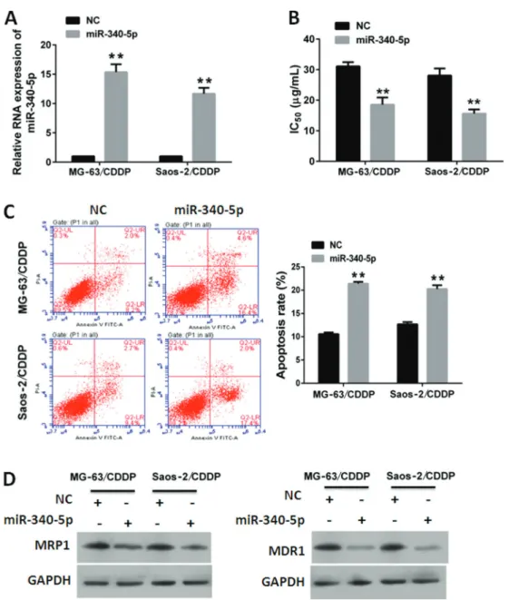
Figure 2.miR-340-5p enhanced sensitivity of cisplatin (CDDP) in osteosarcoma cells.A, Relative RNA expression of miR-340-5p in MG-63/CDDP and Saos-2/CDDP cells transfected with miR-340-5p or normal control (NC).B, miR-340-5p decreased IC50of CDDP in MG-63/CDDP and Saos-2/CDDP cells.C, miR-340-5p increased CDDP-induced apoptosis in MG-63/CDDP and Saos-2/CDDP cells. Data are reported as means±SD. **Po0.01 (Student’st-test).D, miR-340-5p decreased the expression of MRP1 and MDR1 in MG-63/
Luciferase reporter assays
OS cells were seeded onto 96-well plates at 6000 cells per well the day before transfection. A mixture of 100 ng luciferase reporter constructs (pmirGLO-LPAATb-WT and pmirGLO-LPAATb-mutant) and 200 ng of NC or miR-340-5p mimics was transfected into OS cells with Lipofectamine 2000. Forty-eight hours later, Firefly and Renilla lucifer-ase activities were measured using a Dual-Luciferlucifer-ase Re-porter System (Promega) according to the manufacturer’s protocol.
Statistical analysis
Data are reported as means±SD, unless otherwise indicated. Student’st-test was used to analyze statistical differences between groups. A P value o0.05 was
con-sidered to be statistically significant.
Results
MiR-340-5p was down-regulated in CDDP-resistant OS cells
In order to determine the role of miR-340-5p in CDDP resistance, we first examined the expression profile of
miR-340-5p in CDDP-resistant OS cells, MG-63/CDDP and Saos-2/CDDP. Normal OS cells, MG-63 and Saos-2, were used as controls. qRT-PCR was performed to exam-ine the expression of miR-340-5p. Our data showed that the expression of miR-340-5p in CDDP-resistant OS cells was much lower than in OS cells (Figure 1). This suggests that miR-340-5p was down-regulated in CDDP-resistant OS cells.
Effect of miR-340-5p on CDDP resistance in OS cells MG-63/CDDP and Saos-2/CDDP cells were treated with CDDP (15mg/mL) following transfection of miR-340-5p mimics. A scramble RNA sequence was used as a NC. The results of qRT-PCR showed that miR-340-5p was suc-cessfully overexpressed after transfection of miR-340-5p mimics compared to NC (Figure 2A). The IC50of CDDP was calculated based on the CCK8 assays. The IC50 values of CDDP in MG-63/CDDP and Saos-2/CDDP cells transfected with miR-340-5p were lower than those trans-fected with NC (Figure 2B).
We then investigated the effects of miR-340-5p on CDDP-induced apoptosis in MG-63/CDDP and Saos-2/ CDDP cells. Our data showed that the apoptosis rates in
Figure 3.A, Schematic diagram of interaction between miR-340-5p and LPAATb.B, Dual luciferase assay of cells co-transfected with luciferase reporter constructs and miR-340-5p or normal control (NC).C, miR-340-5p did not affect mRNA expression of LPAATbin MG-63/CDDP and Saos-2/CDDP cells. Data are reported as means±SD. **Po0.01 (Student’st-test). D, miR-340-5p decreased
cells transfected with miR-340-5p were higher than in those transfected with NC (Figure 2C).
We also investigated the effects of miR-340-5p on drug transporters, including MRP1 and MDR1 (24–26). Our data showed that the accumulations of MRP1 and MDR1 in cells transfected with miR-340-5p were lower than in those transfected with NC (Figure 2D). These results suggest that miR-340-5p impaired CDDP resis-tance in OS cells.
MiR-340-5p down-regulated LPAATb
In order to explore the mechanism underlying the inhibitory effects of miR-340-5p on CDDP resistance, we identified the target of miR-340-5p using an online pro-gram (www.microrna.org). LPAATb showed high scores and a miR-340-5p binding site in its 30UTR (Figure 3A). In order to investigate whether LPAATb is a target of miR-340-5p, we created luciferase reporter constructs containing wild-type or mutated 30UTRs of LPAATb, and examined whether miR-340-5p binds to the 30UTR of LPAATb.
Our data showed that miR-340-5p expression was decreased relative to luciferase activities in MG-63 cells transfected with pmirGLO-LPAATb-WT, but not in MG-63 cells transfected with the pmirGLO-LPAATb-mutant (Figure 3B). These results indicate that miR-340-5p binds to the 30UTR of LPAATb.
Next, we examined the mRNA and protein expres-sion of LPAATbin MG-63/CDDP and Saos-2/CDDP cells transfected with mimics of miR-340-5p. Our data showed that expression of miR-340-5p did not affect the mRNA expression of LPAATb(Figure 3C). However, protein accu-mulation of LPAATbwas decreased in MG-63/CDDP and Saos-2/CDDP cells (Figure 3D), indicating that miR-340-5p acts as a negative regulator of LPAATb.
To further verify our previous finding, we investigate the expression of LPAATb in CDDP-resistant OS cells, MG-63/CDDP and Saos-2/CDDP, in which miR-340-5p proved to be down-regulated. Normal OS cells, MG-63 and Saos-2, were used as controls. Western blot analysis showed that the expression of LPAATbin CDDP-resistant OS cells was much higher than in OS cells (Figure 4). These data suggested that LPAATb was up-regulated in MG-63/CDDP and Saos-2/CDDP cells, in which miR-340-5p proved to be down-regulated. This result partially supports our previousfinding that miR-340-5p acted as a negative regulator of LPAATb.
Effect of silencing LPAATbon CDDP resistance
We demonstrated that LPAATbis a target of miR-340-5p. In order to verify that miR-340-5p modulated CDDP resistance by down-regulating LPAATb, we investigated the effects of silencing LPAATb on CDDP resistance. If effects of silencing LPAATbwere consistent with effects of 5p, it would be partially proven that miR-340-5p modulated CDDP resistance targeting LPAATb. The
siRNAs targeted LPAATbwere used to suppress LPAATb
in MG-63/CDDP and Saos-2/CDDP cells. A scramble RNA was used as NC. The silencing efficiency of siRNA was confirmed by decreased accumulation of LPAATb in cells transfected with siRNAs (Figure 5A). MG-63/CDDP and Saos-2/CDDP cells were transfected with siRNAs or NC and treated with CDDP for 24 hours. IC50value was determined using CCK8 assays. Our data showed IC50 value of siRNA group was lower than those of NC group (Figure 5B). The effects of silencing LPAATbon CDDP induced-apoptosis was determined by flow cytometry. The results showed the apoptosis rate of siRNA group was higher than those of the NC group (Figure 5C). These results indicate that the effects of silencing LPAATbon CDDP resistance were consistent with those of miR-340-5p.
Enforced expression of LPAATbattenuated effects of
miR-340-5p on CDDP resistance
We demonstrated that LPAATb is a target of miR-340-5p. In order to verify that miR-340-5p modulates CDDP resistance by down-regulating LPAATb, we eval-uated whether up-regulation of LPAATbaffected the role of miR-340-5p in CDDP resistance. We co-transfected expression plasmids of pcDNA-LPAATband miR-340-5p into MG-63/CDDP and Saos-2/CDDP cells. The control group was transfected with the pcDNA3.0 vector and miR-340-5p. Transfection efficiency was confirmed by
Figure 4.LPAATbwas up-regulated in cisplatin (CDDP)-resistant osteosarcoma cells. The expression of LPAATbwas examined in MG-63, Saos-2, MG-63/CDDP, and Saos-2/CDDP cells using western blot analysis. GAPDH served as loading control. Upper: representative image of western blot analysis. Lower: histogram represents the densitometry data for at least 3 independent experiments. Data are reported as means±SD. **Po0.01,
over-expression of LPAATb in cells transfected with pcDNA-LPAATb (Figure 6A). We found that the IC50 of CDDP in cells transfected with pcDNA-LPAATbwas higher than in those transfected with pcDNA3.0 (Figure 6B), suggesting that expression of LPAATb attenuated the effect of miR-340-5p on the IC50of CDDP.
We also investigated whether expression of LPAATb
affected the inhibitory effect of miR-340-5p on CDDP-induced apoptosis. Our data showed that apoptosis in the pcDNA-LPAATb group was lower than that in the control group (Figure 6C), suggesting that LPAATb expression alleviated the promoting effect of miR-340-5p on CDDP-induced apoptosis. In addition, we found that express-ion of MRP1 and MDR1 in the pcDNA-LPAATb group was higher than in the control group (Figure 6D). Taken together, these results suggest that expression of LPAATb
attenuated the effects of miR-340-5p on CDDP resistance.
Discussion
Chemotherapy is an effective treatment for OS. However, many patients develop primary and secondary drug resistance, causing chemotherapy to fail. It is very important to enhance the sensitivity of OS cells to chemo-therapy reagents. However, the molecular mechanism of chemotherapy resistance is not fully understood. Recent studies revealed numerous genes or noncoding RNA mol-ecules involved in the regulation of sensitivity to che-motherapy reagents. In the current study, we found that miR-340 plays an important role in enhancing the sen-sitivity of OS cells to CDDP.
MiR-340 wasfirst reported in melanoma as a regulator of microphthalmia-associated transcription factor (MITF) (27). Recent studies of breast cancer (12,28,29), colorec-tal cancer (13,30), melanoma (15), gastric cancer (16),
Figure 5.Effect of silencing LPAATbon cisplatin (CDDP) resistance.A, Relative protein expression of LPAATbin MG-63/CDDP and Saos-2/CDDP cells transfected with siRNA or normal control (NC).B, Silencing LPAATbdecreased IC50of CDDP in MG-63/CDDP and Saos-2/CDDP cells.C, Silencing LPAATbincreased CDDP-induced apoptosis in MG-63/CDDP and Saos-2/CDDP cells. Data are reported as means±SD. **Po0.01, compared to its respective control (Student’s t-test). D, Silencing LPAATb decreased the
glioblastoma (31,32), hepatocellular carcinoma (33), lung cancer (34), oral squamous cell carcinoma (35), and laryngeal squamous cell carcinoma (36) indicated that miR-340 is involved in the proliferation, metastasis, inva-sion, and apoptosis of cancer cells. Other studies demon-strated that miR-340 serves as a negative regulator in cancer (12,31,37–39). Zhou et al. (40) reported that miR-340 suppresses tumor growth and metastasis in OS. The expression of miR-340 has been associated with tumor progression and prognosis in pediatric OS (14). Our data
indicates that miR-340 regulates CDDP-induced apopto-sis in CDDP-reapopto-sistant OS cells, suggested that miR-340 enhanced the sensitivity of OS cells to CDDP.
The ATP-binding cassette (ABC) transporters trans-port various molecules across extra- and intra-cellular membranes (24). They had been proven by various studies to modulate the development of resistance to anticancer drugs (24). MRP1 (ABCC1) is a member of the MRP subfamily, which is involved in multi-drug resis tance (25). This protein functions as a multispecific organic
Figure 6.Expression of LPAATbattenuated inhibitory effects of miR-340-5p on cisplatin (CDDP) resistance.A, Expression of LPAATbin MG-63/CDDP and Saos-2/CDDP cells co-transfected with miR-340-5p and pcDNA-LPAATbor pcDNA3.0.B, Expression of LPAATb compromised inhibitory effects of miR-340-5p on IC50 of CDDP in MG-63/CDDP and Saos-2/CDDP cells.C, Expression of LPAATb attenuated promoting effects of miR-340-5p on CDDP-induced apoptosis in MG-63/CDDP and Saos-2/CDDP cells. Data are reported as means±SD. **Po0.01 (Student’st-test).D, Expression of LPAATbcompromised inhibitory effects of miR-340-5p on expression of
anion transporter, with oxidized glutathione, cysteinyl leukotrienes, and activated aflatoxin B1 as substrates (25). This protein also transports glucuronides and sulfate conjugates of steroid hormones and bile salts (25). MDR1 (ABCB1) is a member of the MDR/TAP subfamily. Mem-bers of the MDR/TAP subfamily are involved in multidrug resistance. MDR1 is an ATP-dependent drug efflux pump for xenobiotic compounds with broad substrate specificity (26). It is responsible for decreased drug accumulation in multidrug-resistant cells and often mediates the develop-ment of resistance to anticancer drugs (26). It also func-tions as a transporter in the blood-brain barrier (26). Our data showed that miR-340 decreased MRP1 and MDR1 expression. These data support our previous finding that miR-340 modulated CDDP resistance in OS cells.
MiRNAs typically accomplish their function by mod-ulating the expression of their target genes. Identifying target genes is crucial for understanding the role of miRNAs in cancer cells. We identified LPAATbas a novel target of miR-340 in OS. Several genes have been identified as targets of miR-340, including MITF (23), c-Met (12), ROCK1 (36), RhoA (37), Nrf2 (29), p27 (33), MYO10 (34), NRAS (28), MDM2 (35), NF-x03BA, and CTNNB1 (25). MiR-340 binds to the 30UTR of target genes and decreases the accumulation of target genes in cancer cells (12,23,28,30,33,36,38). Our findings are consistent with those of previous studies. We showed that
miR-340 binds to the 30UTR of LPAATb. Mutation in the biding site of miR-340 compromised its binding to the 30UTR of LPAATb. In addition, we found that the effects of LPAATbknockdown on CDDP resistance are similar to the effects of miR-340-5p on CDDP resistance in both MG-63/CDDP and Saos-2/CDDP cells. We also demon-strated that over-expression of LPAATb attenuated the effects of miR-340-5p on CDDP resistance. These results indicate miR-340 affected CDDP resistance by down-regulating LPAATb.
In conclusion, we found that miR-340 enhanced the sensitivity of OS to CDDP by targeting LPAATb. Our study provides insight into CDDP resistance in OS. MiR-340 may serve as a target for chemotherapy of OS. However, there were several limitations to our study. First, in vivo evidence of the role of miR-340 in chemotherapy is required to support the findings of our study. Second, up-stream regulators of the miR-340-LPAATbaxis are un-clear, which affects application of the miR-340-LPAATb
axis in chemotherapy. Third, down-stream signaling path-ways of miR-340-LPAATbaxis have not been determined.
Acknowledgments
This work was supported by the National Natural Science Foundation of China (No. 81302347 and 8140 0418).
References
1. Anninga JK, Gelderblom H, Fiocco M, Kroep JR, Taminiau AH, Hogendoorn PC, et al. Chemotherapeutic adjuvant treat-ment for osteosarcoma: where do we stand?Eur J Cancer 2011; 47: 2431–2445, doi: 10.1016/j.ejca.2011.05.030. 2. De Mattos-Arruda L, Bottai G, Nuciforo PG, Di Tommaso L,
Giovannetti E, Peg V, et al. MicroRNA-21 links epithelial-to-mesenchymal transition and inflammatory signals to confer resistance to neoadjuvant trastuzumab and chemotherapy in HER2-positive breast cancer patients.Oncotarget2015; 6: 37269–37280, doi: 10.18632/oncotarget.5495.
3. Li H, Yang BB. Friend or foe: the role of microRNA in chemotherapy resistance. Acta Pharmacol Sin 2013; 34: 870–879, doi: 10.1038/aps.2013.35.
4. Bockhorn J, Dalton R, Nwachukwu C, Huang S, Prat A, Yee K, et al. MicroRNA-30c inhibits human breast tumour chemotherapy resistance by regulating TWF1 and IL-11. Nat Commun2013; 4: 1393, doi: 10.1038/ncomms2393. 5. Hutvagner G, Zamore PD. A microRNA in a
multiple-turnover RNAi enzyme complex.Science2002; 297: 2056– 2060, doi: 10.1126/science.1073827.
6. Cheng CJ, Bahal R, Babar IA, Pincus Z, Barrera F, Liu C, et al. MicroRNA silencing for cancer therapy targeted to the tumour microenvironment.Nature2015; 518: 107–110, doi: 10.1038/nature13905.
7. Dvinge H, Git A, Graf S, Salmon-Divon M, Curtis C, Sottoriva A, et al. The shaping and functional consequences of the microRNA landscape in breast cancer.Nature2013; 497: 378–382, doi: 10.1038/nature12108.
8. Powers JT, Tsanov KM, Pearson DS, Roels F, Spina CS, Ebright R, et al. Multiple mechanisms disrupt the let-7 microRNA family in neuroblastoma.Nature2016; 535: 246– 251, doi: 10.1038/nature18632.
9. He L, Thomson JM, Hemann MT, Hernando-Monge E, Mu D, Goodson S, et al. A microRNA polycistron as a potential human oncogene. Nature 2005; 435: 828–833, doi: 10.1038/nature03552.
10. Johnson SM, Grosshans H, Shingara J, Byrom M, Jarvis R, Cheng A, et al. RAS is regulated by the let-7 microRNA family. Cell 2005; 120: 635–647, doi: 10.1016/j.cell.2005. 01.014.
11. He L, He X, Lim LP, de Stanchina E, Xuan Z, Liang Y, et al. A microRNA component of the p53 tumour suppressor net-work. Nature 2007; 447: 1130–1134, doi: 10.1038/nature 05939.
12. Wu ZS, Wu Q, Wang CQ, Wang XN, Huang J, Zhao JJ, et al. miR-340 inhibition of breast cancer cell migration and invasion through targeting of oncoprotein c-Met. Cancer 2011; 117: 2842–2852, doi: 10.1002/cncr.25860.
13. Sun Y, Zhao X, Zhou Y, Hu Y. 124, 137 and miR-340 regulate colorectal cancer growth via inhibition of the Warburg effect. Oncol Rep 2012; 28: 1346–1352, doi: 10.3892/or.2012.1958.
15. Poenitzsch Strong AM, Setaluri V, Spiegelman VS. Micro-RNA-340 as a modulator of RAS-RAF-MAPK signaling in melanoma. Arch Biochem Biophys 2014; 563: 118–124, doi: 10.1016/j.abb.2014.07.012.
16. Hou X, Qiao H. Effect of miR-340 on gastric cancer cell proliferation and apoptosis. Int J Clin Exp Pathol 2015; 8: 13108–13113.
17. Agarwal AK, Arioglu E, De Almeida S, Akkoc N, Taylor SI, Bowcock AM, et al. AGPAT2 is mutated in congenital generalized lipodystrophy linked to chromosome 9q34.Nat Genet2002; 31: 21–23, doi: 10.1038/ng880.
18. Burton A. LPAAT-beta identifies aggressive ovarian cancer. Lancet Oncol 2006; 7: 893, doi: 10.1016/S1470-2045(06) 70926-3.
19. Diefenbach CS, Soslow RA, Iasonos A, Linkov I, Hedvat C, Bonham L, et al. Lysophosphatidic acid acyltransferase-beta (LPAAT-beta) is highly expressed in advanced ovarian cancer and is associated with aggressive histology and poor survival. Cancer2006; 107: 1511–1519, doi: 10.1002/cncr.22184. 20. Rastegar F, Gao JL, Shenaq D, Luo Q, Shi Q, Kim SH, J et
al. Lysophosphatidic acid acyltransferase beta (LPAATbeta) promotes the tumor growth of human osteosarcoma.PLoS One2010; 5: e14182, doi: 10.1371/journal.pone.0014182. 21. Song L, Yang J, Duan P, Xu J, Luo X, Luo F, et al.
MicroRNA-24 inhibits osteosarcoma cell proliferation bothin vitro and in vivo by targeting LPAATbeta. Arch Biochem Biophys 2013; 535: 128–135, doi: 10.1016/j.abb.2013. 04.001.
22. Hideshima T, Chauhan D, Ishitsuka K, Yasui H, Raje N, Kumar S, et al. Molecular characterization of PS-341 (bortezomib) resistance: implications for overcoming resis-tance using lysophosphatidic acid acyltransferase (LPAAT)-beta inhibitors.Oncogene2005; 24: 3121–3129, doi: 10.1038/ sj.onc.1208522.
23. Huang H, Jiang Y, Wang Y, Chen T, Yang L, He H, et al. miR-5100 promotes tumor growth in lung cancer by targeting Rab6.Cancer Lett2015; 362: 15–24, doi: 10.1016/j.canlet. 2015.03.004.
24. Chen Z, Shi T, Zhang L, Zhu P, Deng M, Huang C, et al. Mammalian drug efflux transporters of the ATP binding cas-sette (ABC) family in multidrug resistance: A review of the past decade.Cancer Lett2016; 370: 153–164, doi: 10.1016/ j.canlet.2015.10.010.
25. Lu JF, Pokharel D, Bebawy M. MRP1 and its role in anticancer drug resistance. Drug Metab Rev 2015; 47: 406–419, doi: 10.3109/03602532.2015.1105253.
26. Kimura Y, Morita SY, Matsuo M, Ueda K. Mechanism of multidrug recognition by MDR1/ABCB1.Cancer Sci2007; 98: 1303–1310, doi: 10.1111/j.1349-7006.2007.00538.x. 27. Goswami S, Tarapore RS, Teslaa JJ, Grinblat Y, Setaluri V,
Spiegelman VS. MicroRNA-340-mediated degradation of microphthalmia-associated transcription factor mRNA is inhibited by the coding region determinant-binding protein. J Biol Chem 2010; 285: 20532–20540, doi: 10.1074/jbc. M110.109298.
28. Mohammadi Yeganeh S, Vasei M, Tavakoli R, Kia V, Paryan M. The effect of miR-340 over-expression on cell-cycle-related genes in triple-negative breast cancer cells. Eur J Cancer Care2016, doi: 10.1111/ecc.12496.
29. Mohammadi-Yeganeh S, Paryan M, Arefian E, Vasei M, Ghanbarian H, Mahdian R, et al. MicroRNA-340 inhibits the migration, invasion, and metastasis of breast cancer cells by targeting Wnt pathway.Tumour Biol2016; 37: 8993–8900, doi: 10.1007/s13277-015-4513-9.
30. Takeyama H, Yamamoto H, Yamashita S, Wu X, Takahashi H, Nishimura J, et al. Decreased miR-340 expression in bone marrow is associated with liver metastasis of colorectal cancer.Mol Cancer Ther2014; 13: 976–985, doi: 10.1158/ 1535-7163.MCT-13-0571.
31. Huang D, Qiu S, Ge R, He L, Li M, Li Y, et al. miR-340 suppresses glioblastoma multiforme. Oncotarget 2015; 6: 9257–9270, doi: 10.18632/oncotarget.3288.
32. Fiore D, Donnarumma E, Roscigno G, Iaboni M, Russo V, Affinito A, et al.Oncotarget2016; 7: 19531-19547. 33. Shi L, Chen ZG, Wu LL, Zheng JJ, Yang JR, Chen XF, et al.
miR-340 reverses cisplatin resistance of miR-340 predicts glioblastoma survival and modulates key cancer hallmarks through down-regulation of NRAS hepatocellular carcinoma cell lines by targeting Nrf2-dependent antioxidant pathway. Asian Pac J Cancer Prev 2014; 15: 10439–10444, doi: 10.7314/APJCP.2014.15.23.10439.
34. Fernandez S, Risolino M, Verde P. A novel miRNA-mediated STOP sign in lung cancer: miR-340 inhibits the proliferation of lung cancer cells through p27(KIP1).Mol Cell Oncol2015, 2: e977147, doi: 10.4161/23723556.2014.977147. 35. Xu P, Li Y, Zhang H, Li M, Zhu H. MicroRNA-340 Mediates
Metabolic Shift in Oral Squamous Cell Carcinoma by Target-ing Glucose Transporter-1.J Oral Maxillofac Surg2016; 74: 844–850, doi: 10.1016/j.joms.2015.09.038.
36. Yu W, Zhang G, Lu B, Li J, Wu Z, Ma H, et al. MiR-340 impedes the progression of laryngeal squamous cell car-cinoma by targeting EZH2. Gene 2016; 577: 193–201, doi: 10.1016/j.gene.2015.11.045.
37. Fernandez S, Risolino M, Mandia N, Talotta F, Soini Y, Incoronato M, miR-340 et al. inhibits tumor cell proliferation and induces apoptosis by targeting multiple negative reg-ulators of p27 in non-small cell lung cancer.Oncogene2015; 34: 3240–3250, doi: 10.1038/onc.2014.267.
38. Chen CP, Sun ZL, Lu X, Wu WX, Guo WL, Lu JJ, et al. MiR-340 suppresses cell migration and invasion by targeting MYO10 in breast cancer. Oncol Rep 2016; 35: 709–716, doi: 10.3892/or.2015.4411.
39. Huang K, Tang Y, He L, Dai Y. MicroRNA-340 inhibits prostate cancer cell proliferation and metastasis by targeting the MDM2-p53 pathway. Oncol Rep 2016; 35: 887–895, doi: 10.3892/or.2015.4458.