Submitted19 November 2015 Accepted 2 February 2016 Published23 February 2016
Corresponding authors Rafael M. Ferreira, marini64@gmail.com Alessandro M. Varani, amvarani@fcav.unesp.br
Academic editor Gerard Lazo
Additional Information and Declarations can be found on page 20
DOI10.7717/peerj.1734
Copyright 2016 Ferreira et al.
Distributed under
Creative Commons CC-BY 4.0
OPEN ACCESS
Unravelling potential virulence factor
candidates in
Xanthomonas citri. subsp.
citri
by secretome analysis
Rafael M. Ferreira1,*, Leandro M. Moreira2,*, Jesus A. Ferro1, Marcia R.R. Soares3, Marcelo L. Laia4, Alessandro M. Varani1, Julio C.F. de Oliveira5and
Maria Ines T. Ferro1
1Departamento de Tecnologia, Universidade Estadual Paulista ‘‘Júlio de Mesquita Filho’’, Jaboticabal, São Paulo, Brazil
2Departamento de Ciências Biológicas—Núcleo de Pesquisas em Ciências Biológicas-NUPEB, Universidade Federal de Ouro Preto, Ouro Preto, Minas Gerais, Brazil
3Departamento de Bioquímica, Universidade Federal do Rio de Janeiro, Instituto de Química, Rio de Janeiro, Rio de Janeiro, Brazil
4Departamento de Engenharia Florestal, Universidade Federal dos Vales do Jequitinhonha e Mucuri, Diamantina, Minas Gerais, Brazil
5Departamento de Ciências Biológicas, Universidade Federal de São Paulo, Diadema, São Paulo, Brazil
*These authors contributed equally to this work.
ABSTRACT
SubjectsBiochemistry, Genetics, Genomics, Microbiology, Molecular Biology
Keywords Plant–pathogen interaction, Medium inducing pathogenicity, Type III secretion system, Virulence, Asymptomatic mutant, Type II secretion system, Secretomics
INTRODUCTION
Proteins secreted by bacteria are known to have key functions, such as the provision of nutrients, cell–cell communication, detoxification, inhibition of potential competitors, etc. (Tseng, Tyler & Setubal, 2009). The extracellular proteins of pathogenic bacteria also play a critical role in their pathogenicity and adaptation to a compatible host (Kamoun et al., 1993;Qian et al., 2013).
Six types of secretory systems (SS), which have been previously described, are critical for exporting proteins to the external environment or for directly contacting the host (Tseng, Tyler & Setubal, 2009). SS can be classified into two groups: (I) one-step secretion systems, wherein secreted proteins are exported across the inner and outer membranes in a single step, include the type I (T1SS), type III (T3SS), Type IV (T4SS), and type VI (T6SS) secretion systems; or (II) two-step secretion systems, wherein proteins are first exported into the periplasmic space via the general secretion (Sec) or two-arginine (Tat) pathways, and then translocated across the outer membrane via the type II (T2SS), type V (T5SS), or less commonly, the T1 or T4SS systems (Saier, 2006).
Secreted proteins (secretomes) from different organisms cultivated in different physiological conditions have been analysed, which often validates biological information derived from genomic sequencing projects. Previous studies have focused on the secretomes of specialized secretion apparatuses, which include the Tat (Pickering, Yudistira & Oresnik, 2012), T3SS (Deng et al., 2010) and T2SS secretomes (Sikora et al., 2011). In other studies, the overall goal was to characterize the secretome irrespective of the secretion apparatus used (Zijnge, Kieselbach & Oscarsson, 2012). More recently, comparative proteomics, which aims to compare the secretomes of different bacterial strains cultivated in the same physiological conditions (Xu et al., 2013) or the secretomes of the same strain cultivated in different physiological conditions or hosts (Villeth et al., 2009), has gained traction.
The secretomes of plant pathogenic bacteria have also been studied (Kazemi-Pour, Condemine & Hugouvieux-Cotte-Pattat, 2004; Nissinen et al., 2007; Schumacher et al., 2014), including bacteria of the genusXanthomonas(Evans et al., 2007;Wang et al., 2013). Xanthomonas citrisubsp.citri(Xac), strain 306 pathotype A, which causes canker A, affects different citrus species. The study of its general proteome have contributed to a greater understanding of its pathogenicity and adaptation (Yamazaki, Hirata & Tsuyumu, 2008;
Soares et al., 2010;Facincani et al., 2014;Moreira et al., 2015). SinceXac is a quarantine microorganism that is the cause of numerous losses to the citrus industry, characterization of its genome, proteome, and secretome are important for understanding the mechanisms of its interaction with compatible hosts.
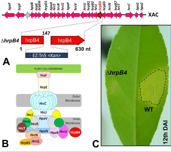
plant hosts. Previous studies have shown that mutations in genes that are part of or regulate this apparatus completely prevent or substantially reduce the classical phenotypes of the disease (Büttner et al., 2002). Random mutations inXac were generated previously by our group using a EZ-Tn5 <Kan>transposition cassette, which were used to identify several genes that might be associated with its virulence (Laia et al., 2009). Of these, one mutation in particular resulted in non-pathogenicity; the mutation was inhrpB4(1hrpB4-XAC0410), located within thehrp(Hypersensitive Response and Pathogenicity) gene cluster and coding for the T3SS apparatus (Fig. 1A). HrpB4 is the inner membrane protein that associates with T3SS, thus a structural protein that plays a role on T3SS apparatus assembly (Fig. 1B). Interestingly, in addition to being unable to induce virulence (Fig. 1C),1hrpB4also fails to develop within the plant despite growing normally in an energy-rich culture medium. At the same time, because1hrpB4was unable to growin planta, it would not be possible to obtain the secreted proteins in infective conditions in concentrations capable of being analysed by proteomics.
To characterize the secretome ofXac, we cultured the wild-type strain and the1hrpB4 mutant in two different culture media: nutrient broth (NB) and the defined medium, XAM1 (Wengelnik & Van den Ackerveken, 1996). The latter medium was chosen based on previous results showing that XAM1 medium simulates conditions experienced by Xac during plant infections (Facincani et al., 2014). The mutant strain, in which the T3SS is non-functional, was chosen to test the dependence of the secretome on the T3SS. In addition, since several authors have previously reported a relationship between xanthan gum production and the pathogenicity of the microorganism (Katzen et al., 1998), assays to determine the production of xanthan gum and the dry weight of cells were performed at each condition.
MATERIALS AND METHODS
Growth conditions
Stocks of wild-typeXac and the1hrpB4mutant strain were stored in phosphate-buffered saline at room temperature. Both were cultured in Nutrient Agar plates prior to the preparation of the pre-inoculum. All maintenance cultures were maintained at 28 ◦C,
and kanamycin was added at 100µg/mL for solid culture media and 50µg/mL for liquid
culture media. Wild-type Xac and1hrpB4 were cultured in a 15-mL liquid Nutrient Broth (NB) pre-inoculum, maintained at 200 rpm and 28 ◦C, until the cultures reached
OD600 nm=1.0 (approximately 108CFU/mL). Cultures were centrifuged at 3,100×g
for 15 min and the supernatant was discarded. The pellet was resuspended in water and added to the culture medium. Both wild-typeXacand1hrpB4were grown in 500 mL NB culture medium (200 rpm, 28 ◦C) until the culture reached an OD
600 nmof at most 1.4
(approximately 109CFU/mL). At OD600 nmless than 1.4, growth is still logarithmic, which
prevents the release of proteins into the culture medium due to cell lysis, thus enabling us to accurately profile the secretome (Alexander Watt et al., 2005). XAM1 media (7.5 mM (NH4)2SO4, 33 mM KH2PO4, 60 mM K2HPO4, 1.7 mM sodium citrate
Figure 1 (A) Cluster ofhrpgenes in theXacgenome, highlighting the mutation of thehrpB4gene in-duced by insertion of the transposition cassette EZ-Tn5 <Kan >. The insertion was made at position 147 in the gene. (B) Structural composition of the corresponding apparatus to the type III secretion system encoded byhrpgenes. The HrpB4 protein is highlighted in red. (C) Phenotype virulence alteration of the mutant1hrpB4(complete absence of symptoms) compared to the wild-type strain (WT) after 12 days of infection inCitrus sinensisplants.
Casamino acids in a final volume of 1 L) was used to induce virulence (Wengelnik & Van den Ackerveken, 1996). Each bacterium was grown in 2 L (4× 500 mL) of this medium, to which 1 mg/mL of Bovine Serum Albumin (BSA) was added. Bacteria were grown at 28 ◦C with shaking at 200 rpm until the culture reached an OD
600of at most 0.57, wherein
growth was still logarithmic.
Wild-typeXacor1hrpB4were grown for 16 or 23 h in NB liquid culture media until it reached an OD600of 1.0 or 1.4, respectively. Both strains were also grown in XAM1 liquid
media for 24 h, which is equivalent in time to the early stages of infectionin vivo. Wild-type bacteria reached an OD600 of 0.57, and the mutant achieved an OD600 of 0.24. Higher
Several replicates were used for each condition to obtain the total pool of proteins used for this study.
Centrifugation and filtration of samples
Cultures were centrifuged in two 60 min steps at 4 ◦C. In the first step, the samples were
centrifuged at 3,100×g, the bacterial pellet was discarded and the supernatant containing secreted proteins was transferred to clean tubes. In the second step, the (first) supernatant was centrifuged at 11,000 ×g and again the supernatant was recovered. For strains cultivated in NB liquid medium, the supernatant was recovered and then filtered using 0.2-µm (MilliporeTM) nitrocellulose membranes shortly after the second centrifugation.
For bacteria cultured in XAM1 medium, the supernatant could not be filtered due to the elevated production of xanthan gum, which caused changes in the membrane filtration profile. All samples were frozen using liquid nitrogen and lyophilized to complete dryness. Each dried sample corresponding to 500 mL of the supernatant was resuspended in 50 mL of double-distilled autoclaved water (ddH2O). To precipitate xanthan gum
produced by cultures grown in XAM1 medium, absolute ethyl alcohol (1:3) was added to the resuspended sample (in ddH2O), and the sample was centrifuged at 3,000×gfor 5 min
at 30 ◦C. These samples were then filtered through 0.2
µm (MilliporeTM) nitrocellulose
membranes and again lyophilized and resuspended in 50 mL of ddH2O.
Precipitation of proteins and SDS-PAGE electrophoresis
Proteins in the filtered supernatant were precipitated with trichloroacetic acid (TCA), with modifications (Hirose et al., 2000). Briefly, TCA was added to the resuspended sample to a final concentration of 10%, and each sample was vigorously mixed and stored at 4 ◦C
for 16 h. Samples were centrifuged at 20,000×gfor 20 min at 4 ◦C. The supernatant was
discarded and the precipitate washed three times with cold acetone to remove residual TCA. Precipitated proteins were then lyophilized to complete dryness and dissolved in 25 mM NH4HCO3buffer, 500 mM urea, pH 8.0. All samples were quantified and normalized. The
proteins were then hydrolysed in solution.
Trypsinization of samples and mass spectrometry
Proteins were reduced by adding 1µg of DTT per 50µg of protein and incubating for 1
h at 37 ◦C. Proteins were then alkylated by adding 5
µg of iodoacetamide per 50µg of
protein and incubating for 1 h at room temperature in the dark. Proteins were trypsinized by adding 20µg of Promega Sequencing Grade Modified Trypsin (Madison, USA) (1:50)
and incubating for 20 h at 37 ◦C. The hydrolysis reaction was terminated by adding
2 µL of formic acid. The samples were then loaded into a WatersR nanoACQUITY
UPLCR capillary chromatography system (Waters, Milford, MA). The digested proteins were desalinized using a Waters Opti-Pak C18 trap column. The volume of the injected sample was 10µL, and liquid chromatography was performed on a reverse-phase C18
column Ease 150 mm×2.1 mm (Waters, Milford, MA). Proteins were eluted with a flow rate of 0.3µL/min using a linear gradient ranging from 5% to 50% acetonitrile containing
The capillary liquid chromatography system (nLC) was coupled to a mass spectrometer with an electrospray ionization (ESI) source and quadrupole/time-of-flight in series analysers (Q-TOF Micro; Waters, Milford, MA) (UPLC-MS/MS), which permitted the direct analysis of eluted peptides in the acetonitrile gradient in Q-TOF. For ESI, a voltage of 3,000 V and a temperature of 80 ◦C was applied in the capillary. A MassLynx data
system (Version 4.1, Waters) was used to control the instrument and acquire data, and the experiments were performed by scanning mass/charge (m/z) ratios between 200 and 2,000 using a first scan time of 1 s, which was applied during the entire chromatographic process. The averages of the mass spectra corresponding to each sign of the total ion current chromatogram (TIC) were calculated, allowing an accurate determination of the molecular mass. Exact mass values were obtained using a LockSprayTM source (Waters, Milford,
MA). This reference mass was used to correct the mass of the analyte (sample) throughout the study. Phosphoric acid was used as the reference in this study, which has an m/z ratio of 588.8692.
The acquisition of data dependent on MS/MS were performed on precursors with charge states 2 or 3 over a mass range of m/z 50–2,000 and at an interval below 2 m/z. At most, 3 ions were selected for MS/MS analysis of a single MS. The masses of Na+ and K+were
automatically excluded. The collision induced dissociation (CID) based MS/MS spectra were obtained by using argon as the collision gas at a pressure of 13 PSI and a collision voltage that ranged from 18 to 45 V, depending on the mass of the precursor. The scanning ratio was 1 s.
Processing of the generated spectra and signal peptide prediction All data were processed with ProteinLynx Global Server (version 2.0, Waters), which automatically corrected values of m/z spectrum of MS and MS/MS according to the mass of the Lockspray reference ion. Proteins were identified using theXacdatabase from NCBI (BioProject PRJNA297) and UniProt databases, and the MASCOT program (Version 2.2.1; Brazilian Synchrotron Light Laboratory). The values of monoisotopic masses of the MS/MS spectra (MS/MS Ion Search) were used, which considered cysteine carbamidomethylation as a fixed modification and oxidation of methionine as a variable modification. In the hydrolysis by trypsin, the possible loss of one cleavage site was considered and the tolerance of masses of peptides and fragments was set to±0.05 Da.
The amino acid sequences of identified proteins were submitted to analysis by PrediSi (Prediction of Signal peptides,www.predisi.de) (Hiller et al., 2004). Identified proteins that lacked signal peptides according to PrediSi analysis were submitted to SecretomeP 2.0 for further analysis (Bendtsen et al., 2005). Comparisons between proteins from different organisms were performed using the bioinformatics tool BLASTP.
Identification of candidate PIP-boxes in the regulatory regions of genes
Several genes regulated by HrpX possess a consensus nucleotide sequence, TTCGC-N15
gene is regulated by HrpX and encodes an effector protein of the T3SS pathway. PIP-box analysis was performed following methods described by Da Silva (Da Silva et al., 2002).
Candidate PIP-box sequences and their genomic locations were identified by searching the completeXac genome for sequences that matched the following PIP-box sequence patterns: 1. Forward orientation (uncomplemented)—TTCGN-N15-TTCGN or
TTCGN-N16-TTCGN or 2. Reverse orientation (complemented)—NCGAA-N15-NCGAA or
NCGAA-N16-NCGAA. Each candidate PIP-box sequence was further analysed to determine
whether it was part of a promoter, which should be between 10 and 1,000 nucleotide bases long and precede the start codon of a gene.
Xanthan gum production
For xanthan gum production, three (triplicate) 250-mL Erlenmeyer flasks containing 100 mL of Gum medium (25 g/L glucose, 3 g/L yeast extract, 2 g/L K2HPO4, 0,1 g/L MgSO4.7H2O, pH 7.0 with 4 M HCl) were inoculated with 2.5 mL of each bacterial strain
grown in NB liquid medium (OD600of 0.3) and incubated at 29 ◦C for 96 h in a rotary
shaker at 178 rpm (Shu & Yang, 1990).
After 96 h, cultures were centrifuged at 9,666×gfor 40 min. The bacterial pellets were stored at−20 ◦C and the supernatants were transferred to 500-mL beakers. Xanthan gum
was recovered from the supernatants by isopropanol precipitation. KCl (4 g) was added to each beaker, followed by stirring at room temperature for 15 min. Two volumes of cold isopropyl alcohol were added and the xanthan gum from each beaker was removed and placed in plastic containers, which were previously weighed, with the aid of a glass rod and a sieve. After 48 h at 37 ◦C, the containers, plus sample, were weighed again and the
amount of xanthan gum calculated. The mass of the bacterial pellet from each culture was also measured. For this, each pellet was resuspended with 1 mL MilliQ autoclaved water and transferred to a pre-weighed beaker and weighed again after 24 h at 70 ◦C. Three
replicates were used for each condition and Statistical Analysis was performed (Unpaired Studentt test).
RESULTS AND DISCUSSION
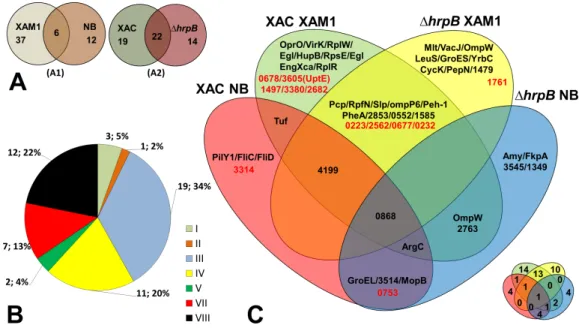
Figure 2 Comparative analysis ofXacsecretome.(A) Comparison highlighting 55 total detected pro-teins, correlating culture mediums (XAM1×NB—A1) and strains (Xacand1hrpB4—A2) analysed. The two circles represent the total proteins detected. (B) Categorization of annotated protein functions, adapted fromDa Silva et al. (2002): I, Intermediary metabolism; II, Biosynthesis of small molecules; III, Metabolism of macromolecules; IV, Cell structure; V, Cellular processes; VI, Mobile genetic elements, VII, Pathogenicity; virulence, and adaptation, VIII, Hypothetical/Conserved hypothetical genes; XI, Without a defined function. (C) Venn Diagram highlighting each of the proteins detected under the four different conditions tested. Proteins annotated as hypothetical are highlighted in red. The same Venn diagram is shown on a smaller scale but with only the numbers of the proteins detected in each condition.
The Venn diagram shown inFig. 2Cunderscores the differences and overlap in proteins secreted under the different conditions tested. Analysis of proteins according to their MASCOT annotated functions revealed that seven were associated with adaptation and virulence (category VII), and 12 were hypothetical proteins (category VIII) (Fig. 2B). The following sections describe in details these main findings.
Proteins secreted by both strains grown in XAM1 medium
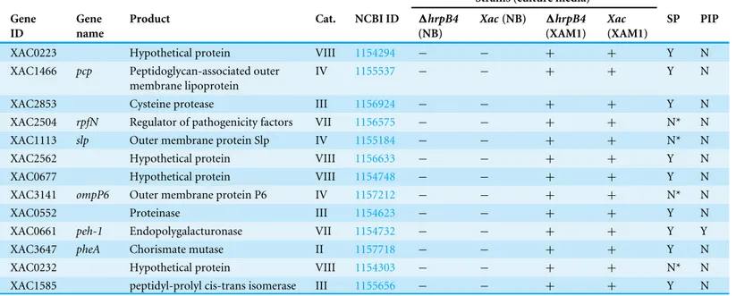
Table 1 Proteins expressed/detected in wild type and1hrpB4strains only in infectious conditions.
Strains (culture media)
Gene ID
Gene name
Product Cat. NCBI ID 1hrpB4
(NB)
Xac(NB) 1hrpB4
(XAM1)
Xac
(XAM1)
SP PIP
XAC0223 Hypothetical protein VIII 1154294 − − + + Y N
XAC1466 pcp Peptidoglycan-associated outer membrane lipoprotein
IV 1155537 − − + + Y N
XAC2853 Cysteine protease III 1156924 − − + + Y N
XAC2504 rpfN Regulator of pathogenicity factors VII 1156575 − − + + N* N
XAC1113 slp Outer membrane protein Slp IV 1155184 − − + + N* N
XAC2562 Hypothetical protein VIII 1156633 − − + + Y N
XAC0677 Hypothetical protein VIII 1154748 − − + + Y N
XAC3141 ompP6 Outer membrane protein P6 IV 1157212 − − + + N* N
XAC0552 Proteinase III 1154623 − − + + Y N
XAC0661 peh-1 Endopolygalacturonase VII 1154732 − − + + Y Y
XAC3647 pheA Chorismate mutase II 1157718 − − + + Y N
XAC0232 Hypothetical protein VIII 1154303 − − + + N* N
XAC1585 peptidyl-prolyl cis-trans isomerase III 1155656 − − + + Y N
Notes.
Gene_ID and Cat (primary category), according toDa Silva et al. (2002); Product, according to Kegg (Ogata et al., 1999); SP, Signal peptide; Y, Yes; N, No; N*, Secreted by non-classical pathways.
other secretory systems, such as Sec, Tat, T2SS, and T5SS. The presence of signal peptide sequences in at least 69% of these proteins reinforces their targeted secretion.
Proteins with annotated functions described bellow appear to have a fundamental role in either the virulence of the genusXanthomonasor their adaptation to the plant. The Pcp protein, encoded by the gene XAC1466, is a peptidoglycan-associated outer membrane lipoprotein homologous to the SlyB protein inXanthomonas campestris. This protein is directly regulated by a two-component system in most bacteria (PhoPQ), which is present in many bacterial pathogens of both animals and vegetables (Perez et al., 2009). This two-component system, which is encoded by the genes XAC4023 and XAC4022, is also expressed inXac and has been shown to be active during the early stages of infection (Moreira et al., 2015). Proteins homologous to Pcp are also essential for maintaining the integrity of the cell envelope inPseudomonas putida(Rodríguez-Herva, Ramos-González & Ramos, 1996), and more recently, were associated with OMV formation inP. aeruginosa (Wessel et al., 2013).
shown to be expressed inXacwhen grown in XAM1 medium, and remains active even in the1hrpB4strain (Laia et al., 2009), which is consistent with its secretion by both strains in our study. Mutants ofXanthomonas campestrisin which the gene encoding the PghAxc protein (homologous to Peh-1 inXac) was inactivated showed attenuated virulence when sprayed ontoArabidopsishosts, demonstrating the importance of this protein in the early stages of infection (Wang, Rong & He, 2008).
A protein with cysteine protease functions, encoded by the XAC2853 gene, was also secreted by both strains grown in XAM1 medium. This protein is homologous to CysP2 inX. oryzaepv.oryzae, has a defective PIP-box sequence upstream of the gene in theXac genome, is secreted by T2SS, and is regulated by HrpG/X (Furutani et al., 2004;Yamazaki, Hirata & Tsuyumu, 2008). Proteins with this function are considered important virulence factors because they hydrolyse peptides in the host cell. Inoculation of a citrus hostin vivo with a mutant deficient in a cysteine protease, encoded by the gene XAC2853, resulted in less virulence (Soares-Costa et al., 2012). In studies involvingPseudomonas, the virulence of the pathogen was directly correlated with the secretion of the AvrRpt2 protein, which has cysteine protease functions, by T3SS (Axtell et al., 2003;Cui et al., 2013).
RpfN, which is characterized as a virulence factor regulator and encoded by the gene XAC2504, has an OprB domain (carbohydrate-selective porin). The OprB domain is a specific porin channel for glucose transport, but can also mediate the transport of other monosaccharides to the inner membrane (Wylie & Worobec, 1995).Pseudomonas aeruginosamutants defective for this protein demonstrated a reduced ability to utilize various types of monosaccharides, indicating its importance in the import of different sugars into the cell (Nikaido, 2003). According to SecretomeP 2.0 analysis, this protein is not predicted to be secreted by the Sec pathway but rather by a non-classical route. The role of this protein in the pathogenicity ofXac is not well understood, but according to Dow and colleagues(2000), endopolygalacturonase levels were increased inXanthomonas campestrismutants, in which this gene was inactivated. Moreover, Moreira and colleagues suggested that the absence of therpfN gene and mutations in the PTS system associated with the internalization of fructose could explain the fastidious growth ofX. fuscansB strain, which causes canker B in citrus plants (Moreira et al., 2010).
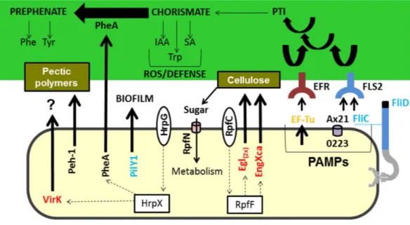
Figure 3 Model highlighting proteins related to virulence characterized in comparative proteomics.
Three of these proteins (A×21, FliC, and Ef-Tu) are characterized as PAMPs capable of inducing a PTI re-sponse, which can consequently lead to the increased synthesis of chorismate. Chorismate is a precursor in the synthesis of IAA, Trp, and SA, which are involved in the ROS response and induction of plant de-fence. However, the secretion of PheA may reduce plant defence responses by shifting the metabolism of chorismate to prephenate synthesis. Also highlighted is the Peh-1 protein, which is an endopolygalactur-onase regulated by HrpX and cellulase (Egl), and endo-1,4-beta-glucanase (EngXca), which is regulated by RpfF. Endopolygalacturonase and cellulase degrade pectin polymers and cellulose in the plant cell wall, re-spectively. Phe, phenylalanine; Tyr, tyrosine; Trp, tryptophan; IAA, indole-3-acetic acid; SA, salicylic acid; PTI, (PAMP) -triggered immunity; PAMPs, pathogen-associated molecular pattern; ROS, reactive oxygen species; EF-Tu, elongation factor Tu, EFR - EF-Tu receiver; A×21, Ax21-triggered immunity; FliC, flag-ellin; FLS2, flagellin sensitive receiver 2; PheA, chorismate mutase; Peh-1, endopolygalacturonase; Egl and EngXca, cellulase. HrpG/HrpX and RpfC/RpfF, two component systems (sensory and regulatory proteins, respectively); Black, detected inXacand1hrpB4grown in XAM1; Red, detected exclusively inXacgrown in XAM1; Orange, detected inXacgrown in BOTH media (NB and XAM1); Blue, detected exclusively in
Xacgrown in NB. Dotted arrows indicate regulation.
Peptidyl-prolyl cis-trans isomerase (PPIase) is a protein encoded by the gene XAC1585. Similar to Slp, Peh-1, and PheA, this protein was identified in the extracellular matrix of both strains when grown in induction medium, confirming data described byYamazaki, Hirata & Tsuyumu (2008). However, a paralogous protein was also secreted only by the mutant 1hrpB4grown in NB medium (Table S1). The PPIases play a pivotal role in catalysing the proper folding of many prokaryotic and eukaryotic proteins that are implicated in a variety of biological functions ranging from cell cycle regulation to bacterial infection (Pissavin & Hugouvieux-Cotte-Pattat, 1997;Zang et al., 2007). In
Pseudomonas syringaeandXanthomonas, proteins orthologous to PPIase were identified in the secretome (Kaffarnik et al., 2009). InXylella fastidiosa, which causes CVC, this protein is part of the composition of mature biofilms (Silva et al., 2011), and is differentially expressed whenXylellais subjected to thermal stress (Da Silva Neto et al., 2007).
The last protein with an annotated function secreted by both strains only when grown in XAM1 medium is a proteinase (XAC0552). The gene encoding this protein has been identified only in species that cause citrus canker; its genomic organization has been maintained in these species. In addition to containing a peptidase_S8 domain in its carboxylterminal region, it contains a Pro_kuma_activ domain (Prokumamolisin -PF09286.6) in its amino-terminal region, whose cleavage results in the activation of the remaining peptide (Comellas-Bigler et al., 2004). Although this domain has not been examined in the context of bacterial pathogen proteins, in the phytopathogenic fungi Mycosphaerella graminicola, expansion of the number of peptidases containing this domain may be associated with enhanced pathogenesis of wheat hosts (Goodwin et al., 2011).
Hypothetical proteins identified in this condition are encoded by the genes: XAC0223, XAC2562, XAC0677, and XAC0232. Of these, a protein worth noting is encoded by the XAC0223 gene (Fig. 3). This protein contains domains associated with the external membrane and a porin domain (Park & Ronald, 2012;Bahar et al., 2014). Orthologues of this gene inX. campestrispv.raphani 756CandX. oryzaepv.oryzicolahave been reported to be activators of XA21-mediated immunity. Ax21 is secreted by a type I secretion system and in association with outer membrane vesicles (Bahar et al., 2014), and can act in a quorum-sensing system, motility, biofilm formation and virulence (Han et al., 2011). In X. oryzae pv.oryzicola, Ax21 was identified in the secretome, and deletion of the Ax21 gene resulted in reduced biofilm formation and extracellular polysaccharide production (Qian et al., 2013). Although classically the rice XA21 receptor directly recognizes Ax21 (Song et al., 1995), recent data have shown that other receptors, such as by Fls2, which is associated with a response in the presence of flagellin in Arabidopsis, can also recognize Ax21 secreted byXanthomonas(Danna et al., 2011). This assumes that there is a dynamic between pathogens and microbe-associated molecular patterns in plants.
Table 2 Proteins expressed/detected only in wild type strain during infectious conditions.
Strains (culture media)
Gene ID
Gene name
Product Cat. NCBI ID 1hrpB4
(NB)
Xac
(NB)
1hrpB4
(XAM1)
Xac
(XAM1)
SP PIP
XAC3472 oprO Polyphosphate-selective porin O IV 1157543 − − − + Y N
XAC0678 Hypothetical protein VIII 1154749 − − − + Y Y
XAC3605 uptE Hypothetical protein IV 1157676 − − − + Y N
XAC0435 virK VirK protein VII 1154506 − − − + Y N
XAC0974 rplW 50S ribosomal protein L23 III 1155045 − − − + N* N
XAC1497 Hypothetical protein VIII 1155568 − − − + Y N
XAC0029 egl Cellulase VII 1154100 − − − + Y N
XAC1081 hupB Histone-like protein III 1155152 − − − + N* N
XAC0989 rpsE 30S ribosomal protein S5 III 1155060 − − − + N N
XAC3380 Hypothetical protein VIII 1157451 − − − + Y N
XAC0028 egl Cellulase VII 1154100 − − − + Y N
XAC2682 Hypothetical protein VIII 1156753 − − − + Y N
XAC0612 engXCA Cellulase VII 1154683 − − − + Y N
XAC0988 rplR 50S ribosomal protein L18 III 1155059 − − − + N N
Notes.
Gene_ID and Cat (primary category), according toDa Silva et al. (2002); Product, according to Kegg (Ogata et al., 1999); SP, Signal peptide; Y, Yes; N, No; N*, Secreted by non-classical pathways.
Proteins secreted only by the wild-type strain grown in XAM1 medium The data so far indicate that the production and secretion of important proteins associated with virulence and adaptation still occur in the1hrpB4mutant. However, the impact of the mutation to the hrpB4gene, which is associated with a loss of function of the T3SS system, is revealed by the identification of 14 unique proteins secreted only by wild-typeXac when cultured in XAM1 medium. Of these, nine had an annotated function (OprO, VirK, ribosomal proteins RplW, RplR and RpsE, HupB and cellulase Egl/Egl, EngXca), whereas the other five were hypothetical proteins (XAC0678, XAC3605, XAC1497, XAC3380 and XAC2682). Ten of the total amount of these proteins have a signal peptide sequence and only one hypothetical protein has a PIP-box sequence upstream of the start of the gene (Table 2). Although lacking annotated functions, all of these hypothetical proteins have a signal peptide sequence, suggesting that their secretion is critical for certain biological processes. Among the proteins with annotated functions, five are worth discussing further.
VirK, encoded by the gene XAC0435, is another protein secreted exclusively by the wild-type strain when cultured in XAM1 medium. This protein has a signal peptide for its secretion through the Sec pathway into the periplasmic space. In E. coli, VirK was characterized as a periplasmic protein required for the efficient secretion of plasmid-encoded toxins (Tapia-Pastrana et al., 2012). An orthologous gene found inRalstonia solanacearum is regulated by HrpG. Microarray studies of the Xac transcriptome demonstrated that the expression of VirK was increased by more than two-fold when grown in XVM2 medium compared to when grown in non-inducing energy-rich culture medium (Astua-monge et al., 2005). The same study reported that a number of genes encoding secreted proteins were robustly expressed by XVM2 medium. Among the enzymes involved in cell wall degradation is cellulase (Egl), which is encoded by the genes XAC0029 and XAC0028. Both proteins were also secreted only by wild-typeXacin XAM1 medium; this was also true for the cellulase, EngXca. These proteins are predicted to be transported via the Sec pathway to the periplasm, and then secreted into the extracellular region by T2SS. Their secretion only by wild-type bacterium indicates that inactivation of T3SS in the mutant could suppress T2SS-secreted enzymes that degrade the cell wall of the host. Furthermore, EngXca has been specifically reported to be controlled byrpfF inXanthomonas campestris. Poplawsky and colleagues(1998)and Siciliano and colleagues
(2006)reported thatXac mutants, in which therpfF andrpfC genes were inactivated, exhibited significantly decreased production of cell wall degrading enzymes and xanthan gum.
During the preparation of samples, the supernatant of the 1hrpB4strain cultured in XAM1 medium could be easily filtered through a 0.2 µm membrane, whereas the
supernatant of the wild-type strain cultured in XAM1 medium could not be easily filtered. This is likely because the excess production of xanthan gum by wild-type bacteria obstructed the pores of the membrane. According to our studies, wild-typeXac is able to produce nearly 10 times more xanthan gum than the pathogenic deficient mutant1hrpB4(mg gum/mg bacteria). Whereas wild-typeXacproduces 46.21± 6.56A mg gum/mg bacteria, 1hrpB4produces only 5.21±1.38B mg gum/mg bacteria (p value=0.0004). These data underscore the importance of these genes to the formation of biofilm, as well as the importance of the hrpB4 gene in maintaining the activities of the T3SS and T2SS secretory systems.
The XAC0678 gene encodes a hypothetical protein but by sequence homology, it is predicted to encode a lipoprotein membrane protein. This hypothetical protein has a consensus PIP-box sequence upstream of the start of the gene, which indicates its probable regulation by HrpX. Furthermore, this protein is predicted to have a signal peptide for the Sec pathway, which could instruct its export into the periplasmic space.
Proteins secreted only by the mutant 1hrpB4grown in XAM1 medium
Table 3 Proteins expressed/detected only in1hrpB4strain during infectious conditions.
Strains (culture media)
Gene ID
Gene name
Product Cat. NCBI ID 1hrpB4
(NB)
Xac
(NB)
1hrpB4
(XAM1)
Xac
(XAM1)
SP PIP
XACb0007 mlt Lytic murein transglycosylase IV 1158494 − − + − Y N
XAC4344 vacJ Lipoprotein III 1158415 − − + − Y N
XAC3664 ompW Outer membrane protein IV 1157735 − − + − Y N
XAC1761 Hypothetical protein VIII 1155832 − − + − Y N
XAC2781 leuS Leucyl-tRNA synthetase III 1156852 − − + − N N
XAC0541 groES Co-chaperonin GroES III 1154612 − − + − N* N
XAC1479 OmpA family protein IV 1155550 − − + − Y N
XAC4342 yrbC Toluene tolerance protein VII 1158413 − − + − Y N
XAC2328 cycK C-type cytochrome biogenesis membrane protein
I 1156399 − − + − Y N
XAC0645 pepN Aminopeptidase III 1154716 − − + − N* N
Notes.
Gene_ID and Cat (primary category), according toDa Silva et al. (2002); Product, according to Kegg (Ogata et al., 1999); SP, Signal peptide; Y, Yes; N, No; N*, Secreted by non-classical pathways.
peptide sequence, and a PIP-box sequence was not present upstream of any of these genes (Table 3).
Among these proteins, one worth noting is Mlt, which is a lytic murein transglycosylase. Xac has two copies of themlt gene, XAC3225 and XACb0007, which are located on the chromosome and on the pXAC64 plasmid, respectively. The chromosomal copy of the gene is flanked by genes that encode the T3SS, XopE3 and XopAI effector proteins and the plasmid copy is flanked by the gene encoding XopE2 (Moreira et al., 2010). Mlt is highly similar to the hopAJ1 protein ofPseudomonas syringae, which was previously annotated as a T3SS helper protein (Oh et al., 2007). Mutations in the chromosomal copy reduced the ability of the mutant to cause canker (Laia et al., 2009).
The proteins encoded by the genes XAC3664 and XAC1479 are predicted to be outer membrane proteins. Although previously annotated as a hypothetical protein, the protein encoded by the gene, XAC1761, is predicted to be a lipoprotein based on its homology with sequences from other organisms.
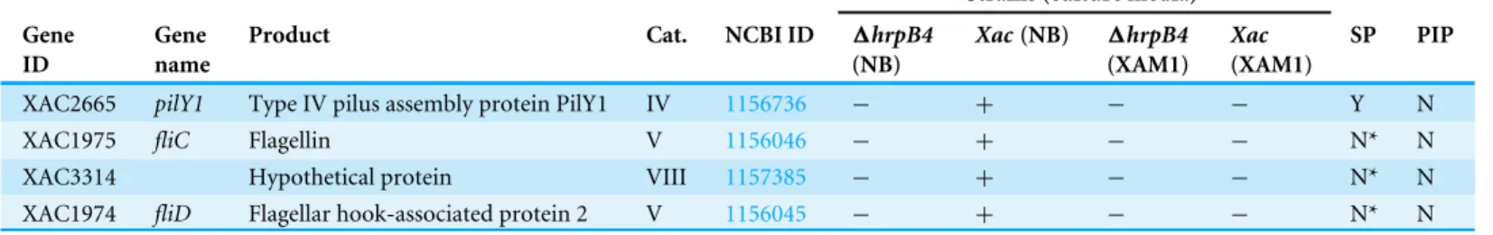
Table 4 Proteins expressed/detected only in wild type strain during non-infectious conditions.
Strains (culture media)
Gene ID
Gene name
Product Cat. NCBI ID 1hrpB4
(NB)
Xac(NB) 1hrpB4
(XAM1)
Xac
(XAM1)
SP PIP
XAC2665 pilY1 Type IV pilus assembly protein PilY1 IV 1156736 − + − − Y N
XAC1975 fliC Flagellin V 1156046 − + − − N* N
XAC3314 Hypothetical protein VIII 1157385 − + − − N* N
XAC1974 fliD Flagellar hook-associated protein 2 V 1156045 − + − − N* N
Notes.
Gene_ID and Cat (primary category), according toDa Silva et al. (2002); Product, according to Kegg (Ogata et al., 1999); SP, Signal peptide; Y, Yes; N, No; N*, Secreted by non-classical pathways.
(Kuchma et al., 2010). InXylella fastidiosa,a mutation in thepilY1gene led to a reduction, but not a complete loss, of type IV pilus apparatus assembly and twitching motility (Li et al., 2007). In Xoo,pilY1, in combination with other genes involved in adhesion and biofilm formation, was upregulated six days after infection in the plant (Soto-Suárez et al., 2010).
FliC and FliD are protein subunits that form the structure of flagella. FliC, also known as flagellin, forms the flagellar filament itself, and is therefore abundantly synthesized by flagellated bacteria. FliC is directly associated with the innate immune response of plants, since it has the ability to bind to specific receptors, such as Toll-like receptor 5 (TLR5) (Hayashi et al., 2001). More recently, part of the N-terminal portion of this protein, corresponding to 22 amino acids (flg22) and conserved across a wide range of pathogens, was shown to activate plant defence mechanisms (Navarro et al., 2004). This 22-amino acid protein fragment is recognized by specific receptors such as by the kinase FLS2, which in turn activate the MAPK signalling cascade (mitogen-activated protein kinase), which is capable of regulating the expression of over 900 genes in theArabidopsis thalianagenome (Asai et al., 2002).
Protein secreted only by the wild-type strain in both growth media The only protein detected exclusively in the secretome of the wild-type strain when grown in either growth media was the EF-Tu protein encoded by the gene XAC0957 (Table S1).
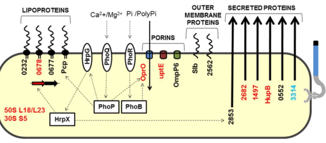
Figure 4 Model highlighting proteins related to adaptation in comparative proteomics.These proteins were grouped into five groups: lipoproteins, porins, outer membrane proteins, secreted proteins, and ri-bosomal proteins. HrpG/HrpX, PhoQ/PhoP and PhoR/PhoB—two component systems (sensory and reg-ulatory proteins, respectively); Black, detected inXacand1hrpB4grown in XAM1; Red, detected exclu-sively inXacgrown in XAM1; Orange, detected inXacgrown in both media (NB and XAM1); Blue, de-tected exclusively inXacgrown in NB. Dotted arrows indicate regulation; Pi/PolyPi, inorganic phosphate and inorganic polyphosphate.
Besides its participation in the production of biofilms, EF-Tu may have another key role in the virulence process, and may represent a potential moonlighting protein (Jeffery, 1999;
Henderson & Martin, 2011).
All other secreted proteins detected in our study
In addition to the proteins described above, 13 other secreted proteins were detected in this study (Table S1). Because these proteins were detected in specific but restricted conditions, we could not exclusively associate them with a specific growth media or specific bacterial strain. The majority (8) of these have enzymatic functions (amylase, proteases, and isomerase). Two membrane-associated proteins (OMPs), and two hypothetical proteins, XAC0753 and XAC0868, were also identified. The latter two proteins have domains with peptidase activity, although XAC0868 has a more representative ricin B lectin domain. Interestingly, the protein encoded by XAC0868 was the only protein detected in all four conditions tested and exists only inXanthomonasstrains that infect citrus plants. A model highlighting proteins related toXacadaptation was devised (Fig. 4).
Pathogenicity ofXaccompared to other plant pathogens
To better understand the importance of secreted proteins in the virulence of plant-specific pathogens of theXanthomonadaceaefamily, a comparative analysis of proteins identified in this study with proteins identified in six other proteomic studies was performed (Table 5). Despite the diversity of model organisms studied and methodologies used, orthologous proteins were identified in these studies.
Table 5 Xacannotated proteins expressed and secreted compared to other studies of Xanthomon-adaceaefamily organisms.
Function/product Mesophyllic Vascular
Xacb Xacc Xacd Xooe Xoof Xccg Xfh
Hydrolitic enzymes
Alpha-amylases X
Aminopeptidases X
Cellulases X X
Endopolygalacturonases X X
Extracellular proteases X
Proteinases X X
Cysteine protease X X X
Serine Proteases X
Motility/chemotaxis
Flagellar associated protein X
Flagellina X X X X
Type IV pilus assembly protein PilY1a X X X
Twiching motility X X
Adaptation/Virulence
Lytic murein transglycosylasea X
RpfNa X X
T3SSe X X
ROS X X
Elongation factor Tua X X X X X X
Membrane associated proteins
TBDRs X X X
Iron/siderophores receptors X X X
ABC transporters X
Lipoproteins X
Outer membrane proteins X X X X X X
Polyphosphate-selective porin Oa X X
Toluene tolerance proteins X
Others
Chorismate mutasea X
Co-chaperonin GroESa X X X
C-type cytochrome biogenesis membrane proteina
X
Histone-like proteina X
Leucyl-tRNA synthetasea X
VirK proteina X X X X
Polyvinyl alcohol dehydrogenasea X
Molecular chaperone GroELa X X X X X
Peptidyl-prolyl cis-trans isomerasea X X X X
Table 5(continued)
Function/product Mesophyllic Vascular
Xacb Xacc Xacd Xooe Xoof Xccg Xfh
Ribosomal proteins X X X X
ATP synthase X X X
Notes.
aUnique proteins. bThis work (reference data). cYamazaki, Hirata & Tsuyumu (2008). dZimaro et al. (2013).
eWang et al. (2013). fGonzález et al. (2012). gSidhu et al. (2008). hSilva et al. (2011).
Zimaro and colleagues compared the proteome of planktonicXac cells andXac biofilm cells, and identified 53 differentially expressed proteins, of which 31 were upregulated in biofilms. Silva and colleagues (2011)identified 456 proteins potentially related to biofilm formation inXylella fastidiosa. Wang and colleagues analysed theXanthomonas oryzae pv.oryzaesecretome grown in culture medium and in plants, and identified 109 unique proteins. Proteins involved in pathogenicity and the induction of host defence were identified only in the secretome of bacteria in contact with the plant (Yamazaki, Hirata & Tsuyumu, 2008). Using the same model study, Gonzalez and colleagues identified 324 different proteins secreted by Xoo in contact with the xylem of the host plant. Of these, 64 were suggested to be associated with the process of virulence (González et al., 2012). Sidhu and colleagues(2008)identified 31 proteins associated with OMV, half of which were associated with the induction of virulence inXanthomonas campestrispv.campestris.
This comparative analysis revealed the correlation of a number of proteins with the life cycle or the infection model of the associated pathogen. For instance,
endopolygalacturonase and proteinases, which seem to be crucial to the virulence process, are induced by mesophilic strains. Proteins associated with motility and chemotaxis have been identified in virtually every proteomics study included in our analysis, which is consistent with the importance of these proteins in the formation of cellular aggregates, biofilms or even for inducing a response in the host plant (Malamud et al., 2011;Dunger et al., 2014). Mlt was detected only in this study, indicating its importance to the induction of virulence inXac. Interestingly, EF-Tu was independently identified in five other studies, including in the Xf strain, whereas GroEL was detected in four other studies, and flagellin (FliC), VirK and peptidyl-prolyl cis-trans isomerase (PPI) were identified in three other studies. It is important to note that FliC is absent in theXylellagenome since it does not have flagellum. The possibility that GroEL, FliC, Ef-Tu and PPI can act as moonlighting proteins in bacteria of the family Xanthomonadaceae is highlighted (Henderson & Martin, 2011).
CONCLUSIONS
identified in total on the secretomes of wild-typeXac and the mutant1hrpBcultured in different growth media. Of these, 37 have a predicted signal peptide for secretion via the general secretory pathway and are potentially related to the pathogenicity and adaptation during infections and plant–pathogen interactions. However, of the 18 proteins lacking a signal peptide, 13 were predicted to be secreted by non-classical routes. This demonstrates that the majority (50) of proteins detected in this study were neither contaminant proteins nor proteins released by cell lysis. Interestingly, the percentage of secreted proteins related to the pathogenicity, virulence and adaptation increased nearly two-fold (from 15 to 29%) when considering only proteins secreted by wild-typeXac bacterium compared to those secreted by its mutant1hrpB4. These results corroborate that thehrpB4gene act as T3SS central player, impacting the emergence of pathogenicity inXanthomonas. For instance, the expression/secretion of several T2SS proteins appears to be regulated by T3SS activity. Our results indicate that this regulation also occurs inXac. The profile of proteins secreted exclusively by wild-typeXacand the increased amount of xanthan gum produced in XAM1 medium suggests that DSFs (diffusible signalling factors) responsible for controlling these factors may be related to the lack of expression of the HrpB4 protein, since the1hrpB4 mutant, which is deficient in the production of this protein, was unable to secrete several of these factors. Further analysis are necessary to confirm that hypothesis.
Overall, the secretome method allows the testing of not only one, but several mutants at a time. This may be a powerful tool to shed light into the biology of Xanthomonas. Indeed, analyses are underway with different mutants generated under different mutagenic methodologies to evaluate others gene targets that might be related to the disease.
ACKNOWLEDGEMENTS
We thank all the members of the Laboratory of Molecular Biology of the Department of Technology (UNESP—Universidade Estadual Paulista—Jaboticabal). We would also like to thank the Brazilian Synchrotron Light Laboratory.
ADDITIONAL INFORMATION AND DECLARATIONS
Funding
This work was supported by grants from CAPES—BIGA (Coordenação de Aperfeiçoamento de Pessoal de Nível Superior) project. RMF was funded by a CAPES post-doctoral fellowship. The funders had no role in study design, data collection and analysis, decision to publish, or preparation of the manuscript.
Grant Disclosures
The following grant information was disclosed by the authors:
CAPES—BIGA (Coordenação de Aperfeiçoamento de Pessoal de Nível Superior). CAPES post-doctoral fellowship.
Competing Interests
Author Contributions
• Rafael M. Ferreira and Leandro M. Moreira conceived and designed the experiments, performed the experiments, analyzed the data, wrote the paper, prepared figures and/or tables, reviewed drafts of the paper.
• Jesus A. Ferro conceived and designed the experiments, analyzed the data, contributed reagents/materials/analysis tools, wrote the paper, reviewed drafts of the paper.
• Marcia R.R. Soares and Julio C.F. de Oliveira conceived and designed the experiments, performed the experiments, analyzed the data, reviewed drafts of the paper.
• Marcelo L. Laia conceived and designed the experiments, analyzed the data, reviewed drafts of the paper.
• Alessandro M. Varani analyzed the data, contributed reagents/materials/analysis tools, wrote the paper, reviewed drafts of the paper.
• Maria Ines T. Ferro analyzed the data, contributed reagents/materials/analysis tools, reviewed drafts of the paper.
Data Availability
The following information was supplied regarding data availability: Zenodo:https://zenodo.org/record/45560#.VrNQg4RSzCc.
Supplemental Information
Supplemental information for this article can be found online athttp://dx.doi.org/10.7717/ peerj.1734#supplemental-information.
REFERENCES
Alexander Watt S, Wilke A, Patschkowski T, Niehaus K. 2005.Comprehensive analysis of the extracellular proteins fromXanthomonas campestrispv.campestrisB100. Proteomics5:153–167DOI 10.1002/pmic.200400905.
Asai T, Tena G, Plotnikova J, Willmann MR, Chiu W-L, Gomez-Gomez L, Boller T, Ausubel FM, Sheen J. 2002.MAP kinase signalling cascade in Arabidopsis innate immunity.Nature415:977–983DOI 10.1038/415977a.
Astua-monge G, Freitas-astua J, Roncoletta J, Carvalho SA, Machado MA, Bacocina G. 2005.Expression profiling of virulence and pathogenicity genes of Xanthomonas axonopodis pv . citri expression profiling of virulence and pathogenicity genes of Xanthomonasaxonopodis pv. citri.Journal of Bacteriology187:1201–1205.
Axtell MJ, Chisholm ST, Dahlbeck D, Staskawicz BJ. 2003.Genetic and molecular evidence that the Pseudomonas syringae type III effector protein AvrRpt2 is a cysteine protease.Molecular Microbiology49:1537–1546
DOI 10.1046/j.1365-2958.2003.03666.x.
Bekal S, Niblack TL, Lambert KN. 2003.A chorismate mutase from the soybean cyst nematode Heterodera glycines shows polymorphisms that correlate with virulence. Molecular Plant-Microbe Interactions16:439–446DOI 10.1094/MPMI.2003.16.5.439.
Bendtsen JD, Kiemer L, Fausbøll A, Brunak S. 2005.Non-classical protein secretion in bacteria.BMC Microbiology5:58DOI 10.1186/1471-2180-5-58.
Büttner D, He SY. 2009.Type III protein secretion in plant pathogenic bacteria.Plant Physiology150:1656–1664DOI 10.1104/pp.109.139089.
Büttner D, Nennstiel D, Klüsener B, Bonas U. 2002.Functional analysis of HrpF , a putative type III translocon protein fromXanthomonas campestrispv. vesicatoria Functional Analysis of HrpF, a putative type III translocon protein from Xan-thomonas campestrispv. vesicatoria.Journal of Bacteriology184:2389–2398.
Caldas TD, El Yaagoubi A, Richarme G. 1998.Chaperone properties of bacterial elongation factor EF-Tu.Journal of Biological Chemistry273:11478–11482
DOI 10.1074/jbc.273.19.11478.
Comellas-Bigler M, Maskos K, Huber R, Oyama H, Oda K, Bode W. 2004.1.2 Å crystal structure of the serine carboxyl proteinase pro-kumamolisin: structure of an intact pro-subtilase.Structure12:1313–1323DOI 10.1016/j.str.2004.04.013.
Cui F, Wu S, Sun W, Coaker G, Kunkel B, He P, Shan L. 2013.The pseudomonas syringae type III effector AvrRpt2 promotes pathogen virulence via stimulating Ara-bidopsis auxin/indole acetic acid protein turnover.Plant Physiology162:1018–1029
DOI 10.1104/pp.113.219659.
Danna CH, Millet YA, Koller T, Han S-W, Bent AF, Ronald PC, Ausubel FM. 2011.The arabidopsis flagellin receptor FLS2 mediates the perception ofXanthomonasAx21 secreted peptides.Proceedings of the National Academy of Sciences of the United States of America108:9286–9291DOI 10.1073/pnas.1106366108.
Da Silva ACR, Ferro JA, Reinach FC, Farah CS, Furlan LR, Quaggio RB, Monteiro-Vitorello CB, Van Sluys MA, Almeida NF, Alves LMC, Do Amaral AM, Bertolini MC, Camargo LEA, Camarotte G, Cannavan F, Cardozo J, Chambergo F, Ciapina LP, Cicarelli RMB, Coutinho LL, Cursino-Santos JR, El-Dorry H, Faria JB, Ferreira AJS, Ferreira RCC, Ferro MIT, Formighieri EF, Franco MC, Greggio CC, Gruber A, Katsuyama AM, Kishi LT, Leite RP, Lemos EGM, Lemos MVF, Locali EC, Machado MA, Madeira AMBN, Martinez-Rossi NM, Martins EC, Meidanis J, Menck CFM, Miyaki CY, Moon DH, Moreira LM, Novo MTM, Okura VK, Oliveira MC, Oliveira VR, Pereira HA, Rossi A, Sena JAD, Silva C, De Souza RF, Spinola LAF, Takita MA, Tamura RE, Teixeira EC, Tezza RID, Trindade dos Santos M, Truffi D, Tsai SM, White FF, Setubal JC, Kitajima JP. 2002.Comparison of the genomes of two Xanthomonaspathogens with differing host specificities.Nature417:459–463
DOI 10.1038/417459a.
Da Silva Neto JF, Koide T, Gomes SL, Marques MV. 2007.The single extracytoplasmic-function sigma factor of Xylella fastidiosa is involved in the heat shock response and presents an unusual regulatory mechanism.Journal of Bacteriology189:551–560
Degrassi G, Devescovi G, Bigirimana J, Venturi V. 2010.Xanthomonas oryzaepv. oryzaeXKK.12 contains an AroQgamma chorismate mutase that is involved in rice virulence.Phytopathology100:262–270 DOI 10.1094/PHYTO-100-3-0262.
Deng W, De Hoog CL, Yu HB, Li Y, Croxen MA, Thomas NA, Puente JL, Foster LJ, Finlay BB. 2010.A comprehensive proteomic analysis of the type III secre-tome of Citrobacter rodentium.Journal of Biological Chemistry285:6790–6800
DOI 10.1074/jbc.M109.086603.
De Souza AA, Takita MA, Coletta-Filho HD, Caldana C, Yanai GM, Muto NH, De Oliveira RC, Nunes LR, Machado MA. 2004.Gene expression profile of the plant pathogen Xylella fastidiosa during biofilm formationin vitro.FEMS Microbiology Letters237:341–353DOI 10.1016/j.femsle.2004.06.055.
Djamei A, Kahmann R. 2012.Ustilago maydis: dissecting the molecular interface between pathogen and plant.PLoS Pathogens8:11–14
DOI 10.1371/journal.ppat.1002955.
Dow JM, Crossman L, Findlay K, He Y-Q, Feng J-X, Tang J-L. 2003.Biofilm dispersal inXanthomonas campestrisis controlled by cell–cell signaling and is required for full virulence to plants.Proceedings of the National Academy of Sciences of the United States of America100:10995–11000DOI 10.1073/pnas.1833360100.
Dow JM, Feng JX, Barber CE, Tang JL, Daniels MJ. 2000.Novel genes involved in the regulation of pathogenicity factor production within the rpf gene cluster of Xanthomonas campestris.Microbiology146(Pt 4):885–891
DOI 10.1099/00221287-146-4-885.
Dunger G, Guzzo CR, Andrade MO, Jones JB, Farah CS. 2014.Xanthomonascitri subsp. citri type IV Pilus is required for twitching motility, biofilm develop-ment, and adherence.Molecular Plant-Microbe Interactions27:1132–1147
DOI 10.1094/MPMI-06-14-0184-R.
Evans FF, Raftery MJ, Egan S, Kjelleberg S. 2007.Profiling the secretome of the marine bacterium Pseudoalteromonas tunicata using amine-specific isobaric tagging (iTRAQ).Journal of Proteome Research6:967–975DOI 10.1021/pr060416x.
Facincani AP, Moreira LM, Soares MR, Ferreira CB, Ferreira RM, Ferro MIT, Ferro JA, Gozzo FC, De Oliveira JCF. 2014.Comparative proteomic analysis reveals that T3SS, Tfp, and xanthan gum are key factors in initial stages of Citrus sinensis infection byXanthomonas citrisubsp. citri.Functional & Integrative Genomics14:205–217
DOI 10.1007/s10142-013-0340-5.
Furutani A, Tsuge S, Ohnishi K, Hikichi Y, Oku T, Tsuno K, Inoue Y, Ochiai H, Kaku H, Kubo Y. 2004.Evidence for HrpXo-dependent expression of type II secretory proteins inXanthomonas oryzaepv.oryzae.Journal of Bacteriology186:1374–1380
DOI 10.1128/JB.186.5.1374-1380.2004.
González JF, Degrassi G, Devescovi G, De Vleesschauwer D, Höfte M, Myers MP, Venturi V. 2012.A proteomic study ofXanthomonas oryzaepv.oryzaein rice xylem sap.Journal of Proteomics75:5911–5919DOI 10.1016/j.jprot.2012.07.019.
J, Bristow J, Van der Burgt A, Canto-Canché B, Churchill ACL, Conde-Ferràez L, Cools HJ, Coutinho PM, Csukai M, Dehal P, De Wit P, Donzelli B, Van de Geest HC, Van Ham RCHJ, Hammond-Kosack KE, Henrissat B, Kilian A, Kobayashi AK, Koopmann E, Kourmpetis Y, Kuzniar A, Lindquist E, Lombard V, Maliepaard C, Martins N, Mehrabi R, Nap JPH, Ponomarenko A, Rudd JJ, Salamov A, Schmutz J, Schouten HJ, Shapiro H, Stergiopoulos I, Torriani SFF, Tu H, De Vries RP, Waal-wijk C, Ware SB, Wiebenga A, Zwiers LH, Oliver RP, Grigoriev IV, Kema GHJ. 2011.Finished genome of the fungal wheat pathogenMycosphaerella graminicola reveals dispensome structure, chromosome plasticity, and stealth pathogenesis.PLoS Genetics7(6):e1002070DOI 10.1371/journal.pgen.1002070.
Guo Y, Figueiredo F, Jones J, Wang N. 2011.HrpG and HrpX play global roles in
coordinating different virulence traits ofXanthomonas axonopodispv. citri.Molecular Plant-Microbe Interactions24:649–661DOI 10.1094/MPMI-09-10-0209.
Han S-W, Sriariyanun M, Lee S-W, Sharma M, Bahar O, Bower Z, Ronald PC. 2011.
Small protein-mediated quorum sensing in a gram-negative bacterium.PLoS ONE
6(12):e29192DOI 10.1371/journal.pone.0029192.
Hayashi F, Smith KD, Ozinsky A, Hawn TR, Yi EC, Goodlett DR, Eng JK, Akira S, Underhill DM, Aderem A. 2001.The innate immune response to bacterial flagellin is mediated by Toll-like receptor 5.Nature410:1099–1103DOI 10.1038/35074106.
Henderson B, Martin A. 2011.Bacterial virulence in the moonlight: multitasking bacterial moonlighting proteins are virulence determinants in infectious disease. Infection and Immunity79:3476–3491DOI 10.1128/IAI.00179-11.
Hiller K, Grote A, Scheer M, Münch R, Jahn D. 2004.PrediSi: Prediction of sig-nal peptides and their cleavage positions.Nucleic Acids Research32:375–379
DOI 10.1093/nar/gkh378.
Hirose I, Sano K, Shioda I, Kumano M, Nakamura K, Yamane K. 2000.Proteome analysis of Bacillus subtilis extracellular proteins: A two-dimensional protein electrophoretic study.Microbiology146:65–75DOI 10.1099/00221287-146-1-65.
Jeffery CJ. 1999.Moonlighting proteins.Trends in Biochemical Sciences24:8–11
DOI 10.1016/S0968-0004(98)01335-8.
Kaffarnik FAR, Jones AME, Rathjen JP, Peck SC. 2009.Effector proteins of the bacterial pathogen Pseudomonas syringae alter the extracellular proteome of the host plant, Arabidopsis thaliana.Molecular & cellular proteomics8:145–156
DOI 10.1074/mcp.M800043-MCP200.
Kamoun S, Young M, Glasscock C, Tyler BM. 1993.Extracellular protein elicitors from phytophthora: host-specificity and induction of resistance to bacterial and fungal phytopathogens.Molecular Plant-Microbe Interactions6:15–25
DOI 10.1094/MPMI-6-015.
Katzen F, Ferreiro DU, Oddo CG, Ielmini MV, Becker A, Pühler A, Ielpi L. 1998. Xan-thomonas campestrispv. campestris gum mutants: Effects on xanthan biosynthesis and plant virulence.Journal of Bacteriology180:1607–1617.
Kazemi-Pour N, Condemine G, Hugouvieux-Cotte-Pattat N. 2004.The secretome of the plant pathogenic bacterium Erwinia chrysanthemi.Proteomics4:3177–3186
DOI 10.1002/pmic.200300814.
Kuchma SL, Ballok AE, Merritt JH, Hammond JH, Lu W, Rabinowitz JD, O& apos;Toole G a. 2010.Cyclic-di-GMP-mediated repression of swarming motility by Pseudomonas aeruginosa: The pilY1 gene and its impact on surface-associated behaviors.Journal of Bacteriology192:2950–2964DOI 10.1128/JB.01642-09.
Laia ML, Moreira LM, Dezajacomo J, Brigati JB, Ferreira CB, Ferro MIT, Silva ACR, Ferro JA, Oliveira JCF. 2009.New genes ofXanthomonascitri subsp. citri involved in pathogenesis and adaptation revealed by a transposon-based mutant library.BMC Microbiology9:12DOI 10.1186/1471-2180-9-12.
Li Y, Hao G, Galvani CD, Meng Y, De La Fuente L, Hoch HC, Burr TJ. 2007.Type I and type IV pili of Xylella fastidiosa affect twitching motility, biofilm formation and cell– cell aggregation.Microbiology153:719–726DOI 10.1099/mic.0.2006/002311-0.
Liu Y, Chatterjee a, Chatterjee AK. 1994.Nucleotide sequence and expression of a novel pectate lyase gene (pel- 3) and a closely linked endopolygalacturonase gene (peh-1) of Erwinia carotovora subsp. carotovora 71.Applied and Environmental Microbiology
60:2545–2552.
Malamud F, Torres PS, Roeschlin R, Rigano LA, Enrique R, Bonomi HR, Castagnaro AP, Marano MR, Vojnov AA. 2011.TheXanthomonas axonopodispv. citri flagellum is required for mature biofilm and canker development.Microbiology157:819–829
DOI 10.1099/mic.0.044255-0.
Moreira LM, Almeida Jr NF, Potnis N, Digiampietri LA, Adi SS, Bortolossi JC, Da Silva AC, Da Silva AM, De Moraes FE, De Oliveira JC, De Souza RF, Facincani AP, Ferraz AL, Ferro MI, Furlan LR, Gimenez DF, Jones JB, Kitajima EW, Laia ML, Leite Jr RP, Nishiyama MY, Rodrigues Neto J, Nociti LA, Norman DJ, Ostroski EH, Pereira Jr HA, Staskawicz BJ, Tezza RI, Ferro JA, Vinatzer BA, Setubal JC. 2010.Novel insights into the genomic basis of citrus canker based on the genome sequences of two strains ofXanthomonasfuscans subsp. aurantifolii.BMC Genomics
11:238 DOI 10.1186/1471-2164-11-238.
Moreira LM, Facincani AP, Ferreira CB, Ferreira RM, Ferro MIT, Gozzo FC, De Oliveira JCF, Ferro JA, Soares MR. 2015.Chemotactic signal transduction and phosphate metabolism as adaptive strategies during citrus canker induction by Xanthomonas citri.Functional & Integrative Genomics15(2):197–210.
Navarro L, Navarro L, Zipfel C, Zipfel C, Rowland O, Rowland O, Keller I, Keller I, Robatzek S, Robatzek S, Boller T, Boller T, Jones JDG, Jones JDG. 2004.The transcriptional innate immune response to g22. Interplay and overlap with Avr gene-dependent defense responses and bacterial pathogenesis.Plant Physiology
Nieves W, Heang J, Asakrah S, Zu Bentrup KH, Roy CJ, Morici LA. 2010. Immunospe-cific responses to bacterial elongation factor Tu during Burkholderia infection and immunization.PLoS ONE5:1–12DOI 10.1371/journal.pone.0014361.
Nikaido H. 2003.Molecular basis of bacterial outer membrane permeability revisited. Microbiology and Molecular Biology Reviews67:593–656
DOI 10.1128/MMBR.67.4.593-656.2003.
Nissinen RM, Ytterberg AJ, Bogdanove AJ, Van Wijk KJ, Beer SV. 2007.Analyses of the secretomes of Erwinia amylovora and selected hrp mutants reveal novel type III secreted proteins and an effect of HrpJ on extracellular harpin levels.Molecular Plant Pathology8:55–67DOI 10.1111/j.1364-3703.2006.00370.x.
Nürnberger T, Kemmerling B. 2006.Receptor protein kinases–pattern recognition receptors in plant immunity.Trends in Plant Science11:519–522
DOI 10.1016/j.tplants.2006.09.005.
Ogata H, Goto S, Sato K, Fujibuchi W, Bono H, Kanehisa M. 1999.KEGG: Ky-oto encyclopedia of genes and genomes.Nucleic Acids Research27:29–34
DOI 10.1093/nar/27.1.29.
Oh H-S, Kvitko BH, Morello JE, Collmer A. 2007.Pseudomonas syringae lytic transg-lycosylases coregulated with the type III secretion system contribute to the translo-cation of effector proteins into plant cells.Journal of Bacteriology189:8277–8289
DOI 10.1128/JB.00998-07.
Otto K, Norbeck J, Larsson T, Karlsson KA, Hermansson M. 2001.Adhesion of type 1-fimbriated escherichia coli to abiotic surfaces leads to altered compo-sition of outer membrane proteins.Journal of Bacteriology183:2445–2453
DOI 10.1128/JB.183.8.2445-2453.2001.
Park C-J, Ronald PC. 2012.Cleavage and nuclear localization of the rice XA21 immune receptor.Nature Communications3:Article 920DOI 10.1038/ncomms1932.
Perez JC, Shin D, Zwir I, Latifi T, Hadley TJ, Groisman EA. 2009.Evolution of a bacterial regulon controlling virulence and mg2+homeostasis.PLoS Genetics5(3): e1000428DOI 10.1371/journal.pgen.1000428.
Pickering BS, Yudistira H, Oresnik IJ. 2012.Characterization of the twin-arginine transport secretome in Sinorhizobium meliloti and evidence for host-dependent phenotypes.Applied and Environmental Microbiology 78:7141–7144
DOI 10.1128/AEM.01458-12.
Pissavin C, Hugouvieux-Cotte-Pattat N. 1997.Characterization of a periplasmic peptidyl-prolyl cis-trans isomerase in Erwinia chrysanthemi.FEMS Microbiology Letters157:59–65DOI 10.1111/j.1574-6968.1997.tb12753.x.
Poplawsky AR, Chun W, Slater H, Daniels MJ, Dow JM. 1998.Synthesis of extracellular polysaccharide, extracellular enzymes , andXanthomonadininXanthomonas campestris? Evidence for the involvement of two intercellular regulatory signals. Molecular Plant-Microbe Interactions11:68–70DOI 10.1094/MPMI.1998.11.1.68.
Qian G, Zhou Y, Zhao Y, Song Z, Wang S, Fan J, Hu B, Venturi V, Liu F. 2013.
functions regulated by the diffusible signal factor (DSF) inXanthomonasoryzae pv. oryzicola.Journal of Proteome Research12:3327–3341DOI 10.1021/pr4001543.
Rigano LA, Siciliano F, Enrique R, Sendín L, Filippone P, Torres PS, Qüesta J, Dow JM, Castagnaro AP, Vojnov AA, Marano MR. 2007.Biofilm formation, epiphytic fitness, and canker development inXanthomonasaxonopodis pv. citri.Molecular Plant-Microbe Interactions20:1222–1230DOI 10.1094/MPMI-20-10-1222.
Rodríguez-Herva JJ, Ramos-González MI, Ramos JL. 1996.The Pseudomonas putida peptidoglycan-associated outer membrane lipoprotein is involved in maintenance of the integrity of the cell envelope.Journal of Bacteriology178:1699–1706.
Saier MH. 2006.Protein secretion systems in gram-negative bacteria.Microbe1:414–419.
Schumacher J, Waite CJ, Bennett MH, Perez MF, Shethi K, Buck M. 2014.Differential secretome analysis of Pseudomonas syringae pv tomato using gel-free MS pro-teomics.Frontiers in Plant Science5:Article 242DOI 10.3389/fpls.2014.00242.
Shu CH, Yang ST. 1990.Effects of temperature on cell growth and xanthan production in batch cultures ofXanthomonas campestris.Biotechnology and Bioengineering
35:454–468DOI 10.1002/bit.260350503.
Siciliano F, Torres P, Sendín L, Bermejo C, Filippone P, Vellice G, Ramallo J, Castag-naro A, Vojnov A, Marano MR. 2006.Analysis of the molecular basis of Xan-thomonasaxonopodis pv. citri pathogenesis in Citrus limon.Electronic Journal of Biotechnology9:199–204.
Sidhu VK, Vorhölter F-J, Niehaus K, Watt SA. 2008.Analysis of outer membrane vesicle associated proteins isolated from the plant pathogenic bacteriumXanthomonas campestrispv. campestris.BMC Microbiology8:87DOI 10.1186/1471-2180-8-87.
Siehnel RJ, Egli C, Hancock RE. 1992.Polyphosphate-selective porin OprO of Pseu-domonas aeruginosa: expression, purification and sequence.Molecular Microbiology
6:2319–2326DOI 10.1111/j.1365-2958.1992.tb01407.x.
Sikora AE, Zielke RA, Lawrence DA, Andrews PC, Sandkvist M. 2011.Proteomic analysis of the Vibrio cholerae type II secretome reveals new proteins, including three related serine proteases.Journal of Biological Chemistry286:16555–16566
DOI 10.1074/jbc.M110.211078.
Silva MS, De Souza AA, Takita MA, Labate CA, Machado MA. 2011.Analysis of the biofilm proteome of Xylella fastidiosa.Proteome Science9:Article 58
DOI 10.1186/1477-5956-9-58.
Soares MR, Facincani AP, Ferreira RM, Moreira LM, De Oliveira JC, Ferro JA, Ferro MI, Meneghini R, Gozzo FC. 2010.Proteome of the phytopathogen Xan-thomonascitri subsp. citri: a global expression profile.Proteome Science8:55
DOI 10.1186/1477-5956-8-55.
Soares-Costa A, Silveira RS, Novo MT, Alves MF, Carmona AK, Belasque J, Henrique-Silva F. 2012.Recombinant expression and characterization of a cysteine peptidase fromXanthomonascitri subsp citri.Genetics and Molecular Research11:4043–4057