T
ABSTRACT
EFFECT OF ACID ETCHING OF GLASS IONOMER
CEMENT SURFACE ON THE MICROLEAKAGE OF
SANDWICH RESTORATIONS
ÁlvaroDELLA BONA1, CarolinePINZETTA2, ViníciusROSA3
1- DDS, MMedSci, PhD, Senior Professor, Department of Restorative Dentistry, Dental School, University of Passo Fundo, Passo Fundo, RS, Brazil.
2- DDS, Dental School, University of Passo Fundo, Passo Fundo, RS, Brazil.
3- DDS, MSc, Graduate student, Department of Dental Materials, University of São Paulo, São Paulo, SP, Brazil.
Corresponding address: - Álvaro Della Bona - Faculdade de Odontologia, Universidade de Passo Fundo, Campus I, BR 285, Km 171, Caixa Postal 611, Passo Fundo, RS, 99001-970, Brasil - e-mail: [email protected]
Received: March 15, 2007- Modification: April 17, 2007 - Accepted: May 15, 2007
he purposes of this study were to evaluate the sealing ability of different glass ionomer cements (GICs) used for sandwich restorations and to assess the effect of acid etching of GIC on microleakage at GIC-resin composite interface. Forty cavities were prepared on the proximal surfaces of 20 permanent human premolars (2 cavities per tooth), assigned to 4 groups (n=10) and restored as follows: Group CIE – conventional GIC (CI) was applied onto the axial and cervical cavity walls, allowed setting for 5 min and acid etched (E) along the cavity margins with 35% phosphoric acid for 15 s, washed for 30 s and water was blotted; the adhesive system was applied and light cured for 10 s, completing the restoration with composite resin light cured for 40 s; Group CIN – same as Group CIE, except for acid etching of the CI surface; Group RME – same as CIE, but using a resin modified GIC (RMGIC); Group RMN – same as Group RME, except for acid etching of the RMGIC surface. Specimens were soaked in 1% methylene blue dye solution at 24ºC for 24 h, rinsed under running water for 1 h, bisected longitudinally and dye penetration was measured following the ISO/TS 11405-2003 standard. Results were statistically analyzed by Kruskal-Wallis and chi-square tests (α=0.05). Dye penetration scores were as follow: CIE – 2.5; CIN – 2.5; RME – 0.9; and RMN – 0.6. The results suggest that phosphoric acid etching of GIC prior to the placement of composite resin does not improve the sealing ability of sandwich restorations. The RMGIC was more effective in preventing dye penetration at the GIC-resin composite-dentin interfaces than CI.
Uniterms: Glass ionomer cements; Composite resins; Dental leakage.
INTRODUCTION
The glass-ionomer cement (GIC) was introduced by Smith26 in the late 1960’s, resulting from the replacement of
phosphoric acid by polyacrylic acid in zinc phosphate cements. The original idea was to unite properties of the silica glass powder, such as high strength, hardness and the capacity to release fluoride, with the biocompatibility and adhesion ability of the polyacrylic acid liquid26. The
anticariogenic property resulting from fluoride release turned out to be the most attractive aspect of this dental material. In addition, GIC adhesion mechanism to tooth structure, thermal compatibility with tooth enamel, biocompatibility and low cytotoxicity render to GIC an interesting clinical option for restorative treatments30.
Since the first commercial appearance of GIC (ASPA, Dentsply De Trey Ltd, Weybridge, UK) in 1976, this material has undergone important modifications in composition to
improve its tensile and fracture strengths, working time, chemical solubility, and polishing appearance4,27,28.
Therefore, new types of GICs have been developed, such as the light-cured resin modified glass ionomer cement (RMGIC)1,27. This material is obtained by adding a resin,
usually the water-soluble polymerizable 2-hydroxyethyl methacrylate (HEMA), to the liquid and its bonding process to tooth structure takes place by micromechanical retention, like in resin composites27. The setting reaction of RMGIC
strength, reduced brittleness, increase of tensile and flexural strengths, resistance to desiccation and acid attack, lower moisture sensitivity and solubility2,9,11,16,27,28. Yet, the
mechanical properties (Table 1) and esthetic appearance still limit its clinical use. Thus, the so-called sandwich restoration or “composite-laminated GIC” technique has been used by clinicians to preserve the fluoride release mechanism and the chemical bond to tooth structure provided by the GIC and RMGIC, and to improve the esthetic and mechanical properties using a resin composite laminate.
Leakage has long been recognized as a problem in restorative dentistry17. Microleakage studies are used to
estimate the resistance of tooth-restoration interface to the passage of bacteria, fluids, chemical substances, molecules and ions3. The absence of a seal at restoration margins
promotes tooth discoloration, adverse pulp response, postoperative sensitivity, and recurrent caries8. Thus, some
in vitro studies developed to predict the clinical marginal sealing ability of several restorative techniques found lesser microleakage when GIC was used as a filling material beneath the composite resin10,14,23. Despite these enthusiastic results,
the clinical performance of sandwich restorations does not seem to be very effective and some failures have been reported, mainly concerning the GIC portion29. Some authors
have stated that this laminate restoration is an interesting alternative to amalgam, especially in high-caries risk patients7. Other authors have suggested that the failures
could be related to an inadequate selection of the GIC-composite system, which should provide high tensile strength, a bonding agent with high wettability and a composite resin with small setting shrinkage17,18.
The bonding mechanisms of restorative materials to tooth tissues are often explained in the literature7. Yet, few studies
have addressed aspects regarding the restorative materials used in sandwich techniques18,20. Although enamel and
dentin pre-treatment before the application of bonding systems and restorative materials is well established in the literature6, the need for GIC surface treatment before the
placement of composite resin in sandwich restorations still
remains debatable. Although acid etching of GIC’s surface increases the tensile bond strength to resin composite12,
the influence of this conditioning step on microleakage has not been reported. The purposes of this study were to evaluate the sealing ability of different GIC materials used for the sandwich restoration technique, and to assess the effect of acid etching of GIC surface on microleakage between the ionomer and composite materials. The tested hypothesis was that acid etching of GIC before placement of the composite resin does not increase the sealing between both restorative materials.
MATERIAL AND METHOD
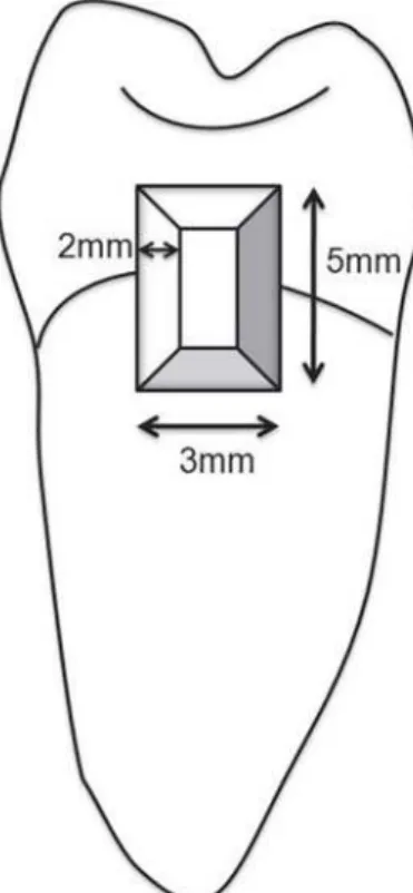
Twenty extracted non-carious human permanent premolars with fully developed roots were selected for this study, which was approved by the local Ethics in Research Committee. The teeth were cleaned of calculus, soft tissue and other debris, and stored in 2% chloramine solution at 5ºC. Two window-like cavities were prepared on both proximal surfaces of each tooth using diamond burs (no. 3145 and FG58L; KG Sorensen, São Paulo, SP, Brazil) at high speed, as shown in Figure 1.
The tested materials included a conventional GIC (CI) (Ketac Fill Plus, 3M-ESPE, St. Paul, MN, USA; lot 178566), a light-cured RMGIC (Vitremer; 3M-ESPE, St. Paul, MN, USA; lot 4JP 2007-12), an adhesive system (AS) (Adper Single Bond; 3M-ESPE, St. Paul, MN, USA; lot 4BU 2007-11) and a composite resin (CR) (Filtek Z250; 3M-ESPE, St. Paul, MN, USA; lot 2LR 2006-05). All materials were handled at room temperature (23ºC) following the manufacturers’ instructions. The 40 cavities were randomly assigned to 4 groups (n=10) and restored according to the sandwich technique18. The
experimental groups were as follows:
Group CIE - CI was applied onto the axial and cervical cavity walls, allowed setting for 5 min and acid etched (E) along the cavity margins with 35% phosphoric acid for 15 s, washed with air-water spray for 30 s, and blotted with an
Property Conventional GIC RMGIC
Working time28 2 min 3 min 45 s
Setting time28 4 min 20 s
Young’s Modulus2 20.5 GPa 55.9 GPa
Compressive strength30 265.3-306.2 MPa 196.5-301.3 MPa
Diametral tensile strength28 16 MPa 37 MPa
Flexural strength30 71-82 MPa 21.2-31.4 MPa
Knoop hardness30 66.4-84.5 KHN 29.7-176.8 KHN
Tensile bond strength to human enamel21 4.9 MPa 11.36 MPa
Tensile bond strength to human dentin21 2.52 MPa 5.55 MPa
Shear bond strength to bovine enamel28 4.6 MPa 11.3 MPa
Shear bond strength to bovine dentin28 4.3 MPa 8.2 MPa
absorbent sponge. The AS was applied and light cured (XL 3000, 3M-ESPE, St. Paul, MN, USA) for 10 s, completing the restoration with a layer of CR that was light cured for 40 s;
Group CIN - same procedures described for Group CIE, except for acid etching of the CI surface; Group: RME -same procedures described for Group CIE, but using the RMGIC. Group RMN - same procedures described for Group RME, except for acid etching of the RMGIC surface.
All restorations were polished with Sof-Lex discs (Polishing Discs, 3M-ESPE, St. Paul, MN, USA). Teeth were coated with 3 layers of nail varnish (Risqué, Niasi S/A, Taboão da Serra, SP, Brazil; lot 331526), except for a window area that included the restoration and 1 mm around it, and soaked in 1% methylene blue dye solution at 24ºC for 24 h, then rinsed under running water for 1 h.
The teeth were adapted to an automatic cutting machine (Struers Minitom, Struers A/S, Denmark) with a water-cooled low-speed (250 rpm) diamond wheel saw, and were bisected longitudinally to separate the mesial from the distal surfaces. Then, at least 2 approximately 1-mm-thick slices were obtained within the restored area. Whenever more than 2 slices were obtained per restoration, only 2 of them were used in the study, being one slice above the cementoenamel junction (CEJ) and another below the CEJ. Therefore, 4 surfaces per restoration were examined in an optical microscope (Meiji EMZ-TR, Meiji Techno Co LTD., Tokyo, Japan; magnification 10x-65x) for marginal sealing and leakage (40 surfaces per group). Representative images were recorded from all specimens and maximum degree of dye penetration was registered according to the following scores (ISO/TS 11405-2003)13: 0 = no dye penetration; 1 = dye
penetration into the enamel portion of the cavity wall; 2 = dye penetration into the dentin portion of the cavity wall but not including the pulpal floor of the cavity; 3 = dye penetration including the pulpal floor of the cavity.
Results were statistically analyzed by Kruskal-Wallis at 5% significance level (α=0.05) and chi-square test for the
two types of GIC and for the acid-etching effect on both GICs (α=0.05).
RESULTS
The sample size, dye penetration scores, dye penetration score means and statistical grouping are summarized in Table 2.
No significant differences were found between Groups CIE and CIN (p>0.05), and between Groups RME and RMN (p>0.05) regarding dye penetration means. When both surface treatments (E and N) were considered, irrespectively of the materials (CI and RMGIC), no statistically significant differences were found (p>0.05). However, significant differences were observed between the GICs (CI and RMGIC), regardless the surface treatments, CI showing a significantly greater dye penetration score mean than that of RMGIC (p<0.05).
Dye penetration at the GIC-CR interface occurred in 4 teeth, being 3 in Group CIE and 1 in Group CIN. This finding was statistically insignificant (p>0.05). Whenever microleakage occurred, the maximum degree of dye penetration was always found within the slice obtained below the CEJ.
DISCUSSION
The polymerization shrinkage developed during the conversion of the monomer molecules into a polymer network is an undesirable resin composite phenomenon and can be considered one of the factors responsible by the lack of adaptation of the restoration to the cavity walls, increasing the susceptibility to caries especially in deep regions of the proximal box of Class II cavities7. The use of GIC as an
underfilling material in conventional sandwich restoration reduces considerably the bulk resin composite used, thus,
Group n 0 1 2 3 Mean*
CIE 10 1 0 2 7 2.5 a
CIN 10 0 0 5 5 2.5 a
RME 10 6 1 1 2 0.9 b
RMN 10 7 0 3 0 0.6 b
TABLE 2- Sample size, dye penetration scores, dye
penetration score means and statistical grouping
*Group means followed by the same letter are not statistically different at p=0.05
FIGURE 1- Schematic image of the window-like cavity
the amount of polymerization shrinkage of the composite resin is decreased and the marginal adaptation may be improved. A further advantage of the sandwich technique is the fluoride-release property of GICs, which is considered to have some inhibitory effect on caries formation and progression around the restoration27.
The GIC is still considered the only material with self-adherence to dental tissue6 and it has been previously
shown that GIC and composite resin can adhere effectively to each other9,12,15,17, regardless of the limitations concerning
this system18. The bond strength between these materials is
influenced by, at least, four factors: 1) the tensile strength of GIC, which is mostly dependent on the powder/liquid ratio; 2) the viscosity of the bonding agent and its ability to wet the GIC’s surface; 3) the volumetric change in the composite resin during polymerization and; 4) the difficulties in packing and adaptation of the composite resin to the GIC without incorporation of voids18. It has been suggested that
the acid etching of GIC would allow a cleaned mildly roughened surface with high surface energy17. It has been
assumed that this procedure would fulfill the requirements to a closer contact and a greater interlocked interface between GIC and composite resin12,15. In spite of these
considerations, the results of the present study indicate that acid etching of CI and RMGIC surfaces did not improve the sealing ability of sandwich restorations.
Considering the tested GIC materials, the sandwich restorations using RMGIC showed significantly less dye penetration than those using CI, which is in agreement with the results of a previous report11. In addition, no significant
differences were found between the surface treatments (E and N) on same material. These findings suggest that the accomplishment of acid etching for GIC is not significantly relevant compared to the type of GIC selected, which should be a material with improved mechanical properties and chemical composition (Table 1).
Previous studies have shown that the inability of conventional GICs to produce an effective seal depends on two factors: 1) the material’s sensitivity to moisture during placement and early set; and 2) the dehydration after setting, resulting in crazing and cracking5,16. Yet, it is assumed that
the better sealing produced by RMGIC is a result of the formation of resin tags into the dentinal tubules allied to the ion exchange process present in the interface between dentin and RMGIC, as previously reported19,20,22. Although some
studies do not testify the presence of these resin tags or even the formation of an hybrid layer24,25, this assumption
stands to be the reason for the superior performance of the RMGIC. In addition, the presence of HEMA in the RMGIC is responsible for the increased bond strengths to resin composite9 and should contribute to prevent dye penetration
through the interface of these materials, as demonstrated by the results of the present study.
CONCLUSION
Within the limitations of this study, the results suggest
that phosphoric acid etching of GIC prior to the placement of composite resin does not improve the sealing ability of sandwich restorations. The resistance to dye penetration at the interfaces (GIC-composite resin-dentin) seems to be primarily controlled by the type of GIC, RMGIC producing significantly less dye penetration than the conventional GIC.
REFERENCES
1- Antonucci JM, McKinney JE, Stansbury JW. Resin-modified glass ionomer cement. USA Patent Application 160856. 1988.
2- Attin T, Vataschki M, Hellwig E. Properties of resin-modified glass-ionomer restorative materials and two polyacid-modified resin composite materials. Quintessence Int. 1996;27:203-9.
3- Bauer JF, Henson JL. Microleakage around dental restorations: a summarizing review. J Am Dent Assoc. 1972;87:1349-57.
4- Bowen RL, Marjenhoff WA. Dental composites/glass ionomers: the materials. Adv Dent Res. 1992;6:44-9.
5- Charbeneau GT. Principles and practice of operative dentistry. Philadelphia: Lea & Febiger; 1988.
6- De Munck J, Van Landuyt K, Peumans M, Poitevin A, Lambrechts P, Braem M, et al. A critical review of the durability of adhesion to tooth tissue: methods and results. J Dent Res. 2005;84:118-32.
7- Dijkenl JWV, Kieri C, Carlen M. Longevity of extensive class II open-sandwich restorations with a resin-modified glass-ionomer cement. J Dent Res. 1999;78:1319-25.
8- Eriksen HM, Pears G. In vitro caries related to marginal leakage around composite resin restorations. J Oral Rehabil. 1978;5:15-20.
9- Fortin D, Vargas MA, Swift EJ Jr. Bonding of resin composites to resin-modified glass ionomers. Am J Dent. 1995;8:201-4.
10- Gupta S, Khinda VI, Grewal N. A comparative study of microleakage below cemento-enamel junction using light cure and chemically cured glass ionomer cement liners. J Indian Soc Pedod Prev Dent. 2002;20:158-64.
11- Hallett KB, Garcia-Godoy F. Microleakage of resin-modified glass ionomer cement restorations: an in vitro study. Dent Mater. 1993;9:306-11.
12- Hinoura K, Moore BK, Phillips RW. Tensile bond strength between glass ionomer cement and composite resin. J Am Dent Assoc. 1987;114:167-72.
13- International Standards Organization. ISO Standard 11405:2003: dental materials-testing of adhesion to tooth structure. Geneva: The Organization; 2003.
14- Loguercio AD, Alessandra R, Mazzocco KC, Dias AL, Busato AL, Singer Jda M, et al. Microleakage in class II composite resin restorations: total bonding and open sandwich technique. J Adhes Dent. 2002;4:137-44.
15- McLean JW, Powis DR, Prosser HJ. The use of glass-ionomer cements in bonding composite resins to dentine. Br Dent J. 1985;158:410-4.
17 Mount GJ. Clinical requirements for a successful ‘sandwich’ -dentine to glass ionomer cement to composite resin. Aust Dent J. 1989;34:259-65.
18- Mount GJ. The tensile strength of the union between various glass ionomer cements and various composite resins. Aust Dent J. 1989;34:136-46.
19- Mount GJ. Adhesion of glass-ionomer cement in the clinical environment. Oper Dent. 1991;16:141-8.
20- Nezu T, Winnik FM. Interaction of water-soluble collagen with poly(acrylic acid). Biomaterials. 2000;21:415-9.
21- Pereira LC, Nunes MC, Dibb RG, Powers JM, Roulet JF, Navarro MF. Mechanical properties and bond strength of glass-ionomer cements. J Adhes Dent. 2002;4:73-80.
22- Pereira PNR, Yamada T, Tei R, Tagami J. Bond strength and interface micromorphology of an improved resin-modified glass ionomer cement. Am J Dent. 1997;10:128-32.
23- Platt JA, Rhodes B. Microleakage of high-strength glass ionomer: resin composite restorations in minimally invasive treatment. J Indiana Dent Assoc. 2001;80:20-2.
24- Ramos JC, Perdigao J. Bond strenght and SEM morphology of dentin-amalgam adhesives. Am J Dent. 1997;10:152-8.
25- Sidhu SK, Schmalz G. The biocompatibility of glass-ionomer cement materials: a status report for the American Journal of Dentistry. Am J Dent. 2001;14:387-96.
26- Smith DC. A new dental cement. Br Dent J. 1968;124:381-4.
27- Tyas MJ. Milestones in adhesion: glass-ionomer cements. J Adhes Dent. 2003;5:259-66.
28- van Noort R. Introduction to dental materials. Saint Louis: Mosby; 1994.
29- Welbury RR, Murray JJ. A clinical trial of the glass-ionomer cement-composite resin “sandwich” technique in class II cavities in permanent premolar and molar teeth. Quintessence Int. 1990;21:507-12.