* Corresponding author.
E-mail: [email protected] (De. Gao).
0102-695X/$ - see front matter © 2014 Sociedade Brasileira de Farmacognosia. Published by Elsevier Editora Ltda. All rights reserved. http://dx.doi.org/10.1016/j.bjp.2014.07.017
Original article
Comparative pharmacognosy of
Pyrrosia petiolosa
and
Pyrrosia davidii
Dandan Cheng
a, Yingying Zhang
b, Xiaowei Xin
a, Demin Gao
a,*
aSchool of Pharmacy, Shandong University of Traditional Chinese Medicine, Jinan, China bSchool of Basic Medicine, Shandong University of Traditional Chinese Medicine, Jinan, China
Introduction
Pyrrosia petiolosa (Christ) Ching, Polypodiaceae, is an important medicinal pteridophyte, which grows in wet places of mountain bare rocks or rock seam in the region of northern China and the middle and lower reaches of Yangtze River (Wang et al., 2006). The dried leaves of
Pyrrosia are used as medicinal materials to treat gonorrhea. At present, three Pyrrosia plants are recorded in the Chinese Pharmacopoeia, including Pyrrosia petiolosa (Christ) Ching, P. sheareri (Baker) Ching and P. lingua (Thunb.) Farw (Chinese Pharmacopoeia, 2010). Several studies have been carried out in recent years (Hsu, 2008; Zhang et al., 2014), to elucidate the chemical constituents and pharmacology of P. petiolosa (Jong et al., 2000). However,
further studies have shown that the efficacy and chemical constituents of Pyrrosia varied between environments and species, resulting in inconsistent clinical effects (Yang et al., 2003; Ma et al., 2006). On the other hand, some non-medicinal Pyrrosia species, such as P. davidii (Giesenh. ex Diels) Ching, have been used to substitute for medicinal
Pyrrosia. These substitutes have not been recorded in the Chinese pharmacopoeia, however, many of them have a wide circulation in the market and display good clinical efficacy (Mi et al., 2012). For pharmaceutical workers, it is difficult to distinguish P. petiolosa from P. davidii, especially from their parts (Shi et al., 2007). Therefore, in the present work, the detailed pharmacognostical studies including morphological and microscopical characteristics, physico-chemical parameters and chemical constituents fingerprints of P. petiolosa and P. davidii.
A R T I C L E I N F O
Article history: Received 23 May 2014 Accepted 30 July 2014
Keywords:
Comparative pharmacognosy HPLC fingerprint
IR spectrum
Phytochemical studies Pyrrosia davidii Pyrrosia petiolosa
A B S T R A C T
Pyrrosia petiolosa (Christ) Ching, Polypodiaceae, is an important medicinal pteridophyte used for the treatment of nephritis and bronchitis, while P. davidii (Giesenhagen. ex Diels) Ching, Polypodiaceae, often substitutes medicinal Pyrrosia in clinic. The present study was aimed to compare the pharmacognosy of P. petiolosa and P. davidii, including plant morphology, microscopic characteristics, physico-chemical parameters, UV and IR spectrum, and HPLC fingerprint. It was revealed that the two herbs had basically similar pharmacognostical characteristics but with certain differences. The present study contributes to the standard-ization and verification of these medicinal materials.
Materials and methods
Materials and reagents
The fresh materials were collected in Mengshan Mountains (N 35° 31′ 30″, E 117° 54′ 46″, Height: 450 m), Jiaodong area (N 36° 48′ 30″, E 121° 18′ 45″, Height: 360 m) in the Shandong province, China. At the Changbai Mountains (N 43° 29′ 98″, E 126° 09′
76″, Height: 480 m) in the Jilin province, China. Some Pyrrosia
medicinal materials were purchased from medicine markets of Anhui, Zhejiang and Yunnan, China. The species were identified by Gao Demin, an associate professor of Shandong University of TCM. The voucher specimens (SDCM 201220, SDCM 201221) were deposited in the Herbarium of Shandong University of TCM (SDCM). All chemical reagents were of analytical grade.
Microscopic identification
Temporary slides of leaf blades, petiole, rhizome and powder of
P. petiolosa and P. davidii (twelve samples from each collection) were prepared and observed under light microscope (Kannan et al., 2012; Rose and Prasad, 2013). For the scanning electron microscopy (SEM) analysis of Pyrrosia samples were fixed in FAA, dehydrated in a graded ethanol series, then dried, mounted and coated, and photographs were taken using an EVO40 SEM (Carl Zeiss, Germany).
Physical and chemical color reaction and TLC analysis
The powders of P. petiolosa and P. davidii leaves and rhizomes were extracted with 80% ethanol under reflux for 4 h, twice. The extracts were combined, filtered, and concentrated for color reaction (Alam and Gupta, 1986). The physical and chemical color reactions were performed as previously described (Chadwick et al., 2006).
Flavonoids and anthraquinones were identified using hydrochloric acid-magnesium reaction and alkali reaction, respectively. Triterpenes were yielded using Rosen-Heimer reaction. Polysaccharides were identified using the periodic acid Schiff reaction. Tannins and saponins were analyzed using ferric trichloride, anisaldehyde-sulfuric acid and Lieberman-Burchard reaction respectively (Mandal and Kumar, 2002; Xiang et al., 2002).
Extracts of P. petiolosa and P. davidii (20 μl) were placed on polyamide-6-layer sheets (Sinopharm Chemical Reagent Co., Ltd), eluted with SDS, C4H9OH, C7H16 (27:63:10, v/v/v) microemulsions (containing 75% water), revealed with anisaldehyde-sulfuric acid, and the retention factors (Rf) were determined.
Histochemical localization
Histochemical localizations of polysaccharides, flavonoids, saponins and anthraquinones were performed according to previous reports (Beaumont et al., 1986; Harborne, 1998).
The determination of physico-chemical parameters
The moisture, total ash and acid-insoluble ash, water-soluble extractives and ethanol-soluble extractives were determined
according to the methods recorded in the Chinese Pharmacopoeia (2010). The experiments were repeated five times.
The determination of UV-VIS spectrum
P. petiolosa and P. davidii extracts were obtained using distilled water, ethanol (70%) and petroleum ether, respectively (Bruni and Tosi, 1982). The obtained samples were treated and further analyzed using UV-VIS absorption spectrophotometry (Kalyuzhny et al., 2000; Liang et al., 2010). The experiments were performed five times. Twelve samples came from previously collected Pyrrosia materials.
HPLC fingerprint
Methanol (50%) extracts of P. petiolosa and P. davidii were prepared (Cai et al., 2012). The HPLC analysis was carried out on Sino Chrom ODS-BP column (4.6 mm×250 mm, 5 μm) using a mobile phase of acetonitrile and phosphoric acid 0.5% solution (gradient elution: 0-10 min, 8%-9%; 10-50 min, 9%-13%) at a flow rate of 1.0 ml/min-1, and a detection wavelength of
326 nm (Zhang et al., 2011). The experiments were repeated five times and twelve samples were obtained from collected
Pyrrosia materials.
The determination of chlorogenic acid
The aqueous, methanol (50%), ethanol (70%), acetone and
n-butanol, ethyl acetate and petroleum ether extracts of roots and leaves were filtered through a 45 μm mesh filter, and used for the determination of chlorogenic acid by HPLC (Zhang et al., 2012). The HPLC analysis was performed as above-mentioned.
IR spectrum
The IR spectra of methanol, acetone extracts, leaves and roots powder of P. petiolosa and P. davidii were performed as previously described (Heneczkowski et al., 2001). The experiments were carried out for six samples from different areas.
Results
Morphological characteristics
Pyrrosia petiolosa and P. davidii are perennial evergreen plants. Their leaves show significant differences in shape, size, thickness and texture.
Pyrrosia petiolosa
Figure 1 – Macroscopic characteristics of Pyrrosia petiolosa and Pyrrosia davidii. A. Macroscopic characteristics of Pyrrosia petiolosa; B. Macroscopic characteristics of Pyrrosia petiolosa; C. Macroscopic characteristics of sporophyll of Pyrrosia petiolosa; D. Macroscopic characteristics of Pyrrosia davidii; E. Macroscopic characteristics of Pyrrosia davidii; F. Macroscopic characteristics of sporophyll of
Pyrrosia davidii.
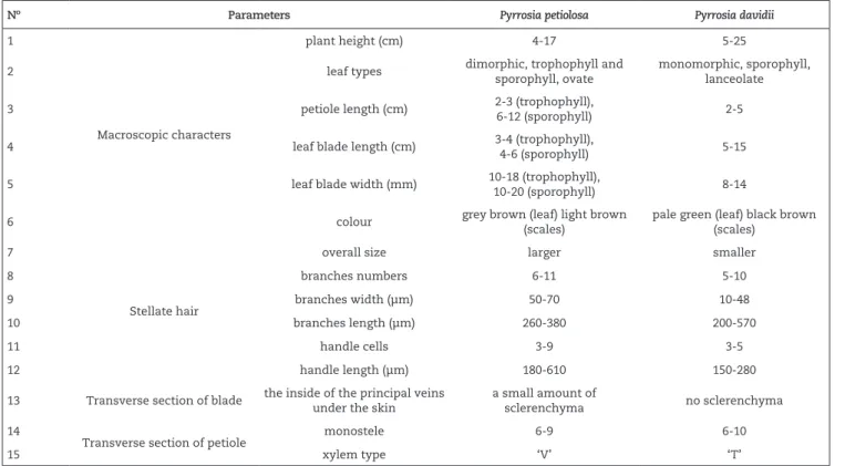
Nº Parameters Pyrrosia petiolosa Pyrrosia davidii
1
Macroscopic characters
plant height (cm) 4-17 5-25
2 leaf types dimorphic, trophophyll and
sporophyll, ovate
monomorphic, sporophyll, lanceolate
3 petiole length (cm) 2-3 (trophophyll),
6-12 (sporophyll) 2-5
4 leaf blade length (cm) 3-4 (trophophyll),
4-6 (sporophyll) 5-15
5 leaf blade width (mm) 10-18 (trophophyll),
10-20 (sporophyll) 8-14
6 colour grey brown (leaf) light brown
(scales)
pale green (leaf) black brown (scales)
7
Stellate hair
overall size larger smaller
8 branches numbers 6-11 5-10
9 branches width (μm) 50-70 10-48
10 branches length (μm) 260-380 200-570
11 handle cells 3-9 3-5
12 handle length (μm) 180-610 150-280
13 Transverse section of blade the inside of the principal veins under the skin
a small amount of
sclerenchyma no sclerenchyma
14
Transverse section of petiole monostele 6-9 6-10
15 xylem type ‘V’ ‘T’
All microscopic characteristics were studied using a biomicroscope (10×40).
Chart 1
Pyrrosia davidii
Presents a short elongated rhizome, 1.6-3.1 mm in diam; the cross-section displays few too many sclerenchyma strands; phyllopodia 0.3-0.7 cm apart. The fronds are monomorphic, with gradually narrowed bases and an acute to acuminate apex. The sori are superficial, without central bundle of paraphyses. The sporangia are contained in long stalks and embedded in capsules (Fig. 1, Chart 1).
Microscopical characteristics
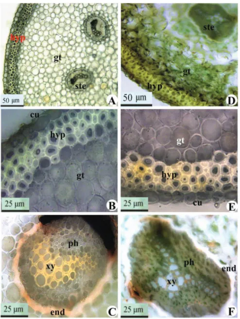
Transverse section of the leaf blade and petiole
The transverse section of the leaf blade and petiole were very similar. The external periclinal cell walls of the upper epidermis cell were wavy. The epidermis was uniseriate and cuticle covered. A 3-4 layered sclerenchymatous hypodermis
was encountered above collenchymatous ground tissue. Palisade tissue was composed of 2-3 layers of closely packed cells and spongy tissue composed of 5-8 columns of closely packed irregular parenchyma. The vessel elements in P. petiolosa were arranged in a ‘V’ shape, while those in P. davidii
were arranged in a ‘T’ shape. A mass of sclerenchyma was found beneath the vascular cylinder in the transverse sections of P. davidii leaf blade and petiole. Phloem surrounds the xylem in both species (Figs. 2 and 3, Chart 1).
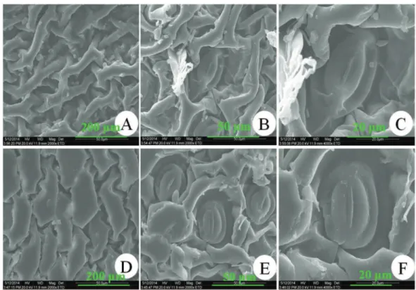
Leaf blade epidermis
The SEM showed that epidermal cells in P. petiolosa were elongated, while those in P. davidii were round or irregularly shaped. Stomata were only present on the abaxial side of the leaf blades in both species, but the distribution pattern differed, with 10-16 per mm2
in P. petiolosa and 15-20 per mm2 in P. davidii (Fig. 3).
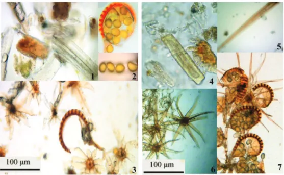
Fronds powder
As are shown in Figs. 4, 5 and 6, powder from the fronds of both species had a large amount of stellate hairs, fibers, stone cells, vessels, spores, sporangia filled with spores, and sporangium bands, but significant differences were found in the morphological characteristics of sporangium bands, spores and stellate hairs. The spores in P. petiolosa were ovoid with a sparse warty surface, while those in P. davidii were kidney-shaped with a dense warty surface (Figs. 4, 5 and 6).
Transverse section of the rhizomes
The structures of the transverse sections of rhizomes were very similar. There were specialized epidermis and scales in two columns of the epidermal cells; 2-5 columns of fiber cells were arranged in a circle inside of the epidermal cells. The amphicribral vascular bundle was composed of 6-11 vascular cylinders. Secretory cells exist in the parenchyma.
Physical and chemical color reactions and TLC
Physical and chemical color reactions indicated the presence of flavonoids, tannins, saponins, anthraquinones, triterpenes and polysaccharides. In addition, the contents of flavonoids, saponins and anthraquinones of P. petiolosa were higher than those in P. davidii. TLC analysis showed significant differences between P. petiolosa and P. davidii. Judged by the brightness of spots, the content of chlorogenic acid in P. petiolosa was significantly higher than that in P. davidii (Fig. 7).
Histochemical localization
P. petiolosa and P. davidii contain similar chemical constituents, including flavonoids, anthraquinones, triterpenes, steroids and tannins, but concentrations were significantly different. The contents of flavonoids, saponins and anthraquinone in P. petiolosa were higher than those in
P. davidii (Table 1).
Physico-chemical parameters
Moisture and total ash content of P. petiolosa were similar to those of P. davidii. However, the contents of acid-insoluble ash, aqueous, ethanol and petroleum ether extracts between P. petiolosa and P. davidii showed significant differences. All parameter values of P. petiolosa were higher than those of P. davidii except in the petroleum ether-soluble extract (Table 2).
UV-VIS spectrum
The UV-VIS spectra and their second derivatives of alcohol and ether extract were similar, while the aqueous extract was different. Therefore, the second derivative UV-VIS spectra of the aqueous extract can be used to distinguish P. petiolosa from
P. davidii. Also, intensity peaks at 220, 242, 263, 302, 249 (from
P. davidii) and 251, 299, 325 (from P. petiolosa) were significantly different (Fig. 8).
Figure 4 – Microstructure characteristics of Pyrrosia petiolosa and Pyrrosia davidii fronds powder. 1. Trachea of Pyrrosia petiolosa; 2. Spores and sporangium band of Pyrrosia petiolosa; 3. Stellate hairs of Pyrrosia petiolosa; 4. Stone cells of Pyrrosia davidii; 5. Fiber of
Pyrrosia davidii; 6. Stellate hairs of Pyrrosia davidii; 7. Spores and sporangium band of Pyrrosia davidii.
Figure 5 – Microscopic characteristics of Pyrrosia petiolosa and Pyrrosia davidii sporangia and spore. A, B and C. Pyrrosia petiolosa; D, E and F. Pyrrosia davidii.
HPLC fingerprint
HPLC fingerprints of P. petiolosa and P. davidii were similar; however, the contents of each ingredient from the peak position in P. petiolosa and P. davidii were different, especially at the retention time of the 11th min (Fig. 9, Table 3).
Chlorogenic acid
The chlorogenic acid mainly is present in the ethanol, methanol, methanol 50% and aqueous extract of leaves. The chlorogenic acid content in the different extracts of P. petiolosa
Figure 6 – Microscopic characteristics of Pyrrosia petiolosa and Pyrrosia davidii stellate hairs. A. Pyrrosia petiolosa stellate hairs; B.
Pyrrosia davidii stellate hairs.
Herbs Nº Location Polysaccharides Flavonoids Saponins Anthraquinone
Pyrrosia petiolosa
1 Epidermis + +++ +++ ++
2 Palisade tissue + ++ +++ +++
3 Spongy tissue + ++ ++ ++
4 Collenchyma ++ +++ ++ –
5 Phloem + ++ ++ ++
6 Xylem + – – –
Pyrrosia davidii
7 Epidermis + ++ + +
8 Palisade tissue + + ++ ++
9 Spongy tissue + + + +
10 Collenchyma ++ ++ + –
11 Phloem + + + +
12 Xylem + – – –
+, present; –, absent; n = 10 per sample. The number of (+) is representative of the degree.
Nº Parameters Pyrrosia petiolosa (%) Pyrrosia davidii (%)
1 Moisture content 8.90 ± 0.078 6.74 ± 0.102
2 Ash content 4.99 ± 0.142 4.62 ± 0.082
3 Acid insoluble ash content 0.53 ± 0.033 1.29 ± 0.031
4 Water-soluble extract content 18.18 ± 0.172 25.49 ± 0.480
5 Alcohol soluble extract content 15.92 ± 0.262 20.64 ± 0.445
6 Ether soluble extract content 9.68 ± 0.872 6.73 ± 0.266
Values are mean % ± SD. The values were from three independent replicates.
Nº Pyrrosia petiolosa (%) Pyrrosia davidii (%) Nº Pyrrosia petiolosa (%) Pyrrosia davidii (%)
1 60.80 ± 3.80 43.70 ± 2.80 8 – 2.34 ± 0.32
2 8.52 ± 0.86 6.84 ± 0.76 9 1.16 ± 0.12 3.01 ± 0.32
3 7.25 ± 0.56 5.65 ± 0.56 10 6.15 ± 0.57 2.60 ± 0.37
4 1.19 ± 0.20 0.42 ± 0.30 11 – 6.87 ± 0.69
5 1.55 ± 0.16 0.97 ± 0.16 12 0.45 ± 0.06 9.12 ± 0.86
6 0.54 ± 0.04 0.80 ± 0.07 13 0.61 ± 0.03 0.80 ± 0.03
7 1.43 ± 0.09 0.61 ± 0.09
The values were expressed as mean ± SD of five independent replicates.
Extract Pyrrosia petiolosa (%) Pyrrosia davidii (%) Leaves Roots Leaves Roots
Aqueous 5.44 ± 0.125 1.07 ± 0.091 2.17 ± 0.082 0.41 ± 0.023
Methanol (50%) 6.90 ± 0.232 0.94 ± 0.038 3.63 ± 0.125 1.04 ± 0.061
Methanol 7.10 ± 0.252 1.27 ± 0.045 4.40 ± 0.116 1.07 ± 0.052
Ethanol (70%) 8.13 ± 0.173 1.60 ± 0.036 5.53 ± 0.093 1.17 ± 0.075
Acetone 0.78 ± 0.009 0.60 ± 0.007 0.62 ± 0.082 0.39 ± 0.021
n-butanol 0.70 ± 0.005 0.13 ± 0.008 0.37 ± 0.035 0.07 ± 0.003
Ethyl acetate 0.44 ± 0.008 0.09 ± 0.005 0.41 ± 0.046 0.16 ± 0.0007
Petroleum ether 0.02 ± 0.001 0.01 ± 0.0009 – –
The values were expressed as mean ± SD of five independent replicates.
Table 1
Parts of morphological and microscopic characteristics of Pyrrosia petiolosa and Pyrrosia davidii.
Table 2
Phytochemical parameters of Pyrrosia petiolosa and Pyrrosia davidii.
Table 3
The compound content of different peaks well defined from Pyrrosia petiolosa and Pyrrosia davidii.
Table 4
Figure 8 – The second derivative of ultraviolet-visible spectra of Pyrrosia petiolosa and Pyrrosia davidii. 1. Pyrrosia petiolosa; 2.
Figure 9 – The HPLC fingerprints of Pyrrosia petiolosa and Pyrrosia davidii. A. Pyrrosia petiolosa; B. Pyrrosia davidii; C. Negative control; D. Chlorogenic acid. Wavelength, 326 nm; Mobile phase, acetonitrile (A) 0.5% phosphoric acid solution; (B) 0-10 min, 8-9% (A); 10-50 min, 9%-13% (A); Flow rate, 1.0 ml.min-1. HPLC fingerprints were representative from five experiments and six specimens.
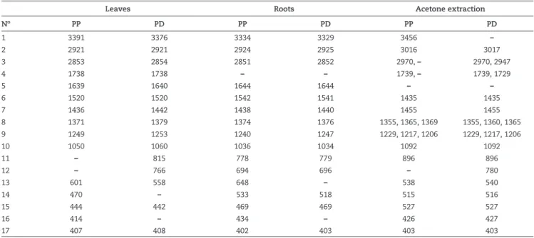
IR spectrum
P. petiolosa and P. davidii contain consistent functional groups (4000-1300 cm-1), such as alcohol hydroxyl, phenolic hydroxyl
and carboxyl, fat hydroxyl, carbonyl, and benzene. However, FTIR spectra fingerprint region (1300-400 cm-1) had significant
differences.
In P. petiolosa, carbohydrates such as glycogen were shown in the spectral output at 1249, 1050 cm-1 in the leaves and 1240,
1036 cm-1 in the roots; while in P. davidii, spectral output at
1253, 1060 cm-1 in the leaves and 1247, 1034 cm-1 in the roots.
These were ascribed to stretching vibrations of hydrogen-bonded C-O groups; esters participated in the spectral output at 1738, 1249 cm-1 in P. petiolosa leaves, 1738, 1253 cm-1 in P.
davidii leaves. These were associated to stretching vibrations of hydrogen-bonded C=O, C-O groups. Amides of the spectral output had peaks at 3391, 1639 cm-1 in P. petiolosa leaves, and
3376, 1640 cm-1 in P. davidii leaves.
The principal differences were the presence of bands at 1544 cm-1, possibly assigned to an unsaturated fat structure, and at
3605 cm-1 associated with an asymmetric O-H, N-H stretching
of P. petiolosa, but neither appeared in P. davidii.
The FTIR spectra fingerprint region (1300-400 cm-1) of P.
petiolosa and P. davidii showed pronounced differences. They displayed only one common peak at 469 cm-1. The peak at
470, 414 cm-1 only appeared in P. petiolosa leaves, while the
peak at 815, 766 cm-1 only appeared in P. davidii leaves. The
peaks at 648, 434 cm-1 only appeared in P. petiolosa roots (Fig.
10, Table 5).
Discussion
The most noticeable differences between P. petiolosa
and P. davidii were the morphological and microscopical characteristics, important factors to guarantee the quality of medicinal materials and clinical efficacy.
The medicinal materials displayed differences in size, shape, texture, and thickness of the leaves, rhizomes, serrated edges, and hair roots, as well as the types of leaves and the size of the petiole. Regarding microscopic characteristics, the wide and short stellate hair branches, long stellate hairs handle and its ‘V’ xylem of P. petiolosa were distinguished from those of P. davidii.
Figure 10 – The comparison of FTIR spectra of Pyrrosia petiolosa and Pyrrosia davidii leaves, roots, methanol extraction. A. leaves; B. roots; C. methanol extract; 1. Pyrrosia petiolosa; 2. Pyrrosia davidii.
Leaves Roots Acetone extraction
Nº PP PD PP PD PP PD
1 3391 3376 3334 3329 3456 –
2 2921 2921 2924 2925 3016 3017
3 2853 2854 2851 2852 2970, – 2970, 2947
4 1738 1738 – – 1739, – 1739, 1729
5 1639 1640 1644 1644 – –
6 1520 1520 1542 1541 1435 1435
7 1436 1442 1438 1440 1455 1455
8 1371 1379 1374 1376 1355, 1365, 1369 1355, 1360, 1365
9 1249 1253 1240 1247 1229, 1217, 1206 1229, 1217, 1206
10 1050 1060 1036 1034 1092 1092
11 – 815 778 779 896 896
12 – 766 694 696 – 780
13 601 558 648 – 538 540
14 470 – 533 518 515 516
15 444 442 469 469 527 527
16 414 – 434 – 426 427
17 407 408 402 403 403 403
The values were expressed as mean ± SD of five independent replicates.
Table 5
Chlorogenic acid is not only an active component, but also an important reference component (Johnston et al., 2003). The content of chlorogenic acid in Pyrrosia leaf was no less than 2% by HPLC method (Chinese Pharmacopoeia, 2010). Although the content of chlorogenic acid in P. petiolosa and P. davidii both reached the standard as medical materials, the amount in the former was significantly higher than that in the latter. This partly explains the fact that P. petiolosa is more widely used in the clinic and circulated in the market.
It has been reported that mangiferin and polyphenols were important active components (Garcia et al., 2003; Sugiyama et al., 2007). The content of flavonoids and polyphenols were higher in P. petiolosa than that in P. davidii by analyzing histochemical locations, UV-VIS spectra and HPLC spectrum, which not only provided a quick and easy method for identification of Pyrrosia materials, but also workes as a standard HPLC and UV-VIS spectra.
In conclusion, the comparative pharmacognosy analysis of P. petiolosa and P. davidii provided a base and standard to quickly identify the two plants, which could ensure the safety of natural medicines for clinical use and further promote the development of Pyrrosia species.
Authors’ contributions
CD carried out most of the experimental work and drafted the manuscript. XX and ZY performed HPLC and UV analysis. GD designed the study, supervised the whole experimental process and edited the manuscript. All the authors have read and approved the final manuscript.
Conflicts of interest
The authors declare no conflicts of interest.
Acknowledgement
The authors thank China Germplasm Bank of wild species (WGB-1204) for financial support. We thank associate professors Zhang Hongmeng and Wang Jihui, from the experimental centre in Shandong University of TCM, who provided the facilities to use high performance liquid chromatography. Moreover, we thank Professor Shi Zhenying (Shanghai Institute of Plant Physiology and Ecology, Chinese Academy of Sciences (CAS), China), Professor Hou Yuantong (College of Life Science, Qufu Normal University, China), Professor Zhang Liqiang and lecturer Kevin Day (Arizona State University Tempe AZ. USA), who carefully revised the manuscript.
R E F E R E N C E S
Alam, N., Gupta, P.C., 1986. Structure of a water-soluble polysaccharide from the seeds of Cassia angustifolia. Planta Med. 52, 308-310.
Beaumont, J., Cutler, D.F., Reynolds, T., Vaughan, J.G., 1986. Secretory tissues in the East African shrubby aloes. Bot. J. Linn. Soc. 92, 399-403.
Bruni, A., Tosi, B., 1982. A method for the pharmacognostic study of Aloe species using fluorescence microscopy. Pharm. Biol. 20, 127-131.
Cai, H., Xu, Z., Luo, S., Zhang, W., Cao, G., Liu, X., Cai, B., 2012. Study on chemical fingerprinting of crude and processed Atractylodes macrocephala from different locations in Zhejiang province by reversed-phase high-performance liquid chromatography coupled with hierarchical cluster analysis. Pharmacogn. Mag. 8, 300-307.
Chadwick, L.R., Pauli, G.F., Farnsworth, N.R., 2006. The pharmacognosy of Humulus lupulus L. (hops) with an emphasis on estrogenic properties. Phytomedicine 13, 119-131.
Chinese Pharmacopoeia, 2010. The Pharmacopoeia Comittee of People’s Republic of China, Beijing.
Garcia, D., Escalante, M., Delgado, R., Ubeira, F.M., Leiro, J., 2003. Anthelminthic and antiallergic activities of Mangifera indica L. stem bark components Vimang and mangiferin. Phytother. Res. 17, 1203-1208.
Harborne, J.B., 1998. Phytochemical methods A Guide to modern techniques of plant analysis. 3rd ed., Springer, 302 p. Heneczkowski, M., Kopacz, M., Nowak, D., Kuźniar, A., 2001.
Infrared spectrum analysis of some flavonoids. Acta. Pol. Pharm. 58, 415-420.
Hsu, C.Y., 2008. Antioxidant activity of Pyrrosia petiolosa. Fitoterapia. 79, 64-66.
Johnston, K.L., Clifford, M.N., Morgan, L.M., 2003. Coffee acutely modifies gastrointestinal hormone secretion and glucose tolerance in humans: glycemic effects of chlorogenic acid and caffeine. Am. J. Clin. Nutr. 78, 728-733.
Jong, H.P., Seong, S.P., Myung, S.W., Chang, H.C., 2000. Pharmacognostical studies on the ‘Suk Wi’. Korean J. Pharmacogn. 31, 288-294.
Kalyuzhny, G., Vaskevich, A., Ashkenasy, G., Shanzer, A., Rubinstein, I., 2000. UV/Vis spectroscopy of metalloporphyrin and metallophthalocyanine monolayers self-assembled on ultrathin gold films. J. Phys. Chem. B. 104, 8238-8244. Kannan, R., Prasant, K., Babu, U.V., 2012. Botanical
pharmacognosy of stem of Gmelina asiatica Linn. Anc. Sci. Life. 31, 190.
Khatoon, S., Rai, V., Rawat, A.K.S., Mehrotra, S., 2006. Comparative pharmacognostic studies of three Phyllanthus species. J. Ethnopharmacol. 104, 79-86.
Liang, X., Zhang, L., Zhang, X., Dai, W., Li, H., Hu, L., Zhang, W., 2010. Qualitative and quantitative analysis of traditional Chinese medicine Niu Huang Jie Du Pill using ultra performance liquid chromatography coupled with tunable UV detector and rapid resolution liquid chromatography coupled with time-of-flight tandem mass spectrometry. J. Pharm. Biomed. Anal. 51, 565-571.
Ma, X.Y., Xie, C.X., Liu, C., Song, J.Y., Yao, H., Luo, K., Chen, S.L., 2010. Species identification of medicinal pteridophytes by a DNA barcode marker, the chloroplast psbA-trnH intergenic region. Biol. Pharm. Bull. 33, 1919-1924.
Mandal, S.C., Ashok Kumar, C.K., 2002. Studies on anti-diarrhoeal activity of Ficus hispida. Leaf extract in rats. Fitoterapia 73, 663-667.
Rose, B.N., Prasad, N.K., 2013. Preliminary phytochemical and pharmacognostical evaluation of Carissa spinarum leaves. Asian J. Pharm. Technol. 3, 30-33.
Shi, Z., Chen, Y.L., Chen, Y.S., Lin, Y.R., Liu, S.W., 2007. Flora of China. Beijing: Science Press & St Louis: Missouri Botanical Garden Press, p. 4786-4795.
Sugiyama, H., Akazome, Y., Shoji, T., Yamaguchi, A., Yasue, M., Kanda, T., Ohtake, Y., 2007. Oligomeric procyanidins in apple polyphenol are main active components for inhibition of pancreatic lipase and triglyceride absorption. J. Agric. Food. Chem. 55, 4604-4609.
Wang, N., Wang, J.H., Li, X., Ling, J.H., Li, N., 2006. Flavonoids from Pyrrosia petiolosa (Christ) Ching: Note. J. Asian. Nat. Prod. Res. 8, 753-756.
Xiang, Z.B., Ren, S.G., Shi, Y.S., Tang, C.H., 2002.
Absorptiophotometric determination of total flavones in stems and leaves of buckwheat. Physical Testing and Chem. Analysis Part B Chemical 38, 436-437.
Yang, C., Shi, J.G., Mo, S.Y., Yang, Y.C., 2003. Chemical constituents of Pyrrosia petiolosa. J. Asian. Nat. Prod. Res. 5, 143-150.
Zhang, H., Zhao, T., Gong, Y., Dong, X., Zhang, W., Sun, S., Li, P., 2014. Attenuation of diabetic nephropathy by Chaihuang-Yishen granule through anti-inflammatory mechanism in streptozotocin-induced rat model of diabetics. J. Ethnopharmacol. 151, 556-564.
Zhang, J.Y., Zhang, J., Jin, H., 2012. Ultraviolet absorption spectrum analysis and identification of medicinal plants of Paris. Spectrosc. Spec. Anal. 32, 2176-2180.