* Corresponding author.
E-mail: denise.endringer@uvv.br (D.C. Endringer); tadeu.andrade@uvv.br (T.U. Andrade).
0102-695X/$ - see front matter © 2014 Sociedade Brasileira de Farmacognosia. Published by Elsevier Editora Ltda. All rights reserved. http://dx.doi.org/10.1016/j.bjp.2014.06.003
Original article
Triterpenes from the
Protium heptaphyllum
resin – chemical
composition and cytotoxicity
Ewelyne M. Lima
a,b, Andrews M. Nascimento
a,b, Dominik Lenz
a, Rodrigo Scherer
a,
Silvana S. Meyrelles
b, Giovanna A.P. Boëchat
a, Tadeu U. Andrade
a,*,
Denise C. Endringer
a,c,*
aDepartamento de Farmácia, Universidade Vila Velha, Vila Velha, ES, Brazil
bDepartamento de Ciências Fisiológicas, Universidade Federal do Espírito Santo, Vitória, ES, Brazil cCampus Vila Velha, Instituto Federal do Espírito Santo, Vila Velha, ES, Brazil
Introduction
Protium heptaphyllum (Aubl.) Marchand, Burseraceae, known as “almécega”, “breu”, and “almíscar”, originated in South America, exudes an oily resin composed of a mixture of triterpenes from the α-amyrin (ursane) and β-amyrin (oleane) series, and an essential oil rich in mono- and sesquiterpenes (Siani et al., 1999a; Maia et al., 2000). Ethnopharmacological studies report the use of this type of resin for ulcer treatment; there are also reports of its use as an analgesic and anti-inflammatory agent (Correa, 1978; Brandão et al., 2008).
The anti-inflammatory effect of the mixture of α-and
β-amyrin, the essential oil, and the crude resin of this plant has been previously reported (Siani et al., 1999b; Aragão et al., 2007). Research aimed at minimizing the effects of breast cancer and the inhibition, reversal or delay of its appearance has received wide interest, and natural products have been used for these purposes (Newman and Cragg, 2012). The role of inflammation in the pathogenesis of human cancers is well established (Allavena et al., 2008). Compounds including inflammatory eicosanoids, reactive oxygen species (ROS) and cytokines are involved in this process, and their levels are endogenously regulated (Basu et al., 2013).
A R T I C L E I N F O
Article history:
Received 19 February 2014 Accepted 30 June 2014
Keywords:
α-Amyrin
β-Amyrin Almíscar
Angiotensin converting enzyme activity Caspase-3 TNF-α
A B S T R A C T
Protium heptaphyllum (Aubl) Marchand, Burseraceae, is popularly used as an analgesic and anti-inflammatory agent. However, the cellular mechanism of action remains unknown. This study aims to evaluate the chemical composition of P. heptaphyllum resin and cytotoxicity on a breast cancer cell line (MCF-7). The chemical composition of the resin was determined by Gas Chromatography coupled to a Mass Spectrometer. The cytotoxicity was evaluated using an MTT assay. Annexin V-FITC, caspase-3, Angiotensin Converting Enzyme activity and Tumor Necrosis Factor alpha (TNF- α) assays were performed to evaluate apoptosis and inflammatory events. The resin consisted of triterpenes, such as α- and β-amyrin. Cytotoxicity was only observed in fractions enriched with α- and β-amyrin. The resin and fractions elicited antiproliferative activity, increased activity of caspase-3 and ACE, and a decrease in the TNF-α
level. Altogether, the resin and fractions enriched with α- and β-amyrin promoted cytotoxicity and apoptosis.
Antitumor activity is possibly a result of the inhibition of inflammation produced by the tumor, the suppression of the expression of certain oncogenes, the activation of a suppressor, and the modulation of defense systems, including antioxidant and immune functions (Liu et al., 1994). The participation of the rennin angiotensin system (RAS) as a pro-apoptotic and anti-apoptotic agent in tumor cells is not well understood (George et al., 2010).
Considering the anti-inflammatory activity of P. heptaphyllum resin, the present study aimed to evaluate the chemical composition, cytotoxicity, and pro-apoptotic nature of the crude resin and other fractions on a mammary adenocarcinoma cell line (MCF-7). The studied fractions were enriched with the isomers α- and β-amyrin from P. heptaphyllum. The participation of ACE and TNF-α in these events was also evaluated.
Materials and methods
Plant material
The resin of the stem of the species of Protium heptaphyllum
(Aubl.) Marchand, Burseraceae, was collected in May 2009 on the Ilha de Guriri, Espírito Santo. A specimen was deposited at the herbarium of the University of Vila Velha (UVV/ES 1802) and identified by botanist Solange Zanotti Schneider.
Fractionation of the resin
An aliquot of the resin obtained from the stem of P. heptaphyllum
(90 g) was dissolved in dichloromethane to eliminate impurities, such as sand and pieces of wood. The clear resin (78.1 g) was subjected to chromatographic separation in a silica gel column (162 g) using a pentane: CH2Cl2 gradient (100:00 ‐ 00:100), followed by a CH2Cl2:EtOAc gradient (10:00 ‐ 00:10). Fractions of 20 ml, eluted with n-hexane: EtOAc (4:1), were collected and then analyzed by thin layer chromatography (TLC) (silica gel 60 F254), yielding 38 fractions which were combined into fifteen primary fractions (FR1-FR15) based on the TLC patterns.
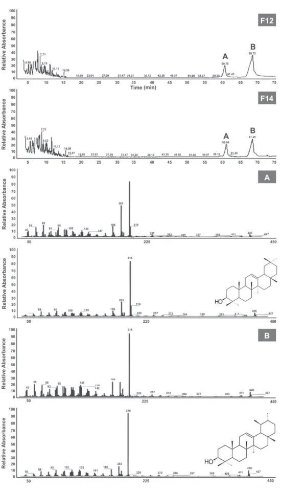
The F2 fraction obtained from the pentane: CH2Cl2 (3:2) gradient was re-chromatographed with pentane and CH2Cl2 (10:00 ‐ 00:10) elution gradient on silica gel (9.9 g), yielding 165 fractions to provide five grouped (FR2-1 to FR2-5) fractions according to their chemical profile by TLC. The F2-3 fraction (7g) obtained from the elution with a pentane: CH2Cl2 (3:2) gradient was re-chromatographed on a column of silica gel (106.3 g) using isocratic elution with CH2Cl2:EtOAc:MeOH (1.8:5.0:0.1), resulting in 44 fractions, combined into eight fractions (FR2-3-1 to FR2-3-8). In an attempt to isolate the isomeric constituent, re-fractionation of the FR2-3-4 fraction (4.1 g) was performed on silica gel, using an elution gradient of pentane:CH2Cl2, CH2Cl2:CHCl3, CHCl3, and CHCl3:MeOH, yielding nineteen fractions (F1 to F19). However, even after subsequent attempts to isolate the isomers, they remained a mixture, as analyzed by GC-MS. The F12 and F14 fractions resulting from the elution of CHCl3 and CHCl3:MeOH, respectively, were mixtures with
majority of two triterpenes (Fig. 1) and were utilized in the bioassays.
Gas chromatography coupled to Mass spectrometry (GC/MS)
The analysis of the chemical constituents of the resin was performed using a gas chromatograph (Trace Ultra, ThermoScientific®) coupled to a mass spectrometer (DSQII,
ThermoScientific®). The volatile substances were separated
on a DB-5 capillary column (30 m × 0.25 mm d.i. × 0.25 µm, J&W Scientific®, Folson, California, USA). The initial temperature
was 70°C for 5 min, and the temperature was then increased to 250°C using a temperature ramp of 3°C/min before reaching and keeping the final temperature for 5 min. We used helium gas with a constant flow of 1 ml/min. The injector temperature was maintained at 220°C, and the temperature of the GC/ MS interface was maintained at 250°C. The mass detector was operated by ionization with electron impact (+70 eV) using the scan mode, held at 35-450 MHz. The samples were diluted with hexane (1 mg/ml) and injected into the GC/MS in duplicate; 1.0 µl was injected with the injector in splitless mode. The identification of the substances contained in the resin was performed by comparing the similarity of the obtained mass spectra obtained with those in the literature (Adams, 2001; NIST/EPA/NIH, 2005) (Fig. 1). The relative percentages of these compounds were calculated from the mean areas of the chromatograms.
Cell line
A mammary adenocarcinoma (MCF-7, ATCC-HTB22) cell line was used and maintained in Dulbecco’s Modified Eagle’s Medium (DMEM) culture medium (Sigma-Aldrich, St. Louis, MO) supplemented with a 10 ml solution of penicillin G, streptomycin and L-glutamine (Sigma-Aldrich, St. Louis, MO) and 20% fetal bovine serum (Gibco, Invitrogen Corporation, Grand Island, NY).
Preparation of samples for assays
The test samples used in the biological assays were fractions containing the isomers α- and β-amyrin (F12 and F14) and the crude resin (RES). These were dissolved in PBS, dimethylsulfoxide (DMSO) (0.09%) and propylene glycol (1%). The final concentration of dichloromethane in the assay was less than 0.003%.
Cellular cytotoxicity assay with colorimetric method of the MTT
Cytotoxicity was determined using the colorimetric MTT (3-bromide-[4.5-dimethyl-thiazol-2-yl]-2.5-diphenyl-tetrazolium) method (Sigma-Aldrich, St. Louis, MO), in which the tetrazolium salt is converted into the formazan salt by living cells, turning the culture blue (Mosmann, 1983). MCF-7 tumor cells were plated in sterile 96 well plates at a concentration of 5×10⁴ cells/ml. Then, 10 µl samples of RES,
St. Louis, MO; 98-102%) was used as a positive control at a final concentration of 0.9 μM (Zheng et al., 2012). Then, 10 µl of MTT solution (5 mg/ml) diluted in DMEM was added, and the plate was incubated for 4 h. Finally, we added 150 µl of isopropanol acidified with 0.04 M HCl and evaluated the samples with an ELISA reader (Thermoplate TP-Reader at 570 nm). The analyses were performed in triplicate, and the percentage of inhibition was calculated using the formula below: % inhibition = (absorbance of negative-control well) × 100 negative control.
Determination of apoptosis by flow cytometry analysis
Apoptosis was assessed using an annexin V-FITC Kit (Sigma Aldrich, St. Louis, MO), according to the manufacturer’s instructions. Briefly, MCF-7 cells were plated in a sterile 24-well plate at a concentration of 5×10⁶ cells/ml. Then, the samples
RES (10 µl), and P. heptaphyllum fractions F12 and F14 (final concentration 40 µg/ml) were added. A solution of hydrogen peroxide (Sigma-Aldrich, St. Louis, MO) (final concentration of 10 µM) was used as positive control. The plate was incubated at 37°C in 5% CO2 for 72 h. After this period, the cells were colored with FITC-conjugated annexin V and propidium iodide for 15 min at room temperature, protected from light, and then analyzed by flow cytometry. The percentage of positive cells determined over 10,000 acquired events was analyzed by a FACSCalibur system equipped with a 488 nm argon laser and
FCS Express 4 FlowCytometry software (De Novo Software, Los Angeles, CA).
Determination of caspase-3 for colorimetric assay
The caspase-3 activity was determined using a colorimetric method in which the presence of caspase-3 lysate produces p-nitroaniline (pNa), which generates a yellow color. MCF-7 cells were plated in a sterile 24-well plate at a concentration of 1×10⁵ cells/ml. Then, samples of RES (10 µl), and the P. heptaphyllum fractions F12 and F14 (final concentration of 40 µg/ml) were added. As a positive control, we used DOX (final concentration of 0.9 µM). The plate was incubated at 37°C in 5% CO2 for 72 h. After this period, the determination of caspase-3 was performed according to the manufacturer’s specification (Sigma-Aldrich, St. Louis, MO) using an ELISA reader (TP-Thermoplate Reader at 405 nm). The analyses were the average of eight replicates, and the results were expressed in ΔmOD405nm/min (Posmantur et al., 1998).
Determination of TNF-α
To quantify the levels of TNF-α, MCF-7 tumor cells were plated in a sterile 24-well plate at a concentration of 1×10⁶ cells/ml.
Then, samples of RES (10 µl), and the P. heptaphyllum fractions F12 and F14 (final concentration 40 µg/ml) were added, and the plate was incubated at 37°C in 5% CO2 for 72 h. As a positive control, we used DOX (final concentration of 0.9 µM). After this period, we proceeded to read the plate in an ELISA reader (TP-Thermoplate Reader at 450 nm) using a commercial kit (Invitrogen, San Jose, USA) according to the manufacturer’s instructions. Analyses were performed in replicates of 8.
ACE activity assay
Angiotensin Converting Enzyme (ACE) activity was determined by measuring the Gly-Gly (glycyl-glycine) cleavage product Hip-Gly-Gly (Hippuryl-glycyl-glycine) with ACE as described by Serra et al. (2005), with modifications. Briefly, the MCF-7 tumor cells were plated in a sterile 24 well plate at a concentration of 5×10⁵ cells/ml, and 10
µl samples of RES, and the P. heptaphyllum fractions F12 and F14 (final concentration of 40 µg/ml) were added. DOX was used as a comparison known compound (final concentration of 0.9 μM). The plate was incubated at 37°C in 5% CO2 for 72 h. For the enzymatic reaction, an aliquot of supernatant was combined with the assay buffer and Gly-Gly substrate solution (100 mM, Sigma Aldrich, St. Louis, MO). After homogenization, the mixture was incubated for 35 min at 37°C. The reaction was stopped by the addition of solutions of sodium tungstate (300 mM) and sulfuric acid (0.33 mM). The system was mixed with 2,4,6-trinitrobenzene sulfonic acid (TNBS) staining reagent (Sigma Aldrich, St. Louis, MO). After 20 min under light, the plate absorbance was read in an ELISA reader (TP-Reader Thermoplate at 405 nm). The blank solution was prepared similarly except the sodium tungstate and sulfuric acid solutions were added before the enzyme solution. The assay was also carried out in the presence of 10 μl of a solution of captopril (final concentration of 64 nM). The assays were performed in replicates of 8. The ACE activity was calculated according to the equation: % activity = (A × 100) / B, where A is the measured absorbance at 405 nm of the well of cells, and B is the absorbance of rabbit lung.
Statistical analysis
The results were expressed as the mean, plus or minus the standard error of the mean (SEM) and ± the standard deviation (SD). They were analyzed by one-way ANOVA. The significance of the difference between the means was determined using the post-hoc Tukey method, adjusted for multiple comparisons with significance above 5% (p < 0.05).
Results and discussion
Identification of the fractions of the P. heptaphyllum
The resin of Protium heptaphyllum (Aubl.) Marchand, Burseraceae, was dissolved in dichloromethane and subjected to a series of chromatographic techniques, producing fractions enriched in triterpenes. Isolation purification was not possible due to the presence of isomers. The fractions F12 and F14, evaluated in the bioassays, were analyzed by GC-MS; the major components identified were the isomers
Other studies have identified and isolated triterpenes from
P. heptaphyllum resin, including the isomers α- and β-amyrin from the resin of this same species (Maia et al., 2000; Susunaga et al., 2001; Vieira Junior et al., 2005).
Cellular cytotoxicity by resin and fractions of the P. heptaphyllum
In the assay for cell viability using MTT, the P. heptaphyllum
resin did not exhibit significant cytotoxicity against MCF-7 cancer cells at a concentration of 40 µg/ml. However, fractions F12 and F14 showed weak cytotoxicity, with IC50 values of 34.8 ± 2.9 and 38.2 ± 3.4 µg/ml, respectively. These values are in accordance with the concentrations recommended by the American National Cancer Institute (NCI) (IC50 < 20 µg/ ml for the extract and fractions) (Boyed, 1997). Furthermore, when those compounds were evaluated separately, each one,
α-amyrin (IC50 2.35 µg) and β-amyrin (IC50 2.48 µg), showed a potent cytotoxic activity on breast cancer cell line, MCF-7 (El-Alfy et al., 2011). Nevertheless, the data El-Alfy et al. (2011) were not depicted as a concentration.
Activation of caspase-3 and apoptosis by resin and fractions of P. heptaphyllum
The inhibition of cell proliferation by activation of caspase-3 and the translocation of phosphatidylserine were observed in the fractions containing α-and β-amyrin (Figs. 2 and 3), suggesting that the induction of cell death by apoptosis is caspase-dependent. Also, increased levels of apoptosis have been associated with cell growth inhibition, indicating that the fractions are potentially oncogenic (Campisi, 2005).
The fraction with a higher percentage of α-amyrin (F12) showed greater apoptotic activity and greater induction of caspase-3. Curiously, at a concentration of 40 µg/ml, the resin showed no significant cytotoxicity, but induced the highest rate of apoptosis observed in all of the fractions, but minimally activated caspase-3 (Figs. 2 and 3).
This result may indicate that the resin promotes apoptosis by caspase-dependent and caspase-independent mechanisms simultaneously. Liang et al. (2001) concluded that MCF-7 cells may undergo apoptosis by sequential activation of effector
Figure 2 – Caspase-3 activity by ELISA in a breast cancer cell line (MCF-7). The cells were treated with essential Protium heptaphyllum crude resin (RES) and its fractions, enriched with α-and β-amyrin (F12 and F14). Ac-DEVD-CHO (caspase-3 inhibitor) and doxorubicin (DOX) were used at 40 μg/ml for 72 h. The values represent the means ± SEM; ap < 0.01 and bp < 0.05 compared
to MCF-7. cp < 0.01 compared to CASP3. dp < 0.01 compared to DOX. ep < 0.01 compared to treatment of fractions with and
caspases-7 and -6. Another proposed mechanism is the modulation of apoptosis through certain signaling proteins, such as NF-κB, Akt, and p53, which act simultaneously through various pathways (Reed, 2003). Furthermore, the resin could induce apoptosis independently from caspase activation through factors such as endonuclease G (EndoG) and apoptosis inducing factor (AIF). EndoG and AIF are two mitochondrial mediators of apoptosis that have the capacity to produce DNA fragmentation on a large scale when translocated into the nucleus (Kroemer et al., 2007).
There are reports that suggest that MCF-7 cells are incapable of activating caspase-3 by genetic deletion (Mooney et al., 2002; Simstein et al., 2003). However, other studies have demonstrated cytotoxicity in MCF-7 cells with substances that interact with caspase-3 during apoptosis (Yang et al., 2006; Abu Bakar et al., 2010), confirming the data obtained in this study.
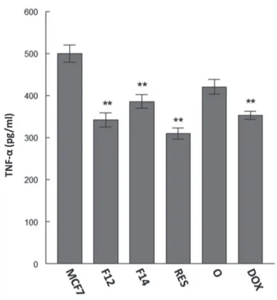
Reduction of of TNF-α level by resin and fractions of P. heptaphyllum
Pinto et al. (2008) demonstrated the anti-inflammatory potential of the isomeric mixture of α- and β-amyrin in an animal model of acute peritonitis showing that TNF-α levels are reduced after treatment. The results of the present study show that treatment with the crude resin and its fractions
is capable to reduce the TNF-α level, thereby inhibiting preexisting inflammatory processes in MCF-7 cells (Fig. 4).
The local administration of low concentrations of TNF-α
shows a potent anti-tumor effect with antiangiogenic action (Lejeune et al., 1998). The combination of doxorubicin with target-TNF-α on H22 allografted tumor showed a stronger antitumor effect than the single doxorubicin agent alone (Jiang et al., 2014). However, its endogenous production increases the development and proliferation of tumor (Ryuto et al., 1996; Samaniego et al., 1997; Yoshida et al., 1997; Leek et al., 1998; Relf et al., 1997). In the present study, doxorubicin decreases the level of endogenous TNF-α.
Alvarez-García et al. (2012) demonstrated that TNF-α may be produced endogenously by MCF-7 breast cancer cells, which was also observed in the results of this study (Fig. 4). Shishodia et al. (2003) suggested that ursolic acid is able to reduce the production of inflammatory cytokines such as TNF-α, thereby inhibiting the activation of nuclear transcription factor (NF-κB), a regulatory factor for several genes that mediate the process of tumorigenesis. Thus, the cytotoxic and pro-apoptotic activities of the doxorubicin, resin and its fractions may be related to the reduction of TNF-α in MCF-7 cells, all of them with the same potency.
Recent research showed that doxorubicin can induce cell death in MCF-7 cell by Akt/ERK-mediated and
Fas/FasL-Figure 3 – Analysis of apoptosis kjby flow cytometry in a breast cancer cell line (MCF-7). The cells were treated with the Protium heptaphyllum crude resin (RES) and the fractions enriched in α-and β-amyrin (F12 and F14) at 40 μg/ml, and hydrogen peroxide (H2O2) for 72 h. After the treatment period of 72 h, the cells were labeled with Annexin V-FITC and propidium iodide (PI) and then analyzed by flow cytometry. Cells in the initial stage of apoptosis were defined as Annexin-V (+) / PI (-), while late apoptotic cells were defined as double positive. A, representative data from three independent experiments. B, percentage of apoptotic cells (the sum of Annexin-V (+) / PI (-) and double positive). The values represent the means ± SEM; **p < 0.01 compared to H2O2. ##p < 0.01
Figure 4 – Quantification of TNF-α by ELISA in a breast cancer cell line (MCF-7). The cells were treated with Protium heptaphyllum
crude resin (RES) and its fractions enriched with α-and β-amyrin (F12 and F14) at 40 μg/ml and doxorubicin (DOX) for 72 h. The values represent the means ± SEM; ** p < 0.01 compared to MCF-7 cells.
mediated caspase-8 activation, this can indicate a probable mechanism in which resin and fractions reduce TNF-α levels (Liu and Chang, 2011), whereas the doxorubicin showed a lower potency than F12, F14 and resin.
Participation of RAS in the pro-apoptotic activity of P. heptaphyllum
The role of the Renin-Angiotensin System (RAS) in the specific context of tumor cells has been discussed in a recent review (George et al., 2010). The role of RAS in angiogenesis, apoptosis and tumor proliferation is large, complex, and sometimes paradoxical (George et al., 2010). Tumor cell lines that possess activated RAS may respond to Angiotensin II (Ang II) stimuli, expressing cytokines that assist in angiogenesis, such as interleukin-8 (IL-8) (George et al., 2010).
However, the tumor cell response to TNF-α requires the action of Ang II to increase apoptosis (Wang et al., 2000). Several studies have reported that cells with high Angiotensin Converting Enzyme (ACE) activity produce higher levels of Ang II, increasing the rate of apoptosis (George et al., 2010). This mechanism may have been verified in the present study because (Herr et al., 2008) showed the expression of RAS components and their receptors in MCF-7 cells.
In this study, ACE activity was evaluated in MCF-7 cells in the presence and absence of treatment with triterpene (Herr et al., 2008). It was also observed that treatment with the resin increases ACE activity (Fig. 5). The high pro-apoptotic rate observed after treatment with the resin and the F12 and F14 fractions may be related to increased enzymatic activity after treatment (Fig. 5), whereas the doxorubicin showed a lower potency than F12, F14 and resin. However, the role of doxorubicin in RAS activation is not yet described.
Altogether, it may be concluded that treatment of MCF-7 cells with resin and the fractions enriched with α- and
β-amyrin was able to promote pro-apoptotic effects, most likely by decreasing the levels of TNF-α and increasing ACE activity. Further studies are required for a detailed evaluation of the mechanisms and pathways to better understand the apoptotic processes under study.
Authors’ contributions
Figure 5 – Percentages of ACE activity determined by ELISA in a breast cancer cell line (MCF-7). The cells were treated with
Protium heptaphyllum crude resin (RES) and its fractions enriched with α- and β-amyrin (F12 and F14) and doxorubicin (DOX) at 40 μg/ml for 72 h. The values represent the means ± SEM; **p < 0.01 and *p < 0.05 compared to MCF-7 cells. ##p < 0.01 and #p < 0.05
compared to DOX.
Conflicts of interest
The authors declare no conflicts of interest.
Acknowledgement
We acknowledge the Fundação de Amparo à Pesquisa do Espírito Santo for financially supporting this work. We also acknowledge University Vila Velha and Tommasi Analítica for providing the knowledge and structure necessary to accomplish this work.
R E F E R E N C E S
Abu Bakar, M.F., Mohamad, M., Rahmat, A., Burr, S.A., Fry, J.R., 2010. Cytotoxicity, cell cycle arrest, and apoptosis in breast cancer cell lines exposed to an extract of the seed kernel of Mangifera pajang (bambangan). Food Chem. Toxicol. 48, 1688-1697.
Adams, R.P., 2001. Identification of essential oil components by gas cromatography/mass spectroscopy. Carol Stream: Allured Publishing Corporation. p. 469.
Allavena, P., Garlanda, C., Borrello, M.G., Sica, A., Mantovani, A., 2008. Pathways connecting inflammation and cancer. Curr. Opin. Genet. Dev. 18, 3-10.
Alvarez-García, V., González, A., Alonso-González, C., Martínez-Campa, C., Cos, S., 2012. Melatonin interferes in the
desmoplastic reaction in breast cancer by regulating cytokine production. J. Pineal. Res. 52, 282-290.
Aragão, G.F., Pinheiro, C.M.C., Bandeira, N.P., Lemos, G.T.L., Viana, B.G.S.J., 2007. Analgesic and anti-inflammatory activities of the isomeric mixture of alpha- and beta-amyrin from Protium heptaphyllum (Aubl.) March. Herb. Pharmacother. 7, 31-47.
Basu, S., Nachat-Kappes, R., Caldefie-Chezet, F., Vasson, M.P., 2013. Eicosanoids and adipokines in breast cancer: from molecular mechanisms to clinical considerations. Antioxid. Redox Sign. 18, 323-360.
Boyed, M.R., 1997. The NCI in vitro anticancer drug discovery screen. In Anticancer drug development guide; preclinical screening, clinical trials and approval; Teicher, B., Ed. Humana Press: Totowa, p. 30.
Brandão, M.G.L., Zanetti, N.N.S., Oliveira, G.R.R., Goulart, L.O., Monte-Mor, R.L.M., 2008. Other medicinal plants and botanical products from the first edition of the Brazilian Official Pharmacopoeia. Rev. Bras. Farmacogn. 18, 127-134. Campisi, J., 2005. Aging, tumor suppression and cancer: high
wire-act! Mech. Ageing. Dev. 126, 51-58.
Correa, P., 1978. Dicionário das plantas úteis do Brasil e das exóticas cultivadas. Imprensa Nacional: Rio de Janeiro.
George, A.J., Thomas, W.G., Hannan, R.D., 2010. The renin– angiotensin system and cancer: old dog, new tricks. Nat. Rev. Cancer. 10, 745-759.
Herr, D., Rodewald, M., Fraser, H.M., Hack, G., Konrad, R., Kreienberg, R., Wulff, C., 2008. Potential role of renin-angiotensin-system for tumor angiogenesis in receptor negative breast cancer. Gynecol. Oncol. 109, 418-425.
Jiang, C., Niu, J., Li, M., Teng, Y., Wang, H., Zhang, Y., 2014. Tumor vasculature-targeted recombinant mutated human TNF-α
enhanced the antitumor activity of doxorubicin by increasing tumor vessel permeability in mouse xenograft models. PLoS One 9. e87036.
Kroemer, G., Galluzzi, L., Brenner, C., 2007. Mitochondrial membrane permeabilization in cell death. Physiol. Rev. 87, 99-163.
Leek, R.D., Landers, R., Fox, S.B., Ng, F., Harris, A.L., Lewis, C.E., 1998. Association of tumour necrosis factor alpha and its receptors with thymidine phosphorylase expression in invasive breast carcinoma. Br. J. Cancer. 77, 2246-2251. Lejeune, F.J., Rüegg, C., Liénard, D., 1998. Clinical applications of
TNF-alpha in cancer. Curr. Opin. Immunol. 10, 573-580. Liang, Y., Yan, C., Schor, N.F., 2001. Apoptosis in the absence of
caspase 3. Oncogene. 20, 6570-6578.
Liu, J., Liu, Y., Mao, Q., Klaassen, C.D., 1994. The effects of 10 triterpenoid compounds on experimental liver injury in mice. Fund. Appl. Toxicol. 22, 34-40.
Liu, W.H., Chang, L.S., 2011. Fas/FasL-dependent and
-independent activation of caspase-8 in doxorubicin-treated human breast cancer MCF-7 cells: ADAM10 down-regulation activates Fas/FasL signaling pathway. Int. J. Biochem. Cell. Biol. 43, 1708-1719.
Maia, R.M., Barbosa, P.R., Cruz, F.G., Roque, N.F., Fascio, M., 2000. Triterpenos da resina de Protium heptaphyllum March (Bourseraceae): caracterização em misturas binárias. Quim. Nova. 23, 623-626.
Mooney, L.M., Al-Sakkaf, K.A., Brown, B.L., Dobson, P.R.M., 2002. Apoptotic mechanisms in T47D and MCF-7 human breast cancer cells. Br. J. Cancer. 87, 909-917.
Mosmann, T., 1983. Rapid colorimetric assay for cellular growth and survival: application to proliferation and cytotoxicity assays. J. Immunol. Methods. 65, 55-63.
Newman, D.J., Cragg, G.M., 2012. Natural products as sources of new drugs over the 30 years from 1981 to 2010. Nat. Prod. 75, 311-335. NIST Mass Spectral Program, NIST/EPA/NIH 2005. National
Institute of Standard Tests/United Stated Environment Protection Agency/National Institute of Health. Mass Spectral Library with Search Program. Data Version: NIST 05, Software Version 2.0d.
Pinto, S.A.H., Pinto, L.M.S., Cunha, G.M.A., Chaves, M.H., Santos, F.A., Rao, V.S., 2008. Anti-inflammatory effect of β, α-amyrin, a pentacyclic triterpene from Protium heptaphyllum in rat model of acute periodontitis. Inflammopharmacology. 16, 48-52.
Posmantur, R., Wang, K.K.W., Gilbertsen, R.B., 1998. Caspase-3-like activity is necessary for il-2 release in activated Jurkat t-cells. Exp. Cell Res. 244, 302-309.
Reed, J.C., 2003. Apoptosis-targeted therapies for cancer. Cancer Cell. 3, 17-22.
Relf, M., LeJeune, S., Scott, P.A., Fox, S., Smith, K., Leek, R., Moghaddam, A., Whitehouse, R., Bicknell, R., Harris, A.L., 1997. Expression of the angiogenic factors vascular endothelial cell growth factor, acidic and basic fibroblast growth factor, tumor growth factor beta-1, platelet-derived endothelial cell growth factor, placenta growth factor, and pleiotrophin in human primary breast cancer and its relation to angiogenesis. Cancer Res. 57, 963-969.
Ryuto, M., Ono, M., Izumi, H., Yoshida, S., Weich, H.A., Kohno, K., Kuwano, M., 1996. Induction of vascular endothelial growth factor by tumor necrosis factor alpha in human glioma cells. Possible roles of SP-1. J. Biol. Chem. 271, 28220-28228. Samaniego, F., Markham, P.D., Gendelman, R., Gallo, R.C., Ensoli,
B., 1997. Inflammatory cytokines induce endothelial cells to produce and release basic fibroblast growth factor and to promote Kaposi’s sarcoma-like lesions in nude mice. J. Immunol. 158, 1887-1894.
Serra, C.P., Cortes, S.F., Lombardi, J.A., Braga De Oliveira, A., Braga, F.C., 2005. Validation of a colorimetric assay for the in vitro screening of inhibitors of angiotensinconverting enzyme (ACE) from plant extracts. Phytomedicine. 12, 424-432. Shishodia, S., Majumdar, S., Banerjee, S., Aggarwal, B.B., 2003.
Ursolic acid inhibits nuclear factor-B activation induced by carcinogenic agents through suppression of IκBα kinase and p65 phosphorylation: correlation with down-regulation of cyclooxygenase 2, matrix metalloproteinase 9, and cyclin D1. Cancer Res. 63, 4375-4383.
Siani, A.C., Ramos, M.F.S., Guimarães, A.C., Susunaga, G.S., Zoghbi, M.G.B., 1999a.Volatile constituents from oleoresin of Protium heptaphyllum (Aubl.) March. J. Essent. Oil Res. 11, 72-74.
Siani, A.C., Ramos, M.F.S., Menezes-de-Lima, O., Soares, R.O.A., Rosas, E.C., Susunaga, G.S., Guimarães, A.C., Zoghbi, M.G.B., Henriques, M.G.M.O., 1999b. Evaluation of anti-inflamatory-related activity of essential oils from the leaves and resin of species of Protium. J. Ethnopharmacol. 66, 57-69.
Simstein, R., Burow, M., Parker, A., Weldon, C., Beckman, B., 2003. Apoptosis, chemoresistance, and breast cancer: insights from the MCF-7 cell model system. Exp. Biol. Med. 228, 995-1003. Susunaga, G.S., Siani, A.C., Pizzolatti, M.G., Yunes, R.A.,
DelleMonache, F., 2001. Triterpenes from the resin of Protium heptaphyllum. Fitoterapia. 72, 709-711.
Vieira Junior, G.M., Souza, C.M.L., Chaves, M.H., 2005. Resina de Protium heptaphyllum: Isolamento, caracterização estrutural e avaliação das propriedades térmicas. Quim. Nova 28, 183-187. Wang, R., Alam, G., Zagariya, A., Gidea, C., Pinillos, H., Lalude,
O., Choudhary, G., Oezatalay, D., Uhal, B.D., 2000. Apoptosis of lung epithelial cells in response to TNF-α requires angiotensin II generation de novo. J. Cell Physiol. 185, 253-259.
Yang, H.L., Chen, C.S., Chang, W.H., Lu, F.J., Lai, Y.C., Chen, C.C., Hseu, T.H., Kuo, C.T., Hseu, Y.C., 2006. Growth inhibition and induction of apoptosis in MCF-7 breast cancer cells by Antrodia camphorate. Cancer Lett. 231, 215-227.
Yoshida, S., Ono, M., Shono, T., Izumi, H., Ishibashi, T., Suzuki, H., Kuwano, M., 1997. Involvement of interleukin-8, vascular endothelial growth factor, and basic fibroblast growth factor in tumor necrosis factor alpha-dependent angiogenesis. Mol. Cell. Biol. 17, 4015-4023.