Gerodontology
Maximiliano Schünke Gomes(a) Patrícia Chagas(b)
Dalva Maria Pereira Padilha(c) Paulo Caramori(d)
Fernando Neves Hugo(c) Carla Helena Augustin Schwanke(e)
Juliana Balbinot Hilgert(c)
(a)Postgraduate Program, School of Dentistry, Univ Federal do Rio Grande do Sul, Porto Alegre, RS, Brazil.
(b)Department of Health Sciences, School of Nutrition, Univ Federal de Santa Maria, Palmeira das Missões, RS, Brazil.
(c)Department of Community Dentistry, School of Dentistry, Univ Federal do Rio Grande do Sul, Porto Alegre, RS, Brazil.
(d)Center for Cardiovascular Diagnosis and Intervention, Hospital São Lucas, Pontifical Catholic Univ of Rio Grande do Sul, Porto Alegre, RS, Brazil.
(e)Geriatrics and Gerontology Institute, Pontifical Catholic Univ of Rio Grande do Sul, Porto Alegre, RS, Brazil.
Corresponding Author: Maximiliano Schünke Gomes E-mail: [email protected]
Association between self-reported oral
health, tooth loss and atherosclerotic
burden
Abstract: Previous studies have suggested that oral diseases may in-luence the development of atherosclerosis. The aim of this study was to test the hypothesis that poor self-reported oral health (SROH) and tooth loss are positively associated with coronary atherosclerotic burden (CAB). 382 consecutive subjects undergoing coronary angiography were included. Socio-demographic characteristics, cardiovascular risk factors and oral health status were collected using a standardized questionnaire, including data on SROH and use of dental prosthesis. Number of teeth and anthropometric measures were collected through clinical examina-tions. CAB at coronary angiography was quantiied using the Friesinger score (FS). Prevalence ratios (PR) were calculated with Poisson regression analyses. Mean age was 60.3 ± 10.8 years, with 63.2% males. In the bivariate analysis, there was a signiicant association (p < 0.05) between CAB and age (≥ 60y) (PR = 1.01, 95% CI = 1.02–1.16), male gender (PR = 1.11, 95% CI = 1.03–1.19), smoking (PR = 1.08, 95% CI = 1.01– 1.16), hypertension (PR = 1.12, 95% CI = 1.03–1.22), diabetes (PR = 1.17, 95% CI = 1.05–1.21), poor SROH (PR = 1.22, 95% CI = 1.02–1.46) and tooth loss (< 20teeth present) (PR = 1.10, 95% CI = 1.02–1.19). The use of dental prosthesis was not associated with CAB. The multivariate mod-els, adjusted for age, gender, smoking, hypertension, diabetes and dyslip-idemia showed that poor SROH (p = 0.03) and tooth loss (p = 0.02) were independently associated with CAB, conirming the study hypothesis.
Descriptors: Atherosclerosis; Risk Factors; Epidemiology; Tooth Loss; Cardiovascular Diseases.
Introduction
Epidemiological studies have suggested that chronic periodontal dis-ease,1-4 lesions of endodontic origin5 and tooth loss6 are associated with
cardiovascular disease (CVD) and mortality.
The triggering of an inlammatory response by infectious agents is a potential mechanism correlating infection to the acceleration of ath-erosclerosis.7 Coronary atherosclerotic burden (CAB) is a term used to
describe the extension of atherosclerosis into coronary vessels.8 Previous
symptomatic atherosclerotic vascular disease (AVD) evaluated by a clini-cal score of CAB was shown to be an independent predictor of early mor-tality in patients with irst-ever ischemic stroke.9
Poor oral health is a major cause of a proinlammatory state and may Declaration of Interests: The authors
certify that they have no commercial or associative interest that represents a conflict of interest in connection with the manuscript.
Submitted: Mar 17, 2012
accelerate the atherosclerotic process or precipitate a plaque rupture.10 Poor oral health may also affect
eating behavior and contribute to poor nutrition, which has been identiied as a risk factor for mor-tality. Potential pathogenic mechanisms linking oral infections and AVD are based on three main path-ways:
• the role of periodontal pathogens and their prod-ucts in the development of endothelial dysfunc-tion;
• the contribution of oral microorganisms to the formation of fatty streaks and atherosclerotic plaques; and
• the role of oral lora in the modulation and matu-ration of atheromatous plaques, facilitating their rupture and vascular thrombosis.10,11
Studies have found associations between tooth loss and carotid12 and aortic13 intima-media
thick-ness, as well as aortic valve sclerosis14. In diabetic
patients, positive correlations between atherogenic factors and oral hygiene, periodontal disease and tooth loss were found.15 In another study, tooth
loss was associated with inlammatory markers and stroke.16
Oral diseases are primarily associated with non-communicable chronic diseases through shared common risk factors such as age, lifestyle, diet, smoking, and low socioeconomic status. Accord-ingly, there is some evidence that, after adjusting for these risk factors, the relationship between oral health and CVD may be weakened.17
Self-reported health status assessing systemic diseases and health-related conditions are widely used in populational investigations. In the last years, self-reported oral health (SROH) status has been in-creasingly implemented in dentistry.18-21
Few studies have tested the relationship between clinical scores of CAB and oral health in humans. The current study tested this association in a group of southern Brazilian patients using a SROH ap-proach,19 supplemented by an oral clinical
examina-tion measuring the number of teeth. The aim of this study was to test the hypothesis that poor SROH status and tooth loss are positively associated with CAB.
Methodology
The research protocol was approved by the Eth-ics and Research Committee of the Pontiical Catho-lic University of Rio Grande do Sul (PUCRS), num-ber 08/04211. All participants provided a signed informed consent form. Consecutive adult patients (≥ 18 years) undergoing coronary angiography to in-vestigate coronary artery disease in the Center for Cardiovascular Diagnosis and Intervention, São Lu-cas Hospital (Porto Alegre, Brazil), were invited to participate. Emergency cases or patients unable to answer the questionnaire due to physical or mental conditions were excluded. Data were collected prior to the angiography, from October 2008 to Decem-ber 2009, including a total of 382 individuals. All participants survived after angiography.
Socio-demographic data (age, gender, marital status, education and occupation) and medical car-diovascular risk factors (smoking, hypertension, dyslipidemia, diabetes, family history of coronary heart disease [CHD] and use of statins) were col-lected using a structured questionnaire. Weight (kg) was measured using an anthropometric calibrated scale (Filizola, São Paulo, Brazil). Height (m) was measured using the stadiometer of the anthropomet-ric scale. The body mass index (BMI) was calculated by dividing the weight by the height squared. Data collection procedures are further described in a pre-vious study.22
Information on oral health was collected using a structured questionnaire. Measures of self-per-ception on oral health included the variables “self-reported oral health (SROH)” (excellent, very good, good, fair or poor)19,23 and “use of dental
prosthe-sis” (yes or no). Total number of natural teeth was measured by simpliied oral clinical examination, performed by a trained non-dentist examiner. Num-ber of teeth (tooth loss) was dichotomized into non-functional dentition (< 20 teeth) and non-functional den-tition (≥ 20 teeth).6
CAB was evaluated through the Friesinger Score (FS)22,24 on the diagnostic coronary
cardiolo-gist, blinded to the oral health data. For analytic purposes, CAB was dichotomized into low (FS ≤ 7) and high (FS > 7), based on the distribution of the FS in the present sample (mean and standard devia-tion = 7.3 ± 4.0; median = 7).
Data were analyzed using SPSS v.17 (IBM, Chi-cago, USA). Descriptive statistics (N and %) ac-cording to CAB were performed. Prevalence ratios (PR) were calculated with bivariate and multivariate Poisson regression analyses with robust variance.25
Associations between CAB and SROH and between CAB and number of teeth were determined sepa-rately and adjusted for the socio-demographic and medical confounders. Spearman’s correlation (rs) was calculated between SROH and number of teeth. The value for rejection of the null hypothesis was set at p ≤ 0.05.
Results
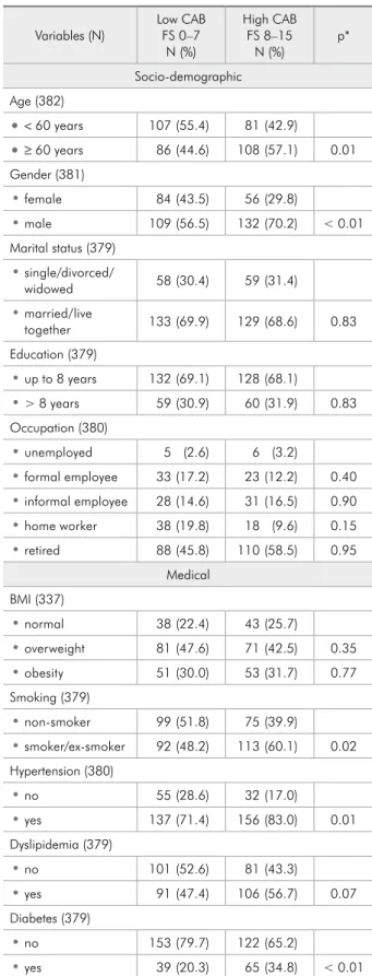
Characteristics of the sample in relation to CAB are shown in Table 1. Mean age was 60.3 (± 10.8), ranging from 23 to 89 years, with males (63.3%) predominating. Dental variables revealed that near-ly 45% of the participants reported poor or fair oral health status, more than 67% wore dental prosthe-sis, and only 33% had ≥ 20 teeth.
In the unadjusted analysis (Table 1), there was a signiicant association between CAB and age (PR = 1.01, 95% CI = 1.02–1.16), gender (PR = 1.11, 95% CI = 1.03–1.19), smoking (PR = 1.08, 95% CI = 1.01–1.16), hypertension (PR = 1.12, 95% CI = 1.03– 1.22), diabetes (PR = 1.17, 95% CI = 1.05–1.21), poor SROH (PR = 1.22, 95% CI = 1.02–1.46) and number of teeth < 20 (PR = 1.10, 95% CI = 1.02– 1.19).
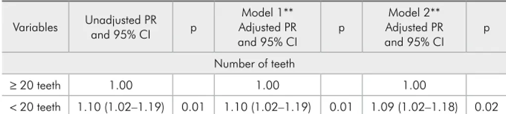
Multivariate models testing the association of SROH and number of teeth with CAB, after adjust-ing for age and gender (Model 1) and age, gender, smoking, hypertension, diabetes and dyslipidemia (Model 2) are shown in Tables 2 and 3. Poor SROH (PR = 1.22, 95% CI = 1.02–1.47) or fair SROH (PR = 1.20, 95% CI = 1.02–1.41) and having < 20 teeth (PR = 1.09, 95% CI = 1.02–1.18) were inde-pendently associated with CAB after adjustments. A signiicant correlation between SROH and number of teeth (rs= 0.23, p < 0.01) was found.
Table 1 - Socio-demographic, medical and dental charac-teristics of participants (N, %), by coronary atherosclerotic burden (CAB) as measured with the Friesinger Score (FS) (continued on next page).
Variables (N) Low CABFS 0–7 N (%)
High CAB FS 8–15
N (%) p* Socio-demographic
Age (382)
• < 60 years 107 (55.4) 81 (42.9)
• ≥ 60 years 86 (44.6) 108 (57.1) 0.01 Gender (381)
• female 84 (43.5) 56 (29.8)
• male 109 (56.5) 132 (70.2) < 0.01 Marital status (379)
• single/divorced/
widowed 58 (30.4) 59 (31.4)
• married/live
together 133 (69.9) 129 (68.6) 0.83 Education (379)
• up to 8 years 132 (69.1) 128 (68.1)
• > 8 years 59 (30.9) 60 (31.9) 0.83 Occupation (380)
• unemployed 5 (2.6) 6 (3.2)
• formal employee 33 (17.2) 23 (12.2) 0.40
• informal employee 28 (14.6) 31 (16.5) 0.90
• home worker 38 (19.8) 18 (9.6) 0.15
• retired 88 (45.8) 110 (58.5) 0.95 Medical
BMI (337)
• normal 38 (22.4) 43 (25.7)
• overweight 81 (47.6) 71 (42.5) 0.35
• obesity 51 (30.0) 53 (31.7) 0.77 Smoking (379)
• non-smoker 99 (51.8) 75 (39.9)
• smoker/ex-smoker 92 (48.2) 113 (60.1) 0.02 Hypertension (380)
• no 55 (28.6) 32 (17.0)
• yes 137 (71.4) 156 (83.0) 0.01 Dyslipidemia (379)
• no 101 (52.6) 81 (43.3)
• yes 91 (47.4) 106 (56.7) 0.07 Diabetes (379)
• no 153 (79.7) 122 (65.2)
Discussion
The results of this cross-sectional study con-irmed the hypothesis that SROH status and num-ber of teeth were signiicantly associated with CAB in this group of Brazilian patients. Most important-ly, this association was independent of other tra-ditional risk factors for CVD, such as age, gender, smoking, hypertension, diabetes and dyslipidemia. To our knowledge, this is one of the irst studies to provide evidence of the connection between CAB and oral diseases based on the assessment of SROH status in combination with tooth loss. Nevertheless, inherent limitations of the study design do not al-low inferences about causality concerning this as-sociation.
The present study evaluated CAB through the FS. Other studies assessed carotid and aortic inti-ma-media thickness or aortic valve sclerosis,12-14
in-stead of luminal obstruction of the main coronary arteries quantiied using the FS. Unlike other sys-tems for evaluating the extent of CHD, the FS was speciically developed for the assessment of parietal AVD, regardless of the area of perfused myocardi-um through the stenosis.22
Potential mechanisms that link oral infections to
Variables Unadjusted PR and 95% CI p Adjusted PR and Model 1*
95% CI p
Model 2* Adjusted PR and
95% CI p
SROH
excellent 1.00 1.00 1.00
very good 1.12 (0.93–1.35) 0.22 1.11 (0.92–1.33) 0.26 1.13 (0.94–1.35) 0.20 good 1.16 (0.99–1.36) 0.07 1.15 (0.98–1.35) 0.08 1.16 (0.99–1.37) 0.07 fair 1.19 (1.01–1.39) 0.04 1.19 (1.02–1.39) 0.03 1.20 (1.02–1.41) 0.03 poor 1.22 (1.02–1.46) 0.03 1.26 (1.05–1.50) 0.01 1.22 (1.02–1.47) 0.03
p = p-value, Poisson regression. (*) adjusted through Poisson regression for: Model 1 - age, gender; Model 2 - age, gender, smoking, hypertension, diabetes, dyslipidemia.
Table 2 - Adjusted models for the association of self-reported oral health (SROH) with coronary atherosclerotic burden (CAB) as measured with the Friesinger Score (FS). N = 378. Family history of CHD (379)
• no 152 (79.2) 145 (77.5)
• yes 40 (20.8) 42 (22.5) 0.70 Use of Statins (374)
• no 110 (57.9) 98 (53.3)
• yes 80 (42.1) 86 (46.7) 0.37 Dental
SROH (378)
• excellent 15 (7.9) 6 (3.2)
• very good 24 (12.6) 19 (10.2) 0.22
• good 74 (38.7) 72 (38.5) 0.07
• fair 61 (31.9) 68 (36.4) 0.04
• poor 17 (8.9) 22 (11.8) 0.03 Use of dental prosthesis (378)
• no 123 (64.4) 132 (70.6)
• yes 68 (35.6) 55 (29.4) 0.20 Number of teeth (374)
• < 20 teeth 117 (61.3) 135 (73.8)
• ≥ 20 teeth 74 (38.7) 48 (26.2) 0.01 Total (382) 193 (50.5) 189 (49.5)
-* p-value for bivariate analysis, Poisson regression; BMI = body mass index; CHD = coronary heart disease; SROH = self-reported oral health.
Table 1 (continued)
Variables Unadjusted PRand 95% CI p Adjusted PRModel 1** and 95% CI p
Model 2** Adjusted PR and 95% CI p Number of teeth
≥ 20 teeth 1.00 1.00 1.00
< 20 teeth 1.10 (1.02–1.19) 0.01 1.10 (1.02–1.19) 0.01 1.09 (1.02–1.18) 0.02
p = p-value, Poisson regression. (*) adjusted through Poisson regression for: Model 1 - age, gender; Model 2 - age, gender, smoking, hypertension, diabetes, dyslipidemia.
atherogenesis are based on the role of periodontal pathogens and their products in the development of endothelial dysfunction, formation of fatty streaks and maturation of atherosclerotic plaques, with their rupture and vascular thrombosis.11 Our results
conirm previous indings in which periodontal dis-ease,15 lesions of endodontic origin5 or tooth loss
12-14,16 were signiicantly associated with AVD,12-14
ath-erogenic risk factors15 or cardiovascular events such
as stroke16 and CHD.5 Nevertheless, other studies
failed to ind this association.17,26
The methodologies of previous studies testing the association between oral health and CVD dif-fered from that of the present study. A cohort study3
measured the mean bone loss and the probing pock-et depth scores per tooth. In other studies, subjects received a periodontal examination4 or a
periodon-tal microbiological evaluation,2 and a carotid scan
using high-resolution ultrasound was the method used to evaluate subclinical AVD. Importantly, there is extensive variability in the literature regarding deinitions of the oral exposure, including salivary low, reported periodontal disease, number of teeth, oral organisms, antibodies to oral organisms, and different periodontal disease parameters.1
This study focused on the assessment of SROH and number of teeth, instead of investigating mi-crobiological, clinical and radiographic parame-ters. The use of SROH measures provides relevant cost and time savings in large epidemiological sur-veys.19,21 SROH is a known Likert-type scale and
previous studies have provided consistent evidence of the construct validity of this model.19,23
Limitations of the SROH approach must be clariied. Self-perceived oral health was shown to be better in individuals with more teeth and recent dental treatment and worse in those with tooth mo-bility, coronal decay and medical problems.19 SROH
measures showed valid estimates for variables such as number of teeth, illings, root canal therapy and prosthesis, but was less accurate for the assessment of dental caries and periodontal disease.20 In fact, it
is known that self-perceived oral health is inluenced not only by dental clinical oral status, but also by social and psychological issues.27
This study includes self-perceived measures of
disease, not only related to dental variables, but also to medical cardiovascular risk factors. Diagnosis of chronic diseases by a physician would result in more reliable information regarding the presence of co-morbidities. Although SROH is a known valid mea-sure, the analysis would beneit from the inclusion of a detailed oral clinical evaluation. Unfortunately, such data were not collected due to hospital service characteristics. However, the signiicant correlation between SROH and number of teeth found here in-dicates that the accuracy of self-perceived oral mea-sures was not divergent from the actual clinical ind-ings in this sample.
Number of teeth is a surrogate variable com-monly used to access history of periodontal disease. Most studies, however, do not consider that tooth loss may occur not only due to periodontitis, but also due to dental caries, endodontic infections or trauma. Periodontal disease is the main reason for tooth loss at the tooth level, but caries/endodontic disease is the most common cause of tooth loss at the individual level.28 Tooth loss is one of the
stron-gest populational oral health indicators, working as a “registry” of the history of both periodontal and endodontic diseases. Tooth loss is not only a biologi-cal process; it can also involve factors such as atti-tudes of patients and providers, access to care issues and dental care delivery systems.
The weak but statistically signiicant associa-tion between oral status and AVD estimated in this study may raise questions about the “statistical signiicance” versus its “clinical relevance”. In this study, the PR of classic risk factors for CVD were similar to those found for dental variables. The low but statistically signiicant PR do not affect the clin-ical relevance of these classic risk factors for CVD. The aim of our study was to test the hypothesis of the association rather than exploring its clinical rel-evance, which may only be considered after future evidence from longitudinal studies.
longitudi-nal studies, which would contribute to a better un-derstanding of this multifactorial relationship.
Conclusion
SROH status and number of teeth were indepen-dently associated with CAB, measured by the FS, in a group of Brazilian patients, thus conirming the study hypothesis.
Acknowledgements
The authors thank Ms. Tatiana P. Galdino for as-sistance during data collection, Dr. Christiano Barcel-los for the FS evaluations, and Dr. Mark A. Reynolds for reviewing the manuscript. This study was sup-ported in part by the CAPES Foundation, Ministry of Education of Brazil, scholarship number 1433/11-3. The authors disclose any conlict of interest.
References
1. Beck JD, Offenbacher S. Systemic effects of periodontitis: epi-demiology of periodontal disease and cardiovascular disease. J Periodontol. 2005 Nov;76(11 Suppl):2089-100.
2. Desvarieux M, Demmer RT, Rundek T, Boden-Albala B, Ja-cobs DR Jr, Sacco RL, et al. Periodontal microbiota and ca-rotid intima-media thickness: the Oral Infections and Vascular Disease Epidemiology Study (INVEST). Circulation. 2005 Feb 8;111(5):576-82.
3. Beck J, Garcia R, Heiss G, Vokonas PS, Offenbacher S. Peri-odontal disease and cardiovascular disease. J Periodontol. 1996 Oct;67(10 Suppl):1123-37.
4. Desvarieux M, Demmer RT, Rundek T, Boden-Albala B, Jacobs DR Jr, Papapanou PN, et al. Relationship between periodontal disease, tooth loss, and carotid artery plaque: the Oral Infections and Vascular Disease Epidemiology Study (INVEST). Stroke. 2003 Sep;34(9):2120-5.
5. Caplan DJ, Chasen JB, Krall EA, Cai J, Kang S, Garcia RI, et al. Lesions of endodontic origin and risk of coronary heart disease. J Dent Res. 2006 Nov;85(11):996-1000.
6. Padilha DM, Hilgert JB, Hugo FN, Bos AJ, Ferrucci L. Number of teeth and mortality risk in the Baltimore Longitudinal Study of Aging. J Gerontol A Biol Sci Med Sci. 2008 Jul;63(7):739-44. 7. Hayashi C, Viereck J, Hua N, Phinikaridou A, Madrigal AG,
Gibson FC 3rd, et al. Porphyromonas gingivalis accelerates in-flammatory atherosclerosis in the innominate artery of ApoE deficient mice. Atherosclerosis. 2011 Mar;215(1):52-9. 8. Guerrero M, Harjai K, Stone GW, Brodie B, Cox D, Boura J,
et al. Usefulness of the presence of peripheral vascular disease in predicting mortality in acute myocardial infarction patients treated with primary angioplasty (from the Primary Angio-plasty in Myocardial Infarction Database). Am J Cardiol. 2005 Sep 1;96(5):649-54.
9. Roquer J, Ois A, Rodriguez-Campello A, Gomis M, Mun-teis E, Jimenez-Conde J, et al. Atherosclerotic burden and early mortality in acute ischemic stroke. Arch Neurol. 2007 May;64(5):699-704.
10. Cotti E, Dessi C, Piras A, Mercuro G. Can a chronic dental infection be considered a cause of cardiovascular disease? A review of the literature. Int J Cardiol. 2011 Apr 1;148(1):4-10.
11. Kebschull M, Demmer RT, Papapanou PN. “Gum bug, leave my heart alone!”--epidemiologic and mechanistic evidence linking periodontal infections and atherosclerosis. J Dent Res. 2010 Sep;89(9):879-902.
12. Chin UJ, Ji S, Lee SY, Ryu JJ, Lee JB, Shin C, et al. Relation-ship between tooth loss and carotid intima-media thickness in Korean adults. J Adv Prosthodont. 2010 Dec;2(4):122-7. 13. Castillo R, Fields A, Qureshi G, Salciccioli L, Kassotis J,
Lazar JM. Relationship between aortic atherosclerosis and dental loss in an inner-city population. Angiology. 2009 Jun-Jul;60(3):346-50.
14. Völzke H, Schwahn C, Hummel A, Wolff B, Kleine V, Rob-inson DM, et al. Tooth loss is independently associated with the risk of acquired aortic valve sclerosis. Am Heart J. 2005;150(6):1198-203.
15. Furukawa T, Wakai K, Yamanouchi K, Oshida Y, Miyao M, Watanabe T, et al. Associations of periodontal damage and tooth loss with atherogenic factors among patients with type 2 diabetes mellitus. Intern Med. 2007;46(17):1359-64. 16. You Z, Cushman M, Jenny NS, Howard G. Tooth loss,
sys-temic inflammation, and prevalent stroke among participants in the reasons for geographic and racial difference in stroke (REGARDS) study. Atherosclerosis. 2009 Apr;203(2):615-9. 17. Tuominen R, Reunanen A, Paunio M, Paunio I, Aromaa A.
Oral health indicators poorly predict coronary heart disease deaths. J Dent Res. 2003 Sep;82(9):713-8.
18. Douglass CW, Berlin J, Tennstedt S. The validity of self-re-ported oral health status in the elderly. J Public Health Dent. 1991 Fall;51(4):220-2.
19. Jones JA, Kressin NR, Spiro A 3rd, Randall CW, Miller DR, Hayes C, et al. Self-reported and clinical oral health in users of VA health care. J Gerontol A Biol Sci Med Sci. 2001 Jan;56(1):M55-62.
20. Pitiphat W, Garcia RI, Douglass CW, Joshipura KJ. Validation of self-reported oral health measures. J Public Health Dent. 2002 Spring;62(2):122-8.
22. Chagas P, Caramori P, Barcellos C, Galdino TP, Gomes I, Schwanke CH. Association of different anthropometric mea-sures and indices with coronary atherosclerotic burden. Arq Bras Cardiol. 2011 Sep;97(5):397-401.
23. Gilbert GH, Duncan RP, Heft MW, Dolan TA, Vogel WB. Multidimensionality of oral health in dentate adults. Med Care. 1998 Jul;36(7):988-1001.
24. Javed F, Aziz EF, Nadkarni GN, Khan SA, Sabharwal MS, Malhan R, et al. The association of the Friesinger score and pulse pressure in an urban South Asian patient population: pulse pressure, an independent predictor of coronary artery disease. J Am Soc Hypertens. 2010 May-Jun;4(3):142-7. 25. Barros AJ, Hirakata VN. Alternatives for logistic regression
in cross-sectional studies: an empirical comparison of models
that directly estimate the prevalence ratio. BMC Med Res Methodol. 2003 Oct 20;3:21.
26. Joshipura KJ, Rimm EB, Douglass CW, Trichopoulos D, Ascherio A, Willett WC. Poor oral health and coronary heart disease. J Dent Res. 1996 Sep;75(9):1631-6.
27. Martins AB, Santos CM, Hilgert JB, Marchi RJ, Hugo FN, Padilha DMP. Resilience and self-perceived oral health: a hier-archical approach. J Am Geriatr Soc. 2011 Apr;59(4):725-31. 28. Phipps KR, Stevens VJ. Relative contribution of caries and