Paula Passalini(a)
Tatiana Kelly da Silva Fidalgo(b) Erika Machado Caldeira(c) Rogério Gleiser(d)
Matilde da Cunha Gonçalves Nojima(d)
Lucianne Cople Maia(d)
(a) DDS; (b)PhD Student in Pediatric Dentistry; (c)MsD Student in Orthodontics – School of Dentistry of Rio de Janeiro, Federal University of Rio de Janeiro, Rio de Janeiro, Brazil.
(d) PhD, Associate Professor, Department of Pediatric Dentistry and Orthodontics, School of Dentistry, Federal University of Rio de Janeiro, Rio de Janeiro, Brazil.
Corresponding author:
Lucianne Cople Maia
Rua Gastão Gonçalves, 47/501 - Santa Rosa
Niterói - RJ - Brazil CEP: 24240-030 E-mail: [email protected]
Received for publication on Oct 05, 2009 Accepted for publication on Mar 29, 2010
Mechanical properties of one and
two-step fluoridated orthodontic resins
submitted to different pH cycling regimes
Abstract: The aim of this study is to assess the in vitro shear bond strength and adhesive remnant index (ARI) of one and two-step luori-dated orthodontic resins under conditions that simulate high cariogenic challenge. Edgewise brackets for maxillary central incisors were random-ly bonded to 80 bovine incisors, using either TransbondTM Plus Color
Change orthodontic resin and a self-etching primer adhesive (G1; n = 40) or Orthodontic Fill Magic with a conventional acid-etch technique (G2; n = 40). Each group of resin (n = 10) was divided into: immediate shear (A- pre-cycling control), immersion in artiicial remineralizing saliva (neutral saliva) for 14 days (B- post-cycling control) and pH cycling with high cariogenic challenge (C- acid saliva with pH 5.5 and D- acid saliva with pH 4.5). After 14 days of pH cycling, the shear bond strength and ARI were evaluated. Considering the shear bond strength, TransbondTM
Plus Color Change resin was stronger than Orthodontic Fill Magic when it was submitted to high cariogenic challenge (p < 0.05). Also Trans-bondTM Plus Color Change resin showed better adhesion to enamel than
Orthodontic Fill Magic, in all situations evaluated (p < 0.05). It could be concluded that TransbondTM Plus Color Change resin presented better
shear bond strength and adhesive remnant index when submitted to high cariogenic challenge, in comparison with Orthodontic Fill Magic.
Descriptors: Orthodontic brackets; Composite resins; Shear strength; Tooth demineralization.
Introduction
In orthodontics practice, white spot lesions are observed around orth-odontic appliances with relative frequency.1-3 Caries lesions adjacent to
brackets can be reduced or even completely inhibited when a luoride dentifrice is used.4 However, its use depends on the patient’s compliance,
which is usually inadequate.5 Therefore, preventive measures that do
not depend on an individual’s compliance were developed to solve this problem, such as bonding dental materials with luoride-releasing prop-erties,6,7 which exhibit an additional source of luoride locally, near the
brackets.2,8
Glass ionomer cement was presented as the irst material with poten-tial cariostatic properties, however, its use in orthodontics has the dis-advantage of low bond strength to dental substrate.2,9,10,11 Orthodontic
with glass ionomer orthodontic cements used for this purpose.12
The development of materials with high bond strength associated with luoride release, such as luoride-releasing composite resins, began to solve these problems.13 The literature has demonstrated
that resins for bonding procedures present satis-factory mechanical properties.14,15 Fluoride release
of bonding materials16,17 and their release
mecha-nisms18,19 have been extensively studied. Few in vitro
studies have evaluated the real inluence of cario-genic challenge that simulates oral environment in shear bond strength tests.
The traditional system of bonding orthodontic brackets is a three-step mechanism that involves three separate agents: an enamel conditioner, a priming agent, and an adhesive resin.20 To reduce
chair time and improve effectiveness, a two-step mechanism, which combines a primer and adhesive agent, and recently, self-etching primers (SEPs) have been developed. These systems combine the condi-tioning and priming agents into a single acidic prim-er solution for simultaneous use on both enamel and dentin.21 However, there are no studies in the
litera-ture that assess bonding material properties under conditions that simulate the oral environment, such as using a pH cycling with a high cariogenic chal-lenge model.
Thus, the purpose of the present study is to eval-uate the shear bond strength and adhesive remnant index of one and two-step luoridated orthodontic resins submitted to two pH cycling regimes with different demineralization potentials, simulating a
high cariogenic challenge.
Materials and Methods
Sample preparation
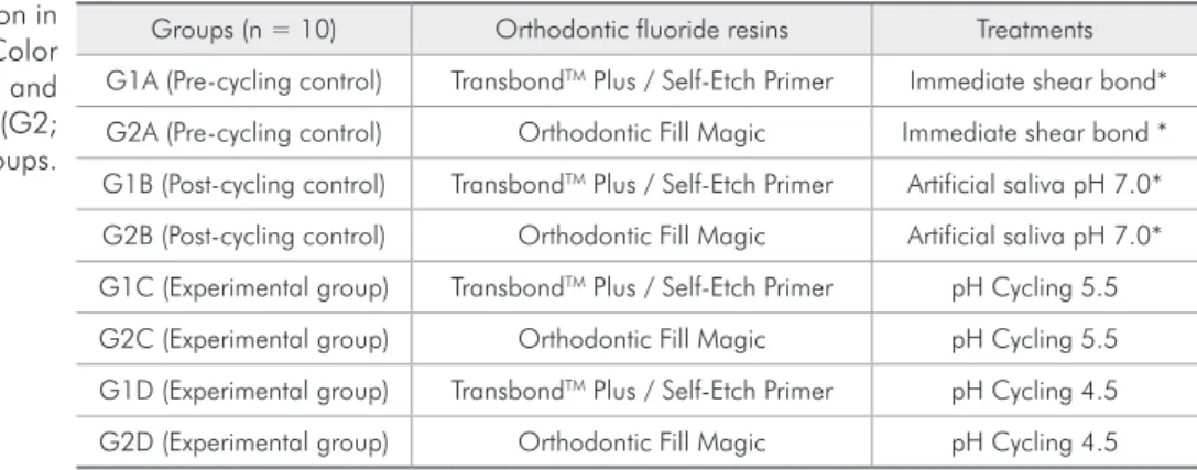
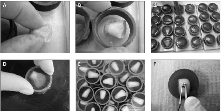
Eighty bovine incisors were randomly divided into 8 groups, n = 10 (Table 1), and sectioned along the cemento-enamel junction (Figure 1A). Next, the crowns were submersed in epoxy resin with the buccal surface facing the glass plate (Figure 1B,C). Silicon carbide abrasive papers with successive grits (180, 400 and 600 - 3M, Rio de Janeiro, RJ, Brazil) were used to expose the bonding area (Figure 1D,E). After this, the coronal portion was submitted to prophylaxis with prophylactic rubber cups (KG So-rensen, Rio de Janeiro, RJ, Brazil) at low speed for 5 seconds. Samples were washed in deionized water and dried using an oil-free air jet and water vapor for 15 seconds.
Maxillary central incisor brackets (Edgewise system – Morelli, Rio de Janeiro, RJ, Brazil) were bonded in the most central area of the middle third of the bovine incisor buccal surface (Figure 1F) with the two different orthodontic light-polymerized lu-oridated resins (Table 1): TransbondTM Plus Color
Change (G1) using a one-step self-etching primer adhesive (TSEP; 3M Unitek, Monrovia, California, USA) and Orthodontic Fill Magic (G2) with a con-ventional acid-etch technique consisting of two steps (Vigodent, Rio de Janeiro, RJ, Brazil). For G1, TransbondTM Plus Self Etching Primer (3M Unitek,
Monrovia, California, USA) was used. For G2, the enamel was previously etched with 37% phosphoric acid (SSWhite, Rio de Janeiro, RJ, Brazil) for 30
sec-Groups (n = 10) Orthodontic fluoride resins Treatments
G1A (Pre-cycling control) TransbondTM Plus / Self-Etch Primer Immediate shear bond*
G2A (Pre-cycling control) Orthodontic Fill Magic Immediate shear bond *
G1B (Post-cycling control) TransbondTM Plus / Self-Etch Primer Artificial saliva pH 7.0*
G2B (Post-cycling control) Orthodontic Fill Magic Artificial saliva pH 7.0*
G1C (Experimental group) TransbondTM Plus / Self-Etch Primer pH Cycling 5.5
G2C (Experimental group) Orthodontic Fill Magic pH Cycling 5.5
G1D (Experimental group) TransbondTM Plus / Self-Etch Primer pH Cycling 4.5
G2D (Experimental group) Orthodontic Fill Magic pH Cycling 4.5
*Groups not submitted to pH cycling.
Table 1 - Sample division in the TransbondTM Plus Color
onds, followed by the application of a one-compo-nent adhesive resin, according to the manufacturer’s instructions. The samples were stored in deionized water at room temperature for one day.
pH cycling
Exactly 24 hours after bonding, negative pre-cy-cling control groups (G1A and G2A) were submitted to immediate shear bond strength testing, without undergoing modiied cariogenic pH cycling accord-ing to Queiroz et al.22 (2008). Negative post-cycling
control groups (G1B and G2B) remained in artiicial remineralizing/neutral saliva (1.54 mmol/L calcium, 1.54 mmol/L phosphate, 20 mmol/L acetic acid and 0.308 g ammonium acetate, adjusted to pH 7.0 with potassium hydroxide; VETEC, Rio de Janeiro, RJ, Brazil)23 for 14 days. Experimental groups (G1C/
G2C and G1D/G2D) were submitted to pH cycling, simulating two different cariogenic challenges, us-ing artiicial remineralizus-ing and demineralizus-ing saliva (3 mmol/L calcium, 3 mmol/L phosphate, 50 ml/L acetic acid and 0.308 g ammonium acetate; VETEC, Rio de Janeiro, RJ, Brazil)24 with medium
and high demineralizing potential (pH adjusted to 5.5 and 4.5 with sodium hydroxide; respectively).
The experimental groups submitted to pH cy-cling remained in demineralizing saliva daily for 22 hours consecutively, and after being washed with deionized water, they were kept in contact with remineralizing saliva for 2 hours, completing a cycle of 24 hours. During the period of pH cycling, the specimens were kept in an incubator (Fanem Ltd., São Paulo, SP, Brazil), at a constant temperature of 37°C in order to simulate the oral environment. These dynamics were reproduced for the period of 14 days, during which the artiicial saliva (neutral and acid) was changed every 2 days.
Shear bond strength
The shear tests were performed in a Universal Test machine (EMIC, São José dos Pinhais, SP, Bra-zil), at a constant speed of 0.5 mm/min. The force required to dislodge the bracket was recorded in Newtons (N) and converted into megapascals (MPa) as a ratio of Newtons to the bracket surface area (MPa = N/mm²).
Figure 1 - Sample preparation. (A) Positioning of buccal surface of bovine incisors facing the glass plate; (B) Insertion of ap-paratus support; (C) Submersion in epoxy resin; (D) Bond area exposure; (E) Washing of the samples; (F) Bonding of incisor brackets.
A B C
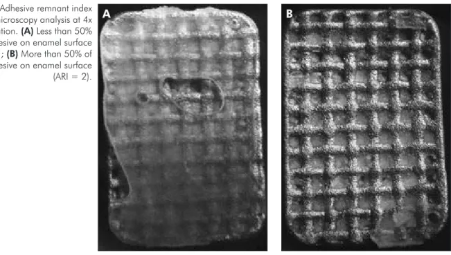
Adhesive Remnant Index (ARI)
The brackets and enamel surfaces were analyzed by two trained and calibrated examiners (Kap-pa = 1.00). An optical microscope (Eclipse E600, Nikon, Melville, NY, USA) was used at 4x magni-ication, with ARI scores according to Artün and Bergland,25 (1984) (Figure 2) as follows: in the range
from 0 to 3, where 0 = No adhesive on enamel sur-face; 1 = Less than 50% of the adhesive on enam-el surface; 2 = More than 50% of the adhesive on enamel surface; 3 = 100% of the adhesive on enamel surface.
Statistical analysis
The shear bond strength test results were insert-ed in the database of the statistical program SPSS 16.0 (SPSS Inc, Chicago, IL, USA) and submitted to analysis of variance (ANOVA) and the Tukey test. For ARI evaluation, Mann-Whitney and Kruskal-Wallis tests (p < 0.05) were applied.
Results
The high cariogenic challenge was able to induce white spot formation around orthodontic brackets (Figure 3).
Shear bond strength
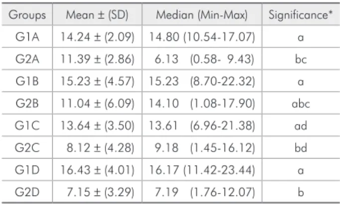
The results showed that G1 had higher shear bond strength than the mean of G2 (Table 2).
There was statistically signiicant differ-ence among the materials when immediate shear bond strength before pH cycling was assessed (G1A > G2A, p = 0.0001). When comparing the pre-cycling and post-cycling control groups, an in-crease in the shear bond strength could be noted when artiicial remineralizing saliva was used, how-ever, without signiicant difference (p > 0.05) for the same material (G1A, G1B and G2A, G2B) (Table 2).
No statistical difference was noted in the be-havior of the experimental groups in both G1 (G1C and G1D) and G2 group (G2C and G2D). Although there was no difference within the same material, G1 presented a higher shear bond strength, showing statistical difference when was compared with G2 group (G1D and G2D, p = 0.0001) under cariogenic challenge with the demineralizing solution at a pH of 4.5. The same was not found for medium cario-genic challenge (G1C and G2C) with demineralizing solution at a pH of 5.5 (Table 2).
Figure 2 - Adhesive remnant index optical microscopy analysis at 4x magnification. (A) Less than 50% of the adhesive on enamel surface (ARI = 1); (B) More than 50% of the adhesive on enamel surface (ARI = 2).
Adhesive remnant index
All specimens in G1 presented the largest amount of resin remnants adhered to the teeth (score 2), while in G2, the greatest amount remained adhered to the brackets (score 1). Under high cariogenic challenge there was an increase in adhesion to the enamel surface (G1D and G2D) (Table 3). Statisti-cal difference was shown (p < 0.05) between the groups G1A and G2A (p = 0.002), G1C and G2C (p < 0.001), G1D and G2D (p = 0.001), G1B and G2B (p < 0.001), G2B and G2D (p = 0.003).
Discussion
Bonding materials used in orthodontics must have ideal physical-chemical and mechanical char-acteristics, as well as suficient bond strength to resist chewing forces.1,26 Representative values of a
satisfactory bonding to the teeth to resist orthodon-tic forces vary from 2.86 to 7.59 MPa.27
Although shear bond strength is usually evaluat-ed under neutral conditions, the high prevalence of white spot lesions around orthodontic appliances2
has aroused interest in the study of this mechani-cal property in the face of the adverse conditions
in the oral environment. In the present study, both bonding materials presented shear bond strength values capable of resisting orthodontic forces,27
nev-ertheless,TransbondTM Plus Color Change presented
higher shear bond strength. It is essential to high-light that high shear bond strength values are im-portant to keep orthodontic bracket adhered to the enamel surface during orthodontic treatment, par-ticularly in patients that present high susceptibility to white spot lesions. Future studies should be con-ducted with regard to this condition.
In our study, shear bond strength values, using a high cariogenic challenge model, varied from 7.15 to 16.43 MPa under all situations analyzed. There was no difference in the adhesive behavior when differ-ent conditions were assessed in the same material. On the other hand, the shear bond strength of one-step was statistically higher than that of two-one-step agent when immediately submitted to shear testing and after high cariogenic challenge (pH 4.5). This result showed the better adhesive stability of one-step when compared with two-one-step agent, under conditions that simulate great luctuations of pH in the oral environment.
Table 2 - Shear Bond Strength values (MPa) of fluoridated orthodontic resins submitted to high cariogenic challenge.
Groups Mean ± (SD) Median (Min-Max) Significance*
G1A 14.24 ± (2.09) 14.80 (10.54-17.07) a
G2A 11.39 ± (2.86) 6.13 (0.58- 9.43) bc
G1B 15.23 ± (4.57) 15.23 (8.70-22.32) a
G2B 11.04 ± (6.09) 14.10 (1.08-17.90) abc
G1C 13.64 ± (3.50) 13.61 (6.96-21.38) ad
G2C 8.12 ± (4.28) 9.18 (1.45-16.12) bd
G1D 16.43 ± (4.01) 16.17 (11.42-23.44) a
G2D 7.15 ± (3.29) 7.19 (1.76-12.07) b
*Equal letters = absence of statistically significant difference (Variance and Tukey test) p < 0.05.
Figure 3 - Representative image of white spot formation after cariogenic challenge.
IRA G1A G2A G1B G2B G1C G2C G1D G2D
Score 0 (%) 0 0 0 0 0 0 0 0
Score 1 (%) 10 90 10 90 10 100 0 70
Score 2 (%) 90 10 90 10 90 0 100 30
Score 3 (%) 0 0 0 0 0 0 0 0
According to the literature,20,21 the bond strength
of self-etching primers is similar to that of the two-step agents. However, the present study exhib-ited higher shear bond strength for TSEP/adhesive, TransbondTM Plus Color Change, when compared
with Orthodontic Fill Magic, showing better prop-erties for the one-step system than for the two-step agents, probably due to being less sensitive to tech-nique.
Furthermore, bonding orthodontic attachments withcomposite resins requires conditioning of the enamel surfacewith phosphoric acid, leading to a substantial loss of enamel by etching.28 The
pro-cedure of debonding orthodontic accessories may result in enamel alterations.25 Several variables
that may inluence bond strength havebeen investi-gated,29 however, high cariogenic challenge has not
been assessed. In the current study, one-step also showed better performance when compared with two-step agents, reinforcing the adhesive properties of the former in comparison with the latter resin.
When the ARI was assessed, one-step had 90% of scores 2, under most of the studied conditions. It is worth emphasizing that high cariogenic challenge at pH 4.5 reached score 2 in most specimens, show-ing that the adverse oral conditions did not interfere negatively in the adhesive quality of the material, as it remained stable in an acidic environment, with ARI equal to that of the controls. Furthermore, the behavior of two-step mechanism tended to improve when the material was exposed to simulated oral conditions, especially high cariogenic challenge. However, most of its scores were 1, showing its higher tendency to adhere to brackets and damage
the dental structure.
With regard to ARI, a previous study21
dem-onstrated that TSEP presented all the adhesive re-mained on the tooth and another study reported that more than 90% of the adhesive remained on the tooth,30 corroborating the present study, which
showed most of the TransbondTM Plus Color Change
specimens with over 50% of the adhesive on enam-el. On the other hand, the two-step agents present-ed less than 50% of the adhesive on enamel. This could be explained by the simultaneous action of phosphoric acid attack and primer that dissolve the enamel surface and allow penetration of the primer monomers into the demineralized enamel.21 This
could reduce the possibility of contamination dur-ing the bonddur-ing procedure, and for this reason, im-prove adhesion on enamel when compared with the two-step agents.
Conclusion
The present study analyzed the mechanical prop-erties of two adhesive systems of luoridated orth-odontic bonding resins, using pH cycling simulating high cariogenic challenge to reproduce in vivo con-ditions. Based on the methods applied, it was shown that TransbondTM Plus Color Change resin
present-ed better shear bond strength and adhesive remnant index when submitted to high cariogenic challenge, in comparison with Orthodontic Fill Magic.
Acknowledgment
The authors thank FAPERJ for their inancial support.
References
1. Ogaard B. Prevalence of white spot lesions in 19-year-olds: a study on untreated and orthodontically treated persons 5 years after treatment. Am J Orthod Dentofacial Orthop. 1989 Nov;96(5):423-7.
2. Benson PE, Shah AA, Millett DT, Dyer F, Parkin N, Vine RS. Fluorides, orthodontics and demineralization: a systematic review. J Orthod. 2005 Jun;32(2):102-14.
3. Gorelick L, Geiger AM, Gwinnett AJ. Incidence of white spot formation after bonding and banding. Am J Orthod. 1982 Feb;81(2):93-8.
4. Ogaard B, Rolla G, Arends J, ten Cate JM. Orthodontic ap-pliances and enamel demineralization. Part 2. Prevention and treatment of lesions. Am J Orthod Dentofacial Orthop. 1988 Aug;94(2):123-8.
5. Geiger AM, Gorelick L, Gwinnett AJ, Benson BJ. Reduc-ing white spot lesions in orthodontic populations with fluoride rinsing. Am J Orthod Dentofacial Orthop. 1992 May;101(5):403-7.
7. Pascotto RC, Navarro MF, Capelozza Filho L, Cury JA. In vivo effect of a resin-modified glass ionomer cement on enamel demineralization around orthodontic brackets. Am J Orthod Dentofacial Orthop. 2004 Jan;125(1):36-41.
8. Gorton J, Featherstone JD. In vivo inhibition of demineraliza-tion around orthodontic brackets. Am J Orthod Dentofacial Orthop. 2003 Jan;123(1):10-4.
9. Rezk-Lega F, Ogaard B, Arends J. An in vivo study on the mer-its of two glass ionomers for the cementation of orthodontic bands. Am J Orthod Dentofacial Orthop. 1991 Feb;99(2):162-7.
10. Twetman S, McWilliam JS, Hallgren A, Oliveby A. Cariostatic effect of glass ionomer retained orthodontic appliances. An in vivo study. Swed Dent J. 1997;21(5):169-75.
11. Bishara SE, Soliman M, Laffoon JF, Warren J. Shear bond strength of a new high fluoride release glass ionomer adhesive. Angle Orthod. 2008 Jan;78(1):125-8.
12. Rezk-Lega F, ∅gaard B. Tensile bond force of glass ionomer cements in direct bonding of orthodontic brackets: An in vitro comparative study. Am J Orthod Dentofacial Orthop. 1991 Oct;100(4):357-61.
13. Wilson RM, Donly KJ. Demineralization around orthodontic brackets bonded with resin-modified glass ionomer cement and fluoride-releasing resin composite. Pediatr Dent. 2001 May-Jun;23(3):255-9.
14. Vicente A, Bravo LA, Romero M, Ortiz AJ, Canteras M. A comparison of the shear bond strength of a resin cement and two orthodontic resin adhesive systems. Angle Orthod. 2005 Jan;75(1):109-13.
15. Tuncer C, Tuncer BB, Ulusoy C. Effect of fluoride-releasing light-cured resin on shear bond strength of orthodontic brack-ets. Am J Orthod Dentofacial Orthop. 2009 Jan;135(1):14 e1-6; discussion -5.
16. Adair SM, Whitford GM, McKnight-Hanes C. Effect of artifi-cial saliva and calcium on fluoride output of controlled-release devices. Caries Res. 1994;28(1):28-34.
17. Cohen WJ, Wiltshire WA, Dawes C, Lavelle CL. Long-term in vitro fluoride release and rerelease from orthodontic bond-ing materials containbond-ing fluoride. Am J Orthod Dentofacial Orthop. 2003 Nov;124(5):571-6.
18. Hallgren A, Oliveby A, Twetman S. Salivary fluoride concen-trations in children with glass ionomer cemented orthodontic appliances. Caries Res. 1990;24(4):239-41.
19. el Mallakh BF, Sarkar NK. Fluoride release from glass-iono-mer cements in de-ionized water and artificial saliva. Dent Mater. 1990 Apr;6(2):118-22.
20. Nakabayashi N. Dentinal bonding mechanisms. Quintessence Int. 1991 Feb;22(2):73-4.
21. Bishara SE, Ajlouni R, Laffoon JF, Warren JJ. Comparison of shear bond strength of two self-etch primer/adhesive systems. Angle Orthod. 2006 Jan;76(1):123-6.
22. Queiroz CS, Hara AT, Paes Leme AF, Cury JA. pH-cycling models to evaluate the effect of low fluoride dentifrice on enamel de- and remineralization. Braz Dent J. 2008;19(1):21-7.
23. Lammers PC, Borggreven JM, Driessens FC. Acid-suscep-tibility of lesions in bovine enamel after remineralization at different pH values and in the presence of different fluoride concentrations. J Dent Res. 1991 Dec;70(12):1486-90. 24. Damato FA, Strang R, Stephen KW. Effect of fluoride
con-centration on remineralization of carious enamel: an in vitro pH-cycling study. Caries Res. 1990;24(3):174-80.
25. Artun J, Bergland S. Clinical trials with crystal growth con-ditioning as an alternative to acid-etch enamel pretreatment. Am J Orthod. 1984 Apr;85(4):333-40.
26. Lowder PD, Foley T, Banting DW. Bond strength of 4 ortho-dontic adhesives used with a caries-protective resin sealant. Am J Orthod Dentofacial Orthop. 2008 Aug;134(2):291-5. 27. Keizer S, ten Cate JM, Arends J. Direct bonding of orthodontic
brackets. Am J Orthod. 1976 Mar;69(3):318-27.
28. Gwinnett AJ, Matsui A. A study of enamel adhesives. The physical relationship between enamel and adhesive. Arch Oral Biol. 1967 Dec;12(12):1615-20.
29. Movahhed HZ, Ogaard B, Syverud M. An in vitro comparison of the shear bond strength of a resin-reinforced glass ionomer cement and a composite adhesive for bonding orthodontic brackets. Eur J Orthod. 2005 Oct;27(5):477-83.