http://dx.doi.org/10.1590/10.1590/2317-6431-2016-1704 ISSN 2317-6431
Oculomotor evaluation in adults: a study of the effect of
age and visual alterations
Avaliação oculomotora em adultos: um estudo do efeito da idade
e de alterações visuais
Vitória Pereira Gonçalves1, Renata Coelho Scharlach2
ABSTRACT
Purpose: To evaluate saccadic and pursuit ocular movements and
optokinetic nystagmus in adults, analyzing the effect of age and visual alterations. Methods: We evaluated 40 subjects of both
genders, aged 20–49 years, with no auditory or vestibular complaints and who presented a normal basic audiology evaluation, absence of spontaneous nystagmus with open eyes, semi-spontaneous nystagmus, and spontaneous nystagmus with eyes closed greater than 6º/s. All participants underwent the tests of spontaneous nystagmus, optokinetic nystagmus, fixed and random saccadic movements, and pendular tracking using computerized vectoelectronystagmography. The findings were analyzed according to age and visual changes (ametropias). The results underwent a descriptive and inferential analysis. Results: There was no difference in the tests of optokinetic nystagmus, fixed and random saccadic movement, and pendular tracking when analyzed with regard to age. As for the variable presence of visual alteration, directional preponderance of nystagmus, observed in the optokinetic nystagmus test, was higher in individuals with visual alterations. In the random saccadic movement, there was also a difference in relation to the maximum velocity, which was higher in individuals with no visual alterations.
Conclusion: The oculomotor tests were not affected by the age factor in the studied age group, but the presence of visual alterations exerted influence on some of the parameters of the oculomotor tests.
Keywords: Nystagmus, physiologic; Nystagmus, optokinetic; Saccades; Postural balance; Reflex, vestibulo-ocular
RESUMO
Objetivo: Avaliar os movimentos oculares de sácadas, perseguição e
o nistagmo optocinético em adultos, analisando o efeito da idade e das alterações visuais. Métodos: Foram avaliados 40 sujeitos de ambos
os gêneros, com faixa etária de 20 a 49 anos de idade, sem queixas auditivas ou vestibulares e que apresentaram avaliação audiológica básica dentro dos padrões da normalidade e ausência de nistagmo espontâneo de olhos abertos, nistagmo semi-espontâneo e nistagmo espontâneo de olhos fechados maior que 6º/s. Todos os participantes foram submetidos às provas de nistagmo espontâneo, nistagmo optocinético, movimentos sacádicos fixos, aleatórios e rastreio pendular, por meio da vectoeletronistagmografia computadorizada. Os achados foram analisados segundo as variáveis idade e presença de alteração visual, do tipo ametropias. Os resultados passaram por análise estatística descritiva e inferencial. Resultados: Não houve diferença nas provas de nistagmo optocinético, sacádico fixo, aleatório e rastreio pendular, quando analisadas com relação à idade. Quanto à variável alteração visual, a preponderância direcional do nistagmo, observada na prova do nistagmo optocinético, foi maior em indivíduos com alterações visuais. Nos movimentos sacádicos aleatórios, também se observou diferença em relação à velocidade máxima, sendo maior em indivíduos sem alterações visuais. Conclusão: As provas oculomotoras não sofreram
influência do fator idade na faixa etária pesquisada, porém, a presença de alterações visuais exerceu influência em alguns dos parâmetros das provas oculomotoras.
Descritores: Nistagmo fisiológico; Nistagmo optocinético; Movimentos sacádicos; Equilíbrio postural; Reflexo vestíbulo-ocular
Study conducted in the Speech and Hearing Therapy Course, Universidade Federal de Santa Catarina – UFSC – Florianópolis (SC), Brazil. (1) Speech and Hearing Therapy Course, Universidade Federal de Santa Catarina – UFSC – Florianópolis (SC), Brazil.
(2) Department of Speech and Hearing Therapy, Universidade Federal de Santa Catarina – UFSC – Florianópolis (SC), Brazil.
Conflict of interests: No
Authors’ contribution: VPG was responsible for data collection and analysis and manuscript preparation; RS was responsible for guiding the elaboration of the
study, data analysis, and manuscript preparation.
Corresponding author: Vitória Pereira Gonçalves. E-mail: [email protected]
INTRODUCTION
Body balance is directly related to the integration between the vestibular, visual, and proprioceptive systems. The infor-mation generated by these receptors is conducted to the central nervous system (CNS), which organizes and processes them, promoting balance maintenance(1).
In the visual system, the retina has a good resolution at its center, i.e., the fovea, and a lower resolution in its periphery.
Therefore, a complex ocular movement is required to maintain a large field of view on the object of interest. In order to maxi-mize foveal vision, the fovea must have the ability to quickly align with the desired objects and maintain this alignment for a sufficient period to allow the visual system to perform a detailed analysis of the image(2).
During natural head movements, the eyes are required to make compensatory movements, keeping a clear vision. This ocular movement is reflexive and known as vestibulo-ocular reflex (VOR), which ensures a stable image(3).
Six systems control these movements: the fixation system, which preserves the image of stationary objects in the fovea; the vestibular system, which maintains objects stable in the center of the retina during short head movements; the optokinetic sys-tem, which preserves the image of the object stable in the retina during sustained head movements; the saccadic system, which performs rapid movements of the fovea toward an object of interest; the smooth pursuit system, which maintains an object in the fovea while this object moves slowly; and the vergence system, which moves the eyes in opposite directions, ensuring that the image is simultaneously maintained in both foveae(2).
The most used test to assess the vestibulo-ocular reflex is electronystagmography (ENG), which captures through electro-des potential corneoretinal variations during eye movement(4,5).
Among the tests that make up the electronystagmography, the random saccadic movement test assesses CNS control in relation to rapid eye movement. The test is composed by visual stimuli presented in a bar with bright points while the ocular movements are captured. The stimuli are presented with ran-dom movements in terms of amplitude and time interval. The test analyzes parameters of latency, accuracy, and velocity(6).
Another test is pendular tracking, which refers to the eye movement made to pursuit a moving target and assesses the integrity of the oculomotor system in controlling slow eye movement(7).
The optokinetic nystagmus test assesses ocular movement, which aims to keep the image on the fovea, triggered by vi-sual stimuli of bright spots moving right and left. The ocular movement comprises a slow component, which refers to the movement of the eyes in pursuit of the object, and a quick com-ponent, which represents the return of the gaze to another end(8).
Through recordings of ocular movements by digital elec-tronystagmography through the capture of corneoretinal va-riation, it is possible to perform an analysis of the oculomotor
movements, which are important for body balance, since we may indirectly obtain information on the vestibulo-ocular re-flex (VOR). In this analysis, we obtain data on the parameters of latency, precision, velocity of the slow component of the nystagmus, velocity of saccadic movements, gain of pendular tracking and optokinetic nystagmus, and symmetry of the latter. The conduction of oculomotor tests during vestibular analysis by means of computerized equipment makes the assessment more sensitive to detect peripheral signals and, mainly, central signals, since it allows for an analysis of parameters such as movement velocity, latency, precision and gain, contributing to a more accurate diagnosis. Therefore, it is important to carry out studies about such tests in different populations with and without vestibular complaints and in different age groups, to define more rigid standard criteria, allowing the establishment of variations that interfere with the results, such as, for example, the presence of visual alterations.
The aim of this study was to study saccadic and pursuit ocular movements and optokinetic nystagmus in adults and to analyze the effect of age and visual disorders.
METHODS
This is a descriptive, quantitative, analytical, cross-sectional study performed in the Vestibulometry Section of the School-Clinic of Speech and Hearing Therapy Pathology of the
Universidade Federal de Santa Catarina (UFSC) in the period
from August to October 2015, after analysis and approval of the research project by the Research Ethics Committee at UFSC, under the number 1,183,096. The sample for this study was composed of 40 subjects (12 men and 28 women). The participants met the following inclusion criteria: 20 to 49 years of age; basic audiological assessment within normal standards(9,10); absence of current or previous complaints of
vestibular alterations; good general health status; absence of spontaneous nystagmus with closed eyes greater than 6°/s; absence of spontaneous nystagmus with eyes open and semi-spontaneous nystagmus(6). The exclusion criteria were: use of
medications acting on the central nervous system and diagnosis of neurological disease. All subjects who met the eligibility criteria were invited to participate in the research. Those who accepted read and signed the Informed Consent Form.
The study used the following procedures: application of a questionnaire and assessment of oculomotor movements.
The questionnaire comprised 18 questions related to socio-demographic aspects, daily habits (smoking, alcohol consump-tion, and practice of physical activity), complaints related to hearing, balance, and the presence of visual disorders.
Oculomotor movements were recorded using a computeri-zed vectoelectronystagmography (VENG) with the Contronic SCE equipment, a BL-99 LED (Light Emitting Diode) bar and the Nistagmus software (version 1.1). Variations in
After skin cleaning, we placed four surface electrodes on the patient’s face, including a ground wire and three active electro-des, two of them located in the periorbital region and one in the medium frontal line. Before collecting the data, we verified the impedance of the electrodes. For the calibration of the ocular movements, we carried out a fixed saccadic movement test in the horizontal and vertical planes using the LED bar positioned one meter away from the patient in the middle line of the eyes, so that the various tests were performed under the same condi-tions. The test was performed in a quiet room with low light. We originally recorded the evaluation of spontaneous nys-tagmus with open and closed eyes, as well as the research on semi-spontaneous nystagmus, since the presence of these nys-tagmi could lead to the exclusion of volunteers from the study. The first test was the fixed saccadic movement test. In this test, the movement of the target was fixed regarding the (hori-zontal) plane, the amplitude, and the velocity of displacement of the point on the luminous bar. The velocity of displacement of the visual stimulus was 0.5 Hz. In the random saccadic move-ment test, the target moved randomly in regards to the amplitude of displacement. Nonetheless, the velocity remained fixed at 1 Hz, as well as the horizontal plane. The parameters analyzed were: maximum dislocation velocity and movement latency.
The pendular tracking test was performed using ocular movement in the pursuit of a target that moved in a sinusoidal fashion. In this test, the velocity of stimulus presentation was 0.4 Hz in the horizontal plane. For this movement, the analysis was qualitative, since the equipment used did not allow for the analysis of parameters such as velocity and gain. Therefore, pendular tracking was classified as type I, type II, type III, or type IV(11).
At the end of the oculomotor tests, we conducted the optoki-netic nystagmus test, which consists in an involuntary ocular movement before continuous visual stimuli. For this evaluation, we presented bright spots in a LED bar, which traveled in the same direction and velocity, clockwise and counter-clockwise in the horizontal plane. The studied velocity was 15°/s. The
parameters analyzed in this test were: measurement of the an-gular velocity of the slow component (AVSC) of the nystagmus obtained with a clockwise and anti-clockwise presentation of the bright stimulus and directional preponderance of nystag-mus (DPN).
For this study, we took into consideration ametropia visual alterations, which are characterized by errors of refraction when the refracted rays in the eye do not converge in the retina. The most frequent ametropias are myopia, hyperopia, and astigma-tism(12). All participants with visual alterations used corrective
lenses during the oculomotor tests.
For data analysis, we used nonparametric Mann-Whitney and Kruskal-Wallis tests. The significance level was set at 5% (p≤0.05).
RESULTS
We analyzed the oculomotor tests in 40 adults, divided into three groups according to age. Group 1 consisted of 16 participants (40%) aged 20 to 29 years, with a mean age of 21 years and 11 months, and including 5 males (31.25%) and 11 females (68.75%). Group 2, comprised 15 adults (37.5%) aged 30 to 39 years, with a mean age of 33 years and one month, and including 7 males (46.66%) and 8 females (53.33%). Finally, group 3 comprised 9 participants (22.5%) aged 40 to 49 years, with a mean age of 43 years and 9 months, all of whom were female (100%).
The presence of spontaneous nystagmus with eyes closed (SNEC) was observed in 7 participants (17.5%); of these, 2 had horizontal nystagmus to the right (28.58%) and 5 had horizontal nystagmus to the left (71.42%). The mean AVSC was 2,7143º/s, the maximum value was 5º/s, and the minimum value was 1°/s.
Initially, we studied the AVSC values of the optokinetic nystagmus to the right, optokinetic nystagmus to the left, and DPN for each group, as well as the comparison between them. According to the Kruskal-Wallis statistical test, no difference was observed between the three age groups (Table 1).
Table 1. Descriptive values and comparison between age groups in the angular velocity of the slow component (°/s) of the optokinetic nystagmus and the directional preponderance of nystagmus
Optokinetic nystagmus AVSC (°/s)
DPN (%)
Right Left
G1 G2 G3 G1 G2 G3 G1 G2 G3
n 16 15 9 16 15 9 16 15 9
Mean 10.68 11 10.11 10.81 10.6 9.4 6.31 5.93 7.88
Median 10 11 10 11.5 10 9 7 7 7
SD 3.07 1.96 1.61 2.95 2.29 1.42 4.61 3.88 4.88
Min value 7 7 7 6 8 8 0 0 0
Max value 18 14 13 16 15 12 14 11 16
p-value 0.483 0.379 0.669
Kruskal-Wallis test (p<0.05)
Regarding fixed and random saccadic movements, we studied the maximum velocity (°/s) and latency (ms) for each group, as well as the comparison between groups. The Kruskal-Wallis statistical test showed no difference between groups (Table 2).
The slow eye movement in pursuit of a target was studied by means of pendular tracking. None of the participants presented type III or type IV tracking. We only observed the presence of type I and type II tracking, which had equal distribution in group 1. In group 2, we were able to verify a greater incidence of type I tracking (60%) and in group 3, we observed a predominance of type II tracking (55.5%) (Figure 1).
Since there were no differences in the values of oculo-motor tests, optokinetic nystagmus, and saccadic movements between the three age groups studied, we decided to combine the samples to analyze the association between the pre-sence of visual disorder (ametropias) and oculomotor tests (Tables 3 and 4).
Therefore, the sample was divided into two groups accor-ding to the presence or absence of visual alterations. The group without visual alterations was composed by 15 participants (37.5%) with a mean age of 29 years and 6 months. Those who had one or more visual alterations comprised the visual disorder
group, which was composed of 25 participants (62.5%) with a mean age of 31 years and 11 months.
In the comparison of the AVSC of the optokinetic nystag-mus to the right and to the left in individuals with and without visual alterations, the Mann-Whitney statistical test showed no difference between the groups. However, it showed differences
Subtitle: Type I = type I pendular tracking; Type II = type II pendular tracking
Figure 1. Distribution of pendular tracking at a velocity of 0.4 Hz according to age group
0% 10% 20% 30% 40% 50% 60% 70%
Group 1 Group 2 Group 3
Type I Type II
Table 2. Descriptive values and comparison between the age groups in maximum velocity (°/s) and latency (ms) of the fixed and random saccadic movements
Fixed saccadic movements Random saccadic movements
Maximum Vel. (°/s) Latency (ms) Maximum Vel. (°/s) Latency (ms)
G1 G2 G3 G1 G2 G3 G1 G2 G3 G1 G2 G3
n 16 15 9 16 15 9 16 15 9 16 15 9
Mean 379.8 357 442.2 71.75 102.6 57 447.5 426.4 371.7 123.6 130.6 185.7
Median 331.5 319 382 117 127 40 375 405 380 147 153 180
SD 167.8 129.7 189 219.7 184.1 172.0 169.1 83.5 37.31 159.8 167.3 39.1
Min value 182 182 166 -453 -293 -180 288 305 295 -453 -447 133
Max value 864 716 696 467 460 433 982 585 438 120 333 253
p-value 0.423 0.522 0.440 0.103
Kruskal-Wallis test (p<0.05)
Subtitle: Maximum Vel. = maximum velocity; G1 = group 1; G2 = group 2; G3 = group 3; SD = standard deviation; Min value = Minimum value; Max value = maximum value
Table 3. Comparison of the angular velocity of the slow component (°/s) of the optokinetic nystagmus and the directional preponderance of nys-tagmus (%) according to the variable presence or absence of visual alteration
Optokinetic nystagmus AVSC (°/s)
DPN (%)
Right Left
No alteration With alteration No alteration With alteration No alteration With alteration
n 15 25 15 25 15 25
Mean 11.13 10.4 11.13 10 4.66 7.64
Median 11 10 12 10 4 8
SD 2.35 2.39 2.94 1.93 4.28 4.10
Min value 7 7 7 6 0 0
Max value 14 18 16 14 12 16
p-value 0.079 0.131 0.034*
*Significant value (p≤0.05) – Mann-Whitney test
in the results of the DPN between the group that had visual alterations and the one that did not have them, observing a higher DPN in the group with visual alterations (p=0.034).
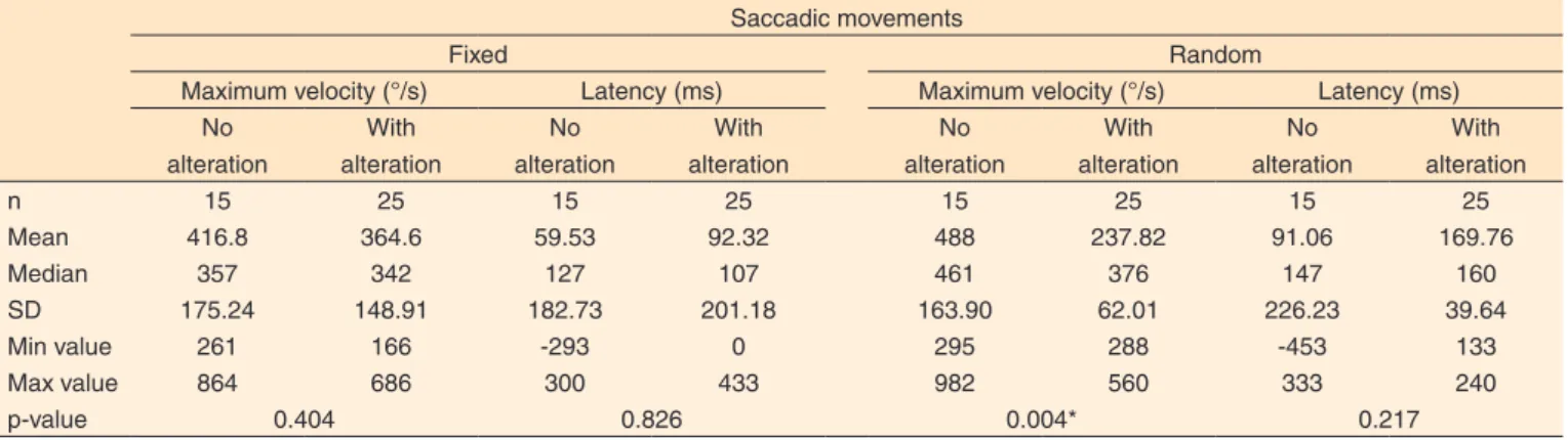
As regards the fixed saccadic movements in individuals with and without visual alterations, no difference was found in relation to the maximum velocity or latency of the movements. However, in the random saccadic movements, we verified that the maximum velocity was higher in individuals without visual alterations (p=0.004).
DISCUSSION
We observed that spontaneous nystagmus with eyes closed was present only in the horizontal direction, with a maximum AVSC of up to 5°/s in 17.5% of the sample; such data are in concordance with the literature, which shows that in normal individuals, the SNEC is equal to or lower than 6°/s(6). A study
conducted with 32 adults without vestibular disorders verified the presence of spontaneous nystagmus in ten participants (31.25%)(4). Another study, also including adults without
vestibular disorders, observed the presence of spontaneous nystagmus in five (12.5%) of 40 participants, including highest AVSC values of 6°/s(13). The presence of SNEC may be related
to the inability of the vestibular system in completely stabilizing the position of the eyes in the absence of visual support, which is deemed to be physiological(14).
Regarding the direction of the nystagmus, a previous stu-dy(13) has found that there was a predominance of horizontal
nystagmus, present in four of the five participants, which also occurred in this study(13). With the aim of assessing the
in-fluence of SNEC in other VENG tests in patients with chronic peripheral vestibulopathy, a national study also observed that the presence of SNEC was prevalent in the horizontal direc-tion(15). Of the seven patients with SNEC, five (71.42%) had
horizontal nystagmus to the left and two (28.58%) to the right, which are results that differ from previous studies(13,15). One of
these studies(13) verified the presence of SNEC in five of the
participants, four of whom had horizontal nystagmus to the right (80%), while the fifth participant (20%) had an oblique nystagmus to the right and upward.
In the optokinetic nystagmus test (Table 1), no difference was found in the AVSC values of nystagmus to the right and to the left and the DPN, when comparing the three age groups. The directional preponderance of nystagmus was 16%. These results were expected, since individuals without alterations present a symmetry of the results and the DPN of up to 16%(16).
A previous study conducted with individuals without alterations also found symmetry in the optokinetic nystagmus test(4).
The age difference did not influence the parameters analyzed in the optokinetic nystagmus test, which differs from a study conducted in adults aged 22 to 82 years, which found that the velocity decreases as age increases(8). However, we may
infer that the age variation analyzed in this study was lower than that necessary to affect the analyzed parameter.
A detailed study of the optokinetic nystagmus test is impor-tant, since the alterations found included asymmetries between the sides of the nystagmus, or alterations in velocity and gain. These alterations indicate a central compromise, or may be present in vestibular dysfunctions(5,17). Therefore, the findings
in this test, during vestibular examination, may assist in the early diagnosis of possible central alterations.
Regarding saccadic movements (Table 2), we verified that the age factor did not affect the velocity of ocular movement, nor its latency. International research comparing the latency of young individuals (18 to 37 years of age) with those of indivi-duals with advanced age (59 to 87 years of age) has observed that the latter had higher latency(18).
Another study has shown the relationship of the latency of saccadic movements in relation to age. The sample comprised individuals aged 5 to 79 years and the results showed that individuals between the ages of 18 and 22 years had a shorter latency time in relation to younger and older subjects. The study also compared a group with individuals aged 20 to 40 years with a group of individuals aged 60 to 80 years and observed
Table 4. Comparison of maximum velocity (°/s) and latency (ms) of the fixed and random saccadic movements according to the variable presence or absence of visual alteration
Saccadic movements
Fixed Random
Maximum velocity (°/s) Latency (ms) Maximum velocity (°/s) Latency (ms)
No alteration
With alteration
No alteration
With alteration
No alteration
With alteration
No alteration
With alteration
n 15 25 15 25 15 25 15 25
Mean 416.8 364.6 59.53 92.32 488 237.82 91.06 169.76
Median 357 342 127 107 461 376 147 160
SD 175.24 148.91 182.73 201.18 163.90 62.01 226.23 39.64
Min value 261 166 -293 0 295 288 -453 133
Max value 864 686 300 433 982 560 333 240
p-value 0.404 0.826 0.004* 0.217
*Significant value (p≤0.05) – Mann-Whitney test
that as age increases, movement latency becomes greater(19).
In the same study, we carried out an analysis of the age of the patients in the same group and observed that the latency alteration in relation to age was higher in the group of indivi-duals aged 60 to 80 years when compared with the variations in the group of individuals aged 20 to 40 years. This shows that, when groups with similar age variations are compared among themselves, the group with more advanced age has a greater alteration in relation to the latency of saccadic movements(19).
A 2005 study showed that the latency of saccadic move-ments was also higher in children when compared with adults(3).
These data show the relevance of studies examining the influen-ce of age in the various age groups, sininfluen-ce reliable parameters of normality are essential during clinical assessments.
Still, regarding the analysis of the saccadic movement test, we were able to observe in the present study a large variation between the maximum and minimum values of the parame-ters of maximum velocity and latency in the same group. The variation in latency was also observed in a study performed in children, in which the authors attributed these findings to an attention deficit at the time of the test(3).
In this study, our aim was not to compare the latency of fixed and random movements, but we observed (Table 2) that the maximum value of the latency of fixed movements was higher than that of random movements; this may be related to the velocity of the stimulus, which was higher in random movements (1 Hz), i.e., when the stimulus is faster, the ocular
movement must also be faster, requiring a shorter latency from the patient.
In the pendular tracking test (Figure 1), we performed a qualitative analysis, because the equipment software did not allow for a quantitative one. The research participants did not present type III or type IV tracking. According to the literature, type I and type II tracking are found in individuals without alterations, while type III tracking is found in individuals with peripheral or central vestibulopathy, and type IV tracking is found in patients with central alterations(11).
As noted, type I and II tracking had an equal distribution in group 1; however, in group 2 we were able to observe that the presence of type I tracking was greater than type II tracking, and in group 3, composed of individuals with a higher age range, there was a predominance of type II tracking (55.55%) over type I tracking. We may infer that individuals with a more advanced age have greater irregularity in tracking than younger individuals when performing a qualitative analysis. We found no studies analyzing the pendular tracking test qualitatively on the basis of age. However, a previous study with individuals without vesti-bular alterations aged 22 and 82 years has verified the influence of age on the test using a quantitative analysis, in which older individuals had a lower velocity during slow eye movements(8).
Finally, we analyzed the effect of visual alterations in eye movements. We did not perform a separate analysis for each type of ametropia due to the small number of participants,
in addition to the fact that some of them had more than one alteration.
When performing the analysis of optokinetic movements in groups with and without visual alterations, we were able to ob-serve that the AVSC showed no difference between the groups,
i.e., the visual alteration did not interfere with the movement.
However, when analyzing the DPN between groups, we obser-ved that it was higher in the group with visual alterations, i.e.
individuals with visual alterations displayed a lower symmetry than those without visual alterations.
In the analysis of the fixed and random saccadic movement test, a difference between groups with and without visual altera-tions was only present for the maximum velocity parameter. In addition, we found that the average latency was shorter for the group without visual alteration (91.06 ms) than for the group with visual alteration (169.76 ms). We may infer that indivi-duals without visual alterations presented a higher saccadic movement velocity, with a shorter latency when the stimulus was not predictable, suggesting a better oculomotor movement.
Based on the results of the study, we observed that even without changing the normality standard there are differences between individuals with and without visual alterations, even with the correction of these alterations through corrective len-ses. This fact emphasizes the importance of studies analyzing the effect of visual alterations on oculomotor tests, with and without the use of corrective lenses, allowing for the definition of normal standards in relation to these variables and contribu-ting to the assessments that do not allow for the use of corrective lenses, as in the case of videonystagmography.
This study is aligned with the literature on the importance of oculomotor movement assessment through computerized vectoelectronystagmography as it has a greater sensitivity in the detection of oculomotor alterations that may suggest central disorders(6,20). With the various analyses that may be performed
by VENG, it is necessary that more studies be carried out in order to establish normal parameters with respect to age va-riation, presence of visual alterations, as well as the analysis of the behavior of the oculomotor system before the variation of velocity and stimuli, increasing at each time the sensitivity of the assessment.
It is essential for professionals who perform vestibular tests to know the parameters of normality. Nonetheless, these parameters may be influenced by some factors, such as age, visual alterations, and stimulus velocity. Therefore, the pro-fessional should be attentive to the disorders that these factors may cause, so that they may interpret test results correctly and not only regarding the normality values, which may also vary between equipment.
CONCLUSION
influenced a few parameters of the oculomotor tests. In the optokinetic nystagmus test, the DPN was higher in individuals with alterations. In the saccadic movement test, individuals without visual alterations displayed a higher maximum velocity.
REFERENCES
1. Albertino S, Albertino RS. Reabilitação vestibular. Revista HUPE. 2012;11(3):42-7.
2. Yacovino DA. Neurociência dos movimentos oculares no envelhecimento e nas doenças neurológicas. In: Maia FCZ, Albernaz PLM, Carmona S, editores. Otoneurologia atual. Rio de Janeiro: Revinter; 2014. p. 53-68.
3. Mezzalira R, Neves LC, Maudonnet OAQ, Bilécki MMC, Ávila FG. Oculomotricidade na infância: o padrão de normalidade é o mesmo do adulto? Rev Bras Otorrinolaringol. 2005;71(5):680-5. http:// dx.doi.org/10.1590/S0034-72992005000500021
4. Costa KCF, Silva SMR, Ganança CF. Estudo das provas oculomotoras e vestibulares por meio da vectonistagmografia digital. Distúrbios Comun. 2005;17(3):315-22.
5. Ganança MM, Caovilla HH, Ganança FF. Eletronistagmografia versus videonistagmografia. Braz J Otorhinolaryngol. 2010;76(3):399-403. http://dx.doi.org/10.1590/S1808-86942010000300021
6. Ganança MM, Caovilla HH, Munhoz MSL, Silva GLM, Frazza MM. As etapas da equilibriometria. In: Caovilla HH, Ganança MM, Munhoz MSL, Silva GLM. Equilibriometria clínica. São Paulo: Atheneu; 1999. (Série Otoneurológica, vol. 1). p. 41-114.
7. Tuma VC, Ganança CF, Ganança MM, Caovilla H. Avaliação oculomotora em pacientes com disfunção vestibular periférica. Rev Bras Otorrinolaringol. 2006;72(3):407-13. http://dx.doi.org/10.1590/ S0034-72992006000300019
8. Simons B, Büttner U. The influence of age on optokinetic nystagmus. Eur Arch Psychiatr Neurol Sci. 1985;234(6):369-73. http://dx.doi. org/10.1007/BF00386053
9. Lloyd LL, Kaplan H. Audiometric interpretation: manual of basic audiometry. Baltimore: University Park Press; 1978.
10. Silmam S, Silvermam CA. Auditory diagnosis: principles and applicacation. San Diego: Singular; 1997. Chapter 2, Basic audiologic testing; p. 10-70.
11. Costa JLR. Estudo da função do sistema vestibular em mulheres com disfunção temporomandibular [dissertação]. Taubaté: Universidade de Taubaté; 2010.
12. Barros EV, Dias VG. Incidência das ametropias no hospital universitário em Campo Grande (MS) entre 1996 e 1998. Arq Bras Oftalmol. 2000;63(3):203-8. http://dx.doi.org/10.1590/S0004-27492000000300006
13. Braga HM, Ito YI, Falsetti HCDF, Caovilla HH, Novo NF et al. Nistagmo espontâneo e semi-espontâneo a vecto-electronistagmografia em individuos normais. Rev Bras Otorrinolaringol. 1981;47(2):127-40.
14. Munaro G, Sleifer P, Pedroso FS. Análise da influência do nistagmo espontâneo e pré-calórico na vectoeletronistagmografia. Rev CEFAC. 2009;11(2):331-7. http://dx.doi.org/10.1590/S1516-18462009000200019
15. Shin E, Manso A, Ganança CF. Influência do nistagmo espontâneo de olhos fechados na vectonistagmografia computadorizada em pacientes com vestibulopatias periféricas crônicas. Arq Int Otorrinolaringol. 2010;14(2):167-73. http://dx.doi.org/10.7162/ S1809-48722010000200004
16. Gonçalves DU, Ganança FF, Bottino MA, Greters ME, Ganança MM, Mezzalira R et al. Otoneurologia clínica. Rio de Janeiro: Revinter; 2014. Capítulo 2, Avaliação clínica; p. 51-96.
17. Franco ES, Panhoca I. Pesquisa da função vestibular em crianças com queixa de dificuldades escolares. Rev Bras Otorrinolaringol. 2008;74(6):815-25. http://dx.doi.org/10.1590/ S0034-72992008000600003
18. Abel LA, Troost BT, Dell’Osso LF. The effects of age on normal saccadic characteristcs and their variability. Vision Res. 1983;23(1):33-7. http://dx.doi.org/10.1016/0042-6989(83)90038-X 19. Munoz DP, Broughton JR, Golgring JE, Armstrong IT. Age-related
performance of human subjects on saccadic eye movement tasks. Exp Brain Res. 1998;121(4):391-400. http://dx.doi.org/10.1007/ s002210050473